ABSTRACT
Interleukin 18 (IL-18) plays an important role in the T-helper-cell type 1 immune response against intracellular parasites, bacteria and viral infections. It has been widely used as an adjuvant for vaccines and as an anticancer agent. However, IL-18 protein lacks a typical signal sequence and requires cleavage into its mature active form by caspase 1. In this study, we constructed mammalian expression vectors carrying cDNA encoding mature canine IL-18 (cIL-18) or mouse IL-18 (mIL-18) fused to the human IL-2 (hIL-2) signal sequence. The expressed proIL-18 proteins were processed to their mature forms in the cells. The supernatants of cells transfected with these plasmids induced high interferon-γ production in canine peripheral blood mononuclear cells or mouse splenocytes, respectively, indicating the secretion of bioactive IL-18. Using reverse genetics, we also generated a recombinant canine distemper virus that expresses cIL-18 or mIL-18 fused to the hIL-2 signal sequence. As expected, both recombinant viruses produced mature IL-18 in the infected cells, which secreted bioactive IL-18. These results indicate that the signal sequence from hIL-2 is suitable for the secretion of mature IL-18. These recombinant viruses can also potentially be used as immunoadjuvants and agents for anticancer therapies in vivo.
Keywords: canine distemper virus, interferon-γ, interleukin 18
Canine distemper virus (CDV) is an enveloped, negative-sense, single-stranded RNA virus of the genus Morbillivirus in the family Paramyxoviridae, which causes a highly contagious and fatal multisystemic infection in domestic and wild carnivores [26]. CDV has ideal features for a live vaccine, because it not only induces an innate immune response, but also induces an adaptive immune response, which elicits life-long immunity in animals. In the 1950s, live attenuated CDV vaccines were introduced to prevent and reduce the incidence of canine distemper (CD) in susceptible animals. However, severe clinical CD outbreaks in immunized dogs have recently been documented [4, 16, 21]. Therefore, it is very important to improve the effects of the current vaccine. The production of highly neutralizing antibodies has always been used as a substitute marker to evaluate the immunogenicity and protective capacity of CDV vaccines, however, the induction of cell-mediated immunity has also been shown to be important for successful vaccination [1, 11, 15, 31].
IFN-γ is an important activator of macrophages and is critical for innate and adaptive immunity against viral and intracellular bacterial infections and for tumor control. IFN-γ is produced by NK cells and by natural killer T cells, CD4+ Th1 cells and CD8+ cytotoxic T lymphocytes, once antigen-specific immunity develops [33]. IL-12 and IL-18 are potent activators of IFN-γ production in NK and T cells and promote the development of T-helper type 1 responses [8, 39]. IL-12 has been extensively tested for its adjuvant activity. Increasing the dose of recombinant human IL-12 in a human cytomegalovirus (CMV) vaccine increased the anti-CMV gB IgG titer and CMV-specific CD4+ T-cell proliferation [17]. DNA expressing IL-12 has also been shown to be an effective adjuvant for the simian immunodeficiency virus gag pDNA vaccine in rhesus macaques [32]. However, IL-12 is encoded by 2 separate genes, IL12A(p35) and IL12B(p40), requiring it to be cloned and expressed as 2 separate subunits in an in vitro study. It has been reported that IL-18 can also augment the IFN-γ-inducing capacity and antitumor activity independently of IL-12 [12]. To date, IL-18 has been used as a vaccine adjuvant and enhanced vaccine efficiency in mouse, cats, pigs and other mammals [20, 25, 28].
IL-18 is synthesized as a biologically inactive precursor protein (proIL-18, 22 kDa) by cells of the immune system and other types of cells [12]. ProIL-18 lacks a typical signal sequence for secretion [27] and is processed into its mature active form (mature IL-18, 18 kDa) by caspase-1 cleavage. Without the caspase-1 cleavage process, the mature IL-18 protein cannot be efficiently secreted across the cell membrane. Several reports have indicated that the addition of a signal sequence derived from various secreted proteins enhances the extracellular secretion of bioactive IL-18 [14, 24, 29]. It has been reported that the fusion of the canine IL12-p40 signal sequence (cIL-12p40ss) to mature canine IL-18 (cIL-18) enhanced the secretion efficiency of bioactive IL-18 [37].
In the present study, we compared the secretion efficiency of signal sequence for bioactive cIL-18. We also generated a recombinant CDV (rCDV) expressing mature IL-18 as a new potential candidate vaccine adjuvant or anticancer reagent [34]. Interestingly, a recent study demonstrated that rCDV expressing rabies virus (RABV) G protein (RV-G) protected both dogs and mice against RABV challenge [39], even though mice are not a susceptible natural host of CDV. Therefore, we also tested mouse IL-18 in a similar manner.
MATERIALS AND METHODS
Cells and viruses: B95a (marmoset lymphoblastoid) cells [19] were maintained in RPMI 1640 medium (Gibco, Carlsbad, CA, U.S.A.), and HEK293 and 293T cells were cultured in Dulbecco’s modified Eagle’s medium (Sigma, St. Louis, MO, U.S.A.) supplemented with 5% FCS (Sigma), 100 U/ml penicillin G and 100 µg/ml streptomycin (Invitrogen, Carlsbd, CA, U.S.A.). The Yanaka strain of CDV [10] and the rCDVs were cultured with B95a cells in RPMI 1640 medium supplemented with 2% FCS. Canine PBMCs were isolated with Ficoll–Paque Plus (GE Healthcare, Amersham, U.K.), according to the manufacturer’s instructions, from freshly drawn venous blood anticoagulated with 0.2 M EDTA, and maintained in RPMI 1640 medium supplemented with 10% FCS. Mouse spleen cells were prepared with a standard method and were cultured in RPMI 1640 medium supplemented with 10% FCS.
Mammalian expression plasmids: Canine PBMCs and mouse splenocytes were lysed with Isogen reagent (Nippon Gene, Tokyo, Japan) for RNA extraction, and the total RNAs isolated were reverse transcribed with SuperScript II Reverse Transcriptase (Invitrogen) and a random 6-mer primer. To construct a mammalian expression plasmid encoding the entire cIL-18, the full-length cIL-18 cDNA was amplified by PCR using canine PBMC cDNA, LA-Taq DNA polymerase (TaKaRa, Otsu, Japan) and a primer pair with SacI restriction sites (SacI-cIL18 F and SacI-cIL18 R, Table 1). This was then inserted into the pCAGGS mammalian expression vector to generate pCAG–cIL18. To construct a mammalian expression plasmid encoding mature cIL-18 fused to the cIL12-p40 signal sequence, the cIL12-p40 cDNA was amplified by PCR from canine PBMC cDNA with a primer pair with AflII and KpnI restriction sites (AflII-cIL12p40 F and KpnI-cIL12p40 R, Table 1) and inserted into the pCAGGS vector, generating pCAG–cIL12p40. The cDNA of mature cIL-18, encoding amino acids 37–193, was amplified by PCR using a primer pair with NdeI and KpnI restriction sites (NdeI-mature-cIL18F and KpnI-mature-cIL18R, Table 1). pCAG–cIL12p40 was digested with NdeI immediately downstream from the cIL12-p40 signal sequence, and with KpnI, and then introduced the mature cIL-18 cDNA digested with NdeI and KpnI, generating pCAG–cIL12ss–cIL18. To construct a mammalian expression plasmid encoding mature cIL-18 or mIL-18 fused to the human IL-2 signal sequence, cDNA encoding mature cIL-18 or mature mIL-18 (amino acids 36–192) was amplified by PCR using primer pairs with NcoI and BglII restriction sites: for cIL-18, NcoI-mature-cIL18 F and BglII-mature-cIL18 R; for mIL-18, NcoI-mature-mIL18 F and BglII-mature-mIL18 R (Table 1). The cDNAs were digested with NcoI and BglII and inserted into the restriction sites in the eukaryotic expression vector pFuse–hIgG2–Fc2 (InvivoGen, San Diego, CA, U.S.A.), which contains the hIL-2 signal sequence for the secretion of fused proteins, generating pFuse–hIL2ss–cIL18 and pFuse–hIL2ss–mIL18.
Table 1. Primers used for PCR amplification.
| Name | Primer sequences |
|---|---|
| SacI-cIL18 F | 5′-GAGCTCATGGCTGCTAACCTAATAGAAG-3 |
| SacI-cIL18 R | 5′-GAGCTCATAGAACGGTTGGTCGGATG-3′ |
| AflII-cIL12p40 F | 5′-CCTTAAGATGCATCCTCAGCAGTTGGTCATCTCC-3′ |
| KpnI-cIL12p40 R | 5′-GGGTACCCTAACTGCAGGACACAGATGCCCAGTC-3′ |
| NdeI-mature-cIL18 F | 5′-GCATATGGTACTTTGGCAAGCTTGAACCTAAAC-3′ |
| KpnI-mature-cIL18 R | 5′-GGTACCCTAGCTCTTGTTTTGAACAGTGAAC-3′ |
| NcoI-mature-cIL18 F | 5′-CCATGGCCTACTTTGGCAAGCTTGAACCTAAAC-3′ |
| BglII-mature-cIL18 R | 5′-AGATCTCTAGCTCTTGTTTTGAACAGTGAAC-3′ |
| NcoI-mature-mIL18 F | 5′-ATAGCCATGGCTAACTTTGGCCGACTTCAC-3′ |
| BglII-mature-mIL18 R | 5′-GGGGAGATCTCTAACTTTGATGTAAGTTAGTG-3′ |
| FseI-cIL18-CDV F | 5′-GGCCGGCCaaactcattataaaaaacttagggctcaggtagtccaacaATGGCTGCTAACCTAATAGAAG-3′ |
| FseI-cIL18-CDV R | 5′-GGCCGGCCTCTACTAGCTCTTGTTTTGAACAGTG-3′ |
| FseI-hIL2ss-CDV F | 5′-GGCCGGCCTCTaaactcattataaaaaacttagggctcaggtagtccaacaATGTACAGGATGCAACTCCT-3′ |
| FseI-hIL2ss-cIL18-CDV R | 5′-GGCCGGCCCTAGCTCTTGTTTTGAACAGTGAAC-3′ |
| FseI-hIL2ss-mIL18-CDV R | 5′-GGCCGGCCCTAACTTTGATGTAAGTTAGTG-3′ |
| CDV-N F | 5′-TGGTTGGTGATCCGAAAATCAACGGACC-3′ |
| CDV-N R | 5′-CCCTCCCATGGAGTTTTCAAGTTCAACACC-3′ |
Restriction sites are underlined. CDV transcription signal unit sequences are described in lowercase letters.
Generation of rabbit polyclonal antibody against cIL-18: The cDNA of cIL-18 was cloned into pET42 (b) E. coli expression vector (Novagen, Darmstadt, Germany) in frame with C-terminal of glutathione-s-transferase (GST) (pET42-cIL18). A 1-liter culture of E. coli (BL21 strain) transformed with pET42-cIL18 was incubated at 37°C. When the optical density at 600 nm (OD600) reached 0.4, expression of the recombinant protein was induced by the addition of 1 mM isopropyl-β-D-thiogalactopyranoside. After a 4-hr incubation, the E. coli was washed with PBS, suspended with sonication buffer (0.5% Triton X-100, 50 mM Tris-HCl [pH 8.0], 1 mM EDTA and 10 mM dithiothreitol) and sonicated with Sonifier450 (Branson, North Olmsted, OH, U.S.A.) for 5 min on ice. The lysate was incubated with 0.2 mg/ml lysozyme (Wako, Osaka, Japan), 10 µg/ml DNase I (Roche, Mannheim, Germany) and 1 mM MgCl2 for 45 min at room temperature. The lysate was added with 7 mM EDTA, incubated for 30 min at 37°C and then centrifugation at 12,000 × g for 20 min. The pellet containing purified inclusion bodies of GST-cIL18 fusion protein (2.5 mg) was mixed with complete Freund’s adjuvant (Difco, Sparks, MD, U.S.A.) and immunized into rabbit. After 2 weeks, the rabbit was immunized with 2.5 mg of GST-cIL18 mixed with incomplete Freund’s adjuvant (Difco). After 2 weeks, the serum was collected. The specificity of the antiserum against cIL-18 was confirmed by immunoblotting (data not shown).
Immunoblotting: 293T cells were transfected with the mammalian expression plasmids using FuGENE®6 Transfection Reagent (Invitrogen), according to the manufacturer’s instructions, and each cell lysate was subjected to immunoblotting analysis. Briefly, the lysates were separated with 12% SDS-PAGE, and the separated proteins were transferred onto an Immobilon-P membrane (Millipore, Billerica, MA, U.S.A.). The membrane was blocked with Block ACE reagent (DS Pharma Biomedical, Osaka, Japan) and then incubated with rabbit anti-cIL-18 antibody, rabbit anti-mIL-18 antibody (Invitrogen) or mouse anti-glyceraldehyde-3-phosphate dehydrogenase (GAPDH) monoclonal antibody (GE Healthcare) overnight at 4°C. The membrane was washed 3 times with 0.05% Tween-PBS and incubated with a 1:2,000 dilution of horseradish-peroxidase-conjugated goat anti-rabbit immunoglobulin antibody or rabbit anti-mouse immunoglobulin antibody (Dako Cytomation, Glostrup, Denmark) for 1 hr at 37°C. The immunoreactive bands were detected with ECL Prime Western Blotting Detection Reagent (GE Healthcare). Chemiluminescence was scanned with a luminescent image analyzer (LAS-1000UV Minisystem; Fujifilm, Tokyo, Japan).
Recovery of recombinant viruses: To generate rCDV, we used reverse genetics to recover the recombinant virus previously established in Paramyxovirus [3, 23]. Plasmid pCDV containing the full-length cDNA of the RNA genome of the CDV Yanaka strain was constructed previously [10]. The entire coding region of cIL-18, hIL-2ss–cIL-18 and hIL-2ss–mIL-18 was amplified with primer pairs containing the CDV transcription signal unit and the FseI restriction site: for entire cIL-18, FseI-cIL18-CDV F and FseI-cIL18-CDV R; for hIL-2ss–cIL-18, FseI-hIL2ss-CDV F and FseI-hIL2ss-cIL18-CDV R; and for hIL2ss-mIL18, FseI-hIL2ss-CDV F and FseI-hIL2ss-mIL18-CDV R (Table 1). The PCR products were inserted into the FseI site in pCDV. The resulting full-genome plasmids were used to generate rCDVs with reverse genetics, as previously described [10]. In brief, HEK293 cells infected with vaccinia virus encoding T7 RNA polymerase were transfected with the full-genome plasmid, together with expression plasmids encoding rinderpest virus nucleoprotein (N), phosphoprotein (P) and large protein (L) (pKS–N, pKS–P and pGEM–L, respectively), using FuGENE®6 Transfection Reagent (Invitrogen). Two days later, transfected HEK293 cells were overlain with B95a cells. Syncytia were observed approximately 10 days later. The viruses were harvested, and their titers were determined with the limiting dilution method and expressed as the 50% tissue culture infective dose (TCID50). To confirm IL-18 mRNA expression, the viral RNAs were extracted with Isogen reagent and reverse transcribed. PCR amplification was performed using the N gene primers (CDV-N F and CDV-N R, Table 1) or the cIL-18 or mIL-18 primers described above. Protein expression was confirmed with an immunoblotting analysis, as described above, using rabbit anti-N antibody [13].
Growth kinetics analysis: Monolayers of B95a cells in a 24-well plate were infected with virus at a multiplicity of infection of 0.01. The infected cells and their supernatants were harvested daily, and the titers of the released viruses and cell-associated viruses were determined as TCID50 using standard methods.
In vitro bioassay for IL-18: Supernatants (100 µl) prepared from 293T cells transfected with the mammalian expression plasmids or B95a cells infected with CDVs were added to 105 canine PBMCs in 100 µl of culture medium in a 96-well plate. After 48 hr, the supernatant from each well was examined for canine IFN-γ with an ELISA (Quantikine Canine IFN-γ Immunoassay; R&D Systems, Minneapolis, MN, U.S.A.), according to the manufacturer’s instructions. Alternatively, 50 µl of the supernatant was added to 106 mouse spleen cells in 50 µl of culture medium. After 48 hr, IFN-γ secretion was detected with the Mouse IFN-gamma ELISpot Kit (R&D Systems), and the frequency of mouse IFN-γ-secreting cells was quantified according to the manufacturer’s instructions.
RESULTS
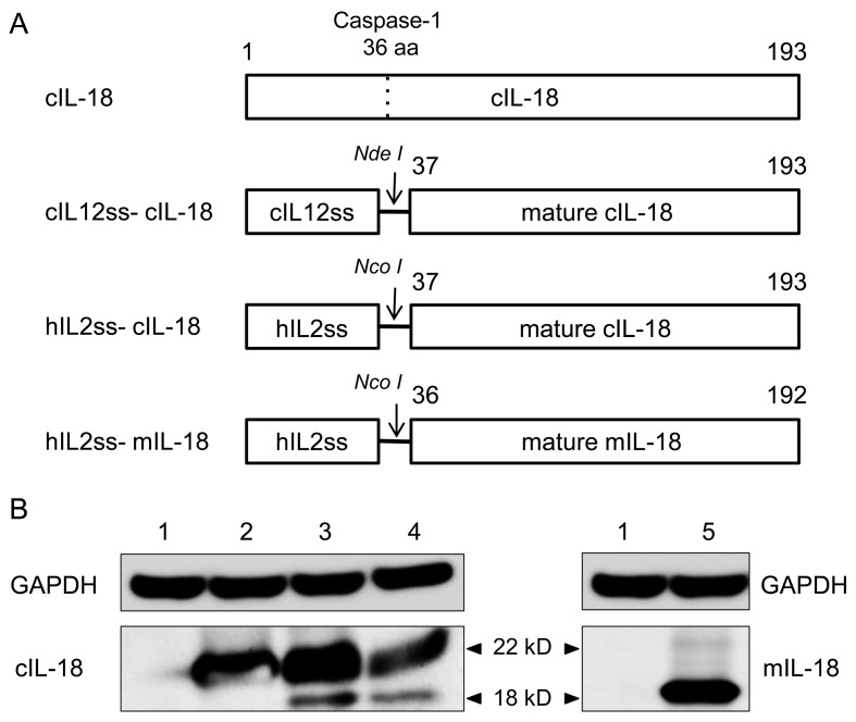
Bioactivity of recombinant IL-18 with different signal sequences in mammalian cells: First, we compared the secretion efficiency of the signal sequence with that of mature cIL-18. 293T cells were transfected with recombinant plasmids encoding entire cIL-18 (pCAG–cIL18) or mature cIL-18 fused to the cIL-12 signal sequence (pCAG–cIL12ss–cIL18) or to the human IL-2 signal sequence (pFuse–hIL2ss–cIL18) (Fig. 1A). The expression of cIL-18 protein in the cells was detected with an immunoblotting analysis. The proIL-18 (22 kDa) protein was detected in all the transfected cells, whereas mature cIL-18 (18 kDa) was detected in the cells transfected with pCAG–cIL12ss–cIL18 or pFuse–hIL2ss–cIL18, but not in cells transfected with pCAG–cIL18 (Fig. 1B).
Fig. 1.
Mammalian expression plasmids for canine and mouse IL-18. (A) Schematic representation of IL-18 constructs. From top to bottom: full-length sequence of canine IL-18, mature canine IL-18 fused to the canine IL-12 signal sequence, mature canine IL-18 fused to the human IL-2 signal sequence and mature mouse IL-18 fused to the human IL-2 signal sequence were inserted into the pCAGGS and pFuse–hIgG2–Fc2 vectors. (B) The expression of IL-18 by transfected 293T cells was analyzed with immunoblotting using anti-GAPDH, anti-cIL-18 and anti-mIL-18 antibodies. 1. Mock; 2. pCAG–cIL18; 3. pCAG–cIL12ss–cIL18; 4. pFuse–hIL2ss–cIL18; 5. pFuse–hIL2ss–mIL18.
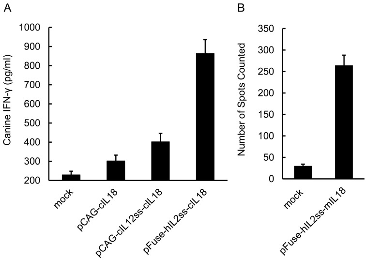
To examine the bioactivity of the cIL-18 secreted from the transfected cells, the culture supernatant was cocultured with canine PBMCs, and the amount of canine IFN-γ produced by the PBMCs was measured. The supernatants of the cells expressing entire cIL-18 induced little IFN-γ production compared with the control, whereas the supernatants of cells expressing cIL-12ss–cIL-18 slightly enhanced the induction efficiency, as in the previous report [37]. Interestingly, the supernatant of cells transfected with pFuse–hIL2ss–cIL18 induced significant IFN-γ production (Fig. 2A). This result suggests that the human IL-2 signal sequence is more effective for expressing mature IL-18 in vitro than the canine IL12-p40 signal sequence. We then used hIL-2ss to express bioactive mouse IL-18 (Fig. 1A). We only detected mature mIL-18 in the pFuse–hIL2ss–mIL18-transfected cells (Fig. 1B). The supernatant from cells transfected with this plasmid also effectively stimulated mouse spleen cells to secrete IFN-γ (Fig. 2B). These results indicate that the human IL-2 signal sequence is suitable for the secretion of bioactive IL-18 protein from cells.
Fig. 2.
Induction of IFN-γ by incubating canine or mouse immune cells with the supernatant from transfected cells. (A) The supernatant from transfected 293T cells or mock-transfected 293T cells was cocultured with canine PBMCs. After 48 hr, the supernatant was examined for IFN-γ production with an ELISA. Three independent experiments were performed (n=3). (B) The supernatant from transfected 293T cells or mock-transfected 293T cells was cocultured with mouse spleen cells in a polyvinylidene difluoride (PVDF)-backed microplate coated with monoclonal antibody specific for mouse IFN-γ in a humidified 37°C incubator for 48 hr. The supernatant was examined for IFN-γ production by counting the individual blue–black spots under a stereomicroscope. Three independent experiments were performed (n=3).
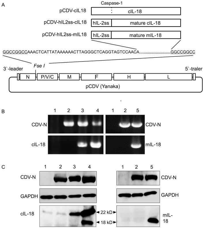
Generation of rCDVs expressing canine or mouse IL-18: To generate rCDVs expressing IL-18, we constructed CDV full-genome plasmids containing cDNA encoding the entire cIL-18, hIL-2ss–cIL-18 or hIL-2ss–mIL-18 sequence (Fig. 3A). The rCDVs were rescued from the plasmids as described in the Methods section, and we successfully generated the rCDVs. The expression of mRNA from the inserted gene in each viral genome was confirmed by RT-PCR (Fig. 3B). To identify the IL-18 proteins expressed by the viruses, infected B95a cells were subjected to an immunoblotting analysis. Two bands corresponding to proIL-18 and mature IL-18 were detected (Fig. 3C), and each sample showed a similar band pattern, corresponding to that of the mammalian expression plasmids (Fig. 1B).
Fig. 3.
Generation and in vitro characterization of rCDVs expressing mature canine or mouse IL-18 fused to the human IL-2 signal sequence. (A) Schematic model of the rCDV genome with the FseI site introduced between the N and P genes and hIL2ss–IL18 inserted at the FseI site. (B) The resulting viruses were harvested and identified by RT-PCR using primers complementary to the CDV-N gene and the canine IL-18 or mouse IL-18 gene. (C) Recombinant viruses were identified by immunoblotting analysis. Cell lysates were examined with anti-CDVN, anti-GAPDH, anti-cIL-18 or anti-mIL-18 antibody. 1: B95a; 2: parental CDV (Yanaka strain); 3: rCDV–cIL18; 4: rCDV–hIL2ss–cIL18; 5: rCDV–hIL2ss–mIL18.
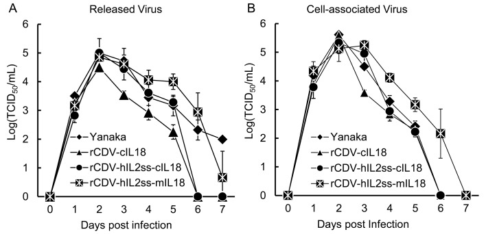
We then examined the growth kinetics of the rCDVs. As shown in Fig. 4, all the recombinant viruses showed slightly higher peaks for cell-associated virus than for released virus, and the maximum titers were similar to that of the parental Yanaka strain (Fig. 4). These results demonstrate that in B95a cells, IL18 inserted into the CDV genome and expressed from recombinant viruses had no obvious effect on viral replication or growth kinetics.
Fig. 4.
Kinetics of recombinant viruses in B95a cells. The infected cells and supernatants were harvested separately every 24 hr after infection for 7 days. The titers of the viruses released into the supernatant (A) and the cell-associated viruses (B) were determined with a TCID50 assay.
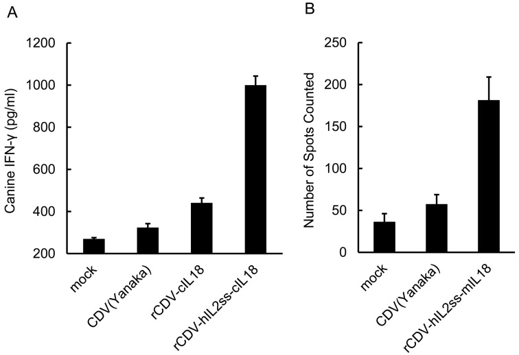
Bioactivity of IL-18 produced by rCDV: To investigate whether the mature IL-18 protein expressed by rCDV was bioactive, we harvested the supernatants of the virus-infected cells and cocultured them with canine PBMCs. The supernatant contained infectious CDV released from the cells, but the parental CDV induced little IFN-γ production in the PBMCs (Fig. 5A) because CDV infection of canine PBMCs is inefficient [10]. The supernatant from the rCDV–cIL18-infected cells induced slight IFN-γ production, and as expected, rCDV–hIL2ss–cIL18 induced significant IFN-γ production (Fig. 5A). Similarly, the supernatant from rCDV–hIL2ss–mIL18-infected cells also stimulated mouse spleen cells and induced IFN-γ production (Fig. 5B). The increased IFN-γ expression in PBMCs and splenocytes confirmed the biological activity of IL-18 secreted by rCDV–hIL2ss–cIL18- and rCDV–hIL2ss–mIL18-infected cells.
Fig. 5.
Induction of IFN-γ production in canine or mouse immune cells after coculture with supernatant harvested from different recombinant-virus-infected B95a cells. (A) B95a cells were infected with parental CDV (Yanaka strain), rCDV–cIL18 or rCDV–hIL2ss–cIL18 for 48 hr. The supernatants were then harvested and cocultured with canine PBMCs for 48 hr. The supernatants were examined for IFN-γ production with an ELISA. Three independent experiments were performed (n=3). (B) B95a cells were infected with parental CDV (Yanaka strain) or rCDV–hIL2ss–mIL18 for 48 hr. The supernatants from the infected B95a cells or mock-infected B95a cells were then cocultured with mouse spleen cells in a PVDF-backed microplate coated with monoclonal antibody specific for mouse IFN-γ in a humidified 37°C incubator for 48 hr. The supernatants were then examined for IFN-γ production by counting the individual blue–black spots under a stereomicroscope. Three independent experiments were performed (n=3).
DISCUSSION
Based on its biological activity, IL-18 has been widely used as a vaccine adjuvant for infectious diseases [5, 6, 12, 18]. Various expression systems and cytokine signal peptides have been used to express bioactive IL-18. For example, feline IL-18 fused to human immunoglobulin κ was constructed as a DNA vaccine adjuvant and enhanced the efficacy of a feline leukemia virus DNA vaccine [14]. The signal sequence from the human IL-1β receptor antagonist protein fused to mature feline or equine IL-18 induced IFN-γ production in a KG-1 assay [24]. Porcine IL-18 fused to the baculovirus gp67-encoded signal sequence displayed bioactivity in a baculovirus system [22]. In the present study, we evaluated the utility of the human IL-2 signal sequence and demonstrated that it is more efficient than the previously reported canine IL12-p40 signal sequence [37] and that it can be used to express both canine and mouse IL-18.
The human IL-2 signal sequence contains 21 amino acids and shares the common characteristics of the signal peptides of other secretory proteins. Intracellular cleavage of the IL-2 signal peptide occurs after Ser20 and leads to the secretion of the antigenic protein. The signal sequences of canine and mouse IL-2 also consist of 20 amino acids, whereas the homology of the canine IL-2 signal sequence with that of the human sequence is 80% and that of the mouse IL-2 signal sequence is only 60%. Meanwhile, the human IL-2 signal sequence derived from the plasmid we used, pFuse–hIgG2–Fc2, can function in a variety of commonly used mammalian cells, from human cells to Chinese hamster cells. Therefore, rCDV–hIL2ss–cIL18 and rCDV–hIL2ss–mIL18 can secrete functional IL-18 after undergoing the proper processing in an appropriate host. The difference in the intracellular processing efficiency of cIL-18 and mIL-18 (Figs. 1B and 3C) may be attributable to their respective primary sequences immediately downstream from the cleavage site (cIL-18: 37-YFGKLEPKLS, mIL-18: 36-NFGRLHCTTA). We will also examine the secretion efficiencies of cIL-18 and mIL-18 at protein level using ELISA in the future.
To date, divalent vaccines based on several recombinant viruses that encode CDV glycoproteins (hemagglutinin [H] and fusion [F]) have been investigated to protect against CDV and other pathogens. For example, vaccines based on recombinant vaccinia viruses or canarypox vectors engineered to express CDV glycoproteins have been tested in dogs and ferrets [30, 35]. These vaccines elicited protective immune responses, but it has yet to be determined whether the duration of these immune responses is equivalent to that of the responses induced with conventional live CDV vaccines [2]. It has been reported that 2 replication-competent canine adenovirus type 2 (CAV2)-based vaccines, expressing the CDV H and F antigens, protected dogs against CDV challenge [9]. It is well known that CAV2 vaccines are easily recovered from the respiratory tracts of sentinel dogs [7].
In the case of CDV, live attenuated vaccines are known to confer long-lasting immunity, and recombination of the CDV genome is impossible during the viral replication cycle. Therefore, the development of CDV-based multivalent vaccines is the most appropriate approach to the development of vaccines against CDV and other pathogens. A recent study demonstrated that an attenuated rCDV vaccine strain expressing RV-G conferred protective immunity against challenge with RABV in mice and dogs, providing particularly effective protection for more than a year in dogs [40]. This result confirms that CDV is a suitable vaccine vector.
Many recent studies have indicated that IL-18 plays an important role not only in host defenses against pathogens but also in immunotherapies for cancer [reviewed in 34]. Oncolytic virotherapy has been used as a novel strategy for cancer treatment. Interestingly, previous reports have shown that CDV and measles virus (MV), a human Morbillivirus, have oncolytic activity. CDV was shown to infect lymphoblasts isolated from canine lymphoma patients, leading to cell death by apoptosis [38]. In our previous study, we also demonstrated that recombinant MV, which was mutated so that it could not bind to the lymphoid-cell receptor SLAM, showed oncolytic activity against breast cancer in vivo [36]. Extrapolating from these data, we surmise that application of rCDV–hIL2ss–cIL18 plays a positive role in the cancer therapy using oncolytic activity of CDV.
In conclusion, we constructed an efficient secretion system of cIL-18 and succeeded in generating rCDV expressing bioactive IL-18. The recombinant CDV, rCDV–hIL2ss–cIL18, could offer a new approach to investigating the role of IL-18 in the host defense mechanism, the pathogenesis of infectious diseases and the treatment of cancer in the host.
Acknowledgments
This work was supported by Grants-in-Aid for Scientific Research (KAKENHI) from the Japan Society for the Promotion of Science (JSPS) and by the Global COE Program Center of Education and Research for Advanced Genome-Based Medicine: for Personalized Medicine and the Control of Worldwide Infectious Diseases (Ministry of Education, Culture, Sports, Science and Technology, Japan).
References
- 1.Appel M. J. G., Shek W. R., Shesberadaran H., Norrby E.1984. Measles virus and inactivated canine distemper virus induce incomplete immunity to canine distemper. Arch. Virol. 82: 73–82. doi: 10.1007/BF01309369 [DOI] [PubMed] [Google Scholar]
- 2.Barrett T.1999. Morbillivirus infection, with special emphasis on morbilliviruses of carnivores. Vet. Microbiol. 69: 3–13. doi: 10.1016/S0378-1135(99)00080-2 [DOI] [PubMed] [Google Scholar]
- 3.Billeter M. A., Naim H. Y., Udem S. A.2009. Reverse genetics of measles virus and resulting multivalent recombinant vaccines: applications of recombinant measles viruses. Curr. Top. Microbiol. Immunol. 329: 129–162 [DOI] [PMC free article] [PubMed] [Google Scholar]
- 4.Blixenkrone-Møller M., Svansson V., Have P., Orvell C., Appel M., Pedersen I. R., Dietz H. H., Henriksen P.1993. Studies on manifestations of canine distemper virus infection in an urban dog population. Vet. Microbiol. 37: 163–173. doi: 10.1016/0378-1135(93)90190-I [DOI] [PubMed] [Google Scholar]
- 5.Bohn E., Sing A., Zumbihl R., Bielfeldt C., Okamura H., Kurimoto M., Heesemann J., Autenrieth I. B.1998. IL-18 (IFN-gamma-inducing factor) regulates early cytokine production in, and promotes resolution of, bacterial infection in mice. J. Immunol. 160: 299–307 [PubMed] [Google Scholar]
- 6.Brummer E.1998–1999. Human defenses against Cryptococcus neoformans: An update. Mycopathologia 143: 121–125. doi: 10.1023/A:1006905331276 [DOI] [PubMed] [Google Scholar]
- 7.Curtis R., Jemmett J. E., Furminger I. G.1978. The pathogenicity of an attenuated strain of canine adenovirus type 2 (CAV2). Vet. Rec. 103: 380–381. doi: 10.1136/vr.103.17.380 [DOI] [PubMed] [Google Scholar]
- 8.Dinarello C.A., Novick N., Puren A.J., Fantuzzi G., Shapiro L., Mühl H., Yoon D.Y., Reznikov L.L., Kim S.H., Rubinstein M.1998. Overview of interleukin-18: more than an interferon-g inducing factor. J. Leukoc. Biol. 63: 658–664 [PubMed] [Google Scholar]
- 9.Fischer L., Tronel J. P., Pardo-David C., Tanner P., Colombet G., Minke J., Audonnet J. C.2002. Vaccination of puppies born to immune dams with a canine adenovirus based vaccine protects against a canine distemper virus challenge. Vaccine 20: 3485–3497. doi: 10.1016/S0264-410X(02)00344-4 [DOI] [PubMed] [Google Scholar]
- 10.Fujita K., Miura R., Yoneda M., Shimizu F., Sato H., Muto Y., Endo Y., Tsukiyama-Kohara K., Kai C.2007. Host range and receptor utilization of canine distemper virus analyzed by recombinant viruses: Involvement of heparin-like molecule in CDV infection. Virology 359: 324–335. doi: 10.1016/j.virol.2006.09.018 [DOI] [PubMed] [Google Scholar]
- 11.Ghosh S., Walker J., Jackson D. C.2001. Identification of canine helper T-cell epitopes from the fusion protein of canine distemper virus. Immunology 104: 58–66. doi: 10.1046/j.0019-2805.2001.01271.x [DOI] [PMC free article] [PubMed] [Google Scholar]
- 12.Gracie J. A., Robertson S. E., Mclnnes I. B.2003. Interleukin-18. J. Leukoc. Biol. 73: 213–224. doi: 10.1189/jlb.0602313 [DOI] [PubMed] [Google Scholar]
- 13.Hagiwara K., Sato H., Inoue Y., Watanabe A., Yoneda M., Ikeda F., Fujita K., Fukuda H., Takamura C., Kozuka-Hata H., Oyama M., Sugano S., Ohmi S., Kai C.2008. Phosphorylation of measles virus nucleoprotein upregulates the transcriptional activity of minigenomic RNA. Proteomics 8: 1871–1879. doi: 10.1002/pmic.200701051 [DOI] [PubMed] [Google Scholar]
- 14.Hanlon L., Argyle D., Bain D., Nicolson L., Dunham S., Golder M. C., McDonald M., McGillivray C., Jarrett O., Neil J. C., Onions D. E.2001. Feline Leukemia Virus DNA Vaccine Efficacy Is Enhanced by Coadministration with Interleukin-12 (IL-12) and IL-18 Expression Vectors. J. Virol. 75: 8424–8433. doi: 10.1128/JVI.75.18.8424-8433.2001 [DOI] [PMC free article] [PubMed] [Google Scholar]
- 15.Hirama K., Togashi K., Wakasa C., Yoneda M., Nishi T., Endo Y., Miura R., Tuskiyama-kohara K., Kai C.2003. Cytotoxic T-lymphocyte activity specific for hemagglutinin (H) protein of canine distemper virus in dogs. J. Vet. Med. Sci. 65: 109–112. doi: 10.1292/jvms.65.109 [DOI] [PubMed] [Google Scholar]
- 16.Iwatsuki K., Tokiyoshi S., Hirayama N., Nakamura K., Ohashi K., Wakasa C., Mikami T., Kai C.2000. Antigenic differences in the H proteins of canine distemper viruses. Vet. Microbiol. 71: 281–286. doi: 10.1016/S0378-1135(99)00172-8 [DOI] [PubMed] [Google Scholar]
- 17.Jacobson M. A., Sinclair E., Bredt B., Agrillo L., Black D., Epling C. L., Carvidi A., Ho T., Bains R., Girling V., Adler S. P.2006. Safety and Immunogenicity of Towne Cytomegalovirus Vaccine with or without Adjuvant Recombinant Interleukin-12. Vaccine 24: 5311–5319. doi: 10.1016/j.vaccine.2006.04.017 [DOI] [PubMed] [Google Scholar]
- 18.Kobayashi K., Kai M., Gidoh M., Nakata N., Endoh M., Singh R. P., Kasama T., Saito H.1998. The possible role of interleukin (IL)-12 and interferon-gamma-inducing factor/IL-18 in protection against experimental Mycobacterium leprae infection in mice. Clin. Immunol. Immunopathol. 88: 226–231. doi: 10.1006/clin.1998.4533 [DOI] [PubMed] [Google Scholar]
- 19.Kobune F., Sakata H., Sugiura A.1990. Marmoset lymphoblastoid cells as a sensitive host for isolation of measles virus. J. Virol. 64: 700–705 [DOI] [PMC free article] [PubMed] [Google Scholar]
- 20.Kong N., Wang X. H., Zhao J. W., Hu J. D., Zhang H., Zhao H. K.2011. A recombinant baculovirus expressing VP2 protein of infectious bursal disease virus (IBDV) and chicken Interleukin-18 (ChIL-18) protein protects against very virulent IBDV. J. Anim. Vet. Adv. 10: 2706–2715 [Google Scholar]
- 21.Latha D., Srinivasan S. R., Thirunavukkarasu P. S., Gunaselan L., Ramadass P., Narayanan R. B.2007. Assessment of canine distemper virus infection in vaccinated and unvaccinated dogs. Indian J. Biotechnol. 6: 35–40 [Google Scholar]
- 22.Muneta Y., Mori Y., Shimoji Y., Yokomizo Y.2000. Porcine interleukin 18: cloning, characterization of the cDNA and expression with the baculovirus system. Cytokine 12: 566–572. doi: 10.1006/cyto.1999.0648 [DOI] [PubMed] [Google Scholar]
- 23.Nagai Y.1999. Paramyxovirus replication and pathogenesis. Reverse genetics transforms understanding. Rev. Med. Virol. 9: 83–99. doi: [DOI] [PubMed] [Google Scholar]
- 24.O’Donovan L. H., McMonagle E. L., Taylor S., Argyle D. J., Nicolson L.2004. Bioactivity and secretion of interleukin-18 (IL-18) generated by equine and feline IL-18 expression constructs. Vet. Immunol. Immunopathol. 102: 421–428. doi: 10.1016/j.vetimm.2004.08.003 [DOI] [PubMed] [Google Scholar]
- 25.O’Donovan L. H., McMonagle E. L., Taylor S., Bain D., Pacitti A. M., Golder M. C., McDonald M., Hanlon L., Onions D. E., Argyle D. J., Jarrett O., Nicolson L.2005. A vector expressing feline mature IL-18 fused to IL-1 antagonist protein signal sequence is an effective adjuvant to a DNA vaccine for feline leukaemia virus. Vaccine 23: 3814–3823. doi: 10.1016/j.vaccine.2005.02.026 [DOI] [PMC free article] [PubMed] [Google Scholar]
- 26.Ohishi K., Ando A., Suzuki R., Takishita K., Kawato M., Katsumata E., Ohtsu D., Okutsu K., Tokutake K., Miyahara H., Nakamura H., Murayama T., Maruyama T.2010. Host-virus specificity of morbilliviruses predicted by structural modeling of the marine mammal SLAM, a receptor. Comp. Immunol. Microbiol. Infect. Dis. 33: 227–241. doi: 10.1016/j.cimid.2008.10.003 [DOI] [PubMed] [Google Scholar]
- 27.Okamura H., Tsutsi H., Komatsu T., Yutsudo M., Hakura A., Tanimoto T., Torigoe K., Okura T., Nukada Y., Hattori K., Akita K., Namba M., Tanabe F., Konishi K., Fukuda S., Kurimoto M.1995. Cloning of a new cytokine that induces IFN-gamma production by T cells. Nature 378: 88–91. doi: 10.1038/378088a0 [DOI] [PubMed] [Google Scholar]
- 28.Okano F., Satoh M., Ido T., Yamada K.1999. Cloning of cDNA for Canine Interleukin-18 and Canine Interleukin-1beta Converting Enzyme and Expression of Canine Interleukin-18. J. Interferon Cytokine Res. 19: 27–32. doi: 10.1089/107999099314388 [DOI] [PubMed] [Google Scholar]
- 29.Osaki T., Hashimoto W., Gambotto A., Okamura H., Robbins P. D., Kurimoto M., Lotze M. T., Tahara H.1999. Potent antitumor effects mediated by local expression of the mature form of the interferon-gamma inducing factor, interleukin-18 (IL-18). Gene Ther. 6: 808–815. doi: 10.1038/sj.gt.3300908 [DOI] [PubMed] [Google Scholar]
- 30.Pardo M. C., Bauman J. E., Mackowiak M.1997. Protection of dogs against canine distemper by vaccination with a canarypox virus recombinant expressing canine distemper virus fusion and hemagglutinin glycoproteins. Am. J. Vet. Res. 58: 833–836 [PubMed] [Google Scholar]
- 31.Salerno-Gonçalves R., Sztein M. B.2006. Cell-mediated immunity and the challenges for vaccine development. Trends Microbiol. 14: 536–542. doi: 10.1016/j.tim.2006.10.004 [DOI] [PubMed] [Google Scholar]
- 32.Schadeck E. B., Sidhu M., Egan M. A., Chong S. Y., Piacente P., Masood A., Garcia-Hand D., Cappello S., Roopchand V., Megati S., Quiroz J., Boyer J. D., Felber B. K., Pavlakis G. N., Weine D. B., Eldridge J. H., Israel Z. R.2006. A dose sparing effect by plasmid encoded IL-12 adjuvant on a SIV gag-plasmid DNA vaccine in rhesus macaques. Vaccine 24: 4677–4687. doi: 10.1016/j.vaccine.2005.10.035 [DOI] [PubMed] [Google Scholar]
- 33.Schoenborn J. R., Wilson C. B.2007. Regulation of interferon-γ during innate and adaptive immune. Adv. Immunol. 96: 41–101. doi: 10.1016/S0065-2776(07)96002-2 [DOI] [PubMed] [Google Scholar]
- 34.Srivastava S., Salim N., Robertson M. J.2010. Interleukin-18: biology and role in the immunotherapy of cancer. Curr. Med. Chem. 17: 3353–3357. doi: 10.2174/092986710793176348 [DOI] [PubMed] [Google Scholar]
- 35.Stephensen C. B., Welter J., Thaker S. R., Taylor J., Tartaglia J., Paoletti E.1997. Canine distemper virus (CDV) infection of ferrets as a model for testing morbillivirus vaccine strategies: NYVAC- and ALVAC-based CDV recombinants protect against symptomatic infection. J. Virol. 71: 1506–1513 [DOI] [PMC free article] [PubMed] [Google Scholar]
- 36.Sugiyama T., Yoneda M., Kuraishi T., Hattori S., Inoue Y., Sato H., Kai C.2013. Measles virus selectively blind to signaling lymphocyte activation molecule as a novel oncolytic virus for breast cancer treatment. Gene Ther. 20: 338–347. doi: 10.1038/gt.2012.44 [DOI] [PubMed] [Google Scholar]
- 37.Sugiura K., Akazawa T., Fujimoto M., Wijewardana V., Mito K., Hatoya S., Taketani S., Komori M., Inoue N., Inaba T.2008. Construction of an expression vector for improved secretion of canine IL-18. Vet. Immunol. Immunopathol. 126: 388–391. doi: 10.1016/j.vetimm.2008.07.011 [DOI] [PubMed] [Google Scholar]
- 38.Suter S. E., Chein M. B., von Messling V., Yip B., Cattaneo R., Vernau W., Madewell B. R., London C. A.2005. In vitro canine distemper virus infection of canine lymphoid cells: a prelude to oncolytic therapy for lymphoma. Clin. Cancer Res. 11: 1579–1587. doi: 10.1158/1078-0432.CCR-04-1944 [DOI] [PubMed] [Google Scholar]
- 39.Trinchieri G., Gerosa F.1996. Immunoregulation by interleukin-12. J. Leukoc. Biol. 59: 505–511 [DOI] [PubMed] [Google Scholar]
- 40.Wang X., Feng N., Ge J. Y., Shuai L., Peng L. Y., Gao Y. W., Yang S. T., Xia X. Z., Bu Z. G.2012. Recombinant canine distemper virus serves as bivalent live vaccine against rabies and canine distemper. Vaccine 30: 5067–5072. doi: 10.1016/j.vaccine.2012.06.001 [DOI] [PubMed] [Google Scholar]


