Figure 2.
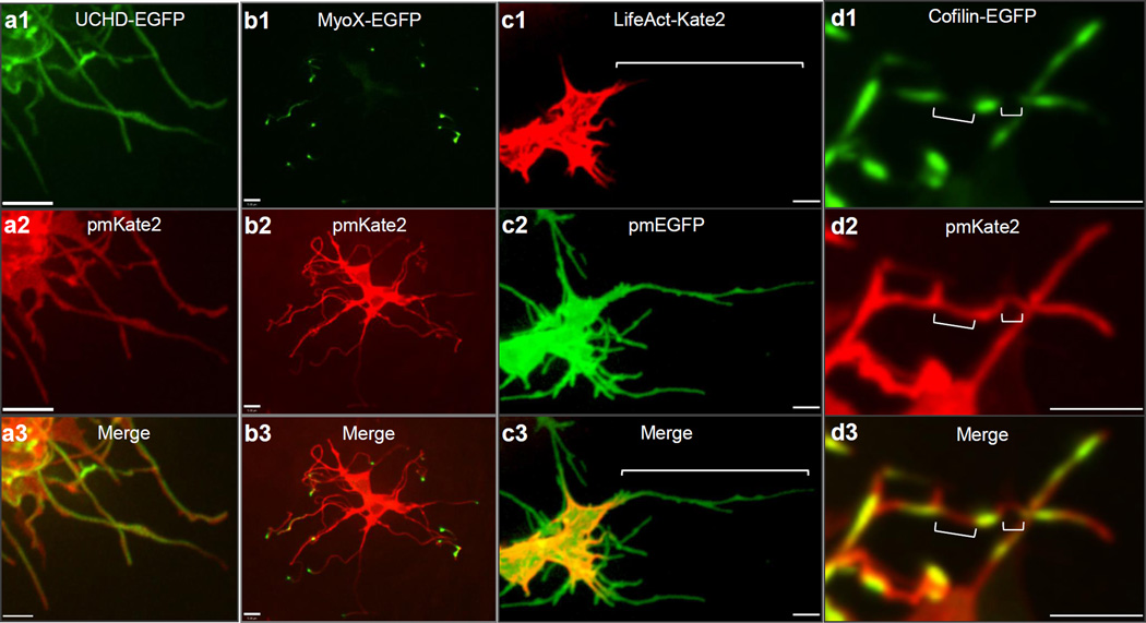
Limb mesenchymal cytoplasmic extensions are a class of specialized actin-based filopodia. (a1–3) UCHD-EGFP demonstrating that membrane labeled pmKate2 filopodia extensions contain actin filaments. Scale = 3µm. (b1–3) Myosin X-EGFP is localized to each pmKate2 labeled filopodium and is concentrated at the distal tip. (c1–3) LifeAct-Kate2 marks only the proximal aspect of pmEGFP labeled filopodia and does not label the entire extension, shown by bracket. (d1–3) Cofilin-EGFP is present in interrupted domains along the filopodia, negative regions shown with brackets. Scale = 5µm.

