

Abstract
Differentiating left and right hand sides during embryogenesis represents a major event in body patterning. Left–Right (L/R) asymmetry in bilateria is essential for handed positioning, morphogenesis and ultimately the function of organs (including the brain), with defective L/R asymmetry leading to severe pathologies in human. How and when symmetry is initially broken during embryogenesis remains debated and is a major focus in the field. Work done over the past 20 years, in both vertebrate and invertebrate models, has revealed a number of distinct pathways and mechanisms important for establishing L/R asymmetry and for spreading it to tissues and organs. In this review, we summarize our current knowledge and discuss the diversity of L/R patterning from cells to organs during evolution.
Keywords: L/R asymmetry; symmetry breaking; directional morphogenesis; evolution, invertebrates; vertebrates
Introduction
The first mutation affecting the whole body plan was isolated a century ago and was shown to invert shell coiling in a small aquatic snail (Lymnaea peregra) 1,2. Despite this early finding and important work describing genetic and cellular aspects of L/R asymmetry 3–11, the first molecular study of L/R asymmetry was described only recently, showing for the first time asymmetric expression of the nodal gene in vertebrates 12. A possible reason for this lag is the fact that in contrast to A/P and D/V asymmetries, laterality is not obvious at first sight, when looking at the external body shape, with snail shell coiling being an exception. Indeed, despite looking mostly bilaterally symmetrical, metazoa also differentiate along the “invisible” L/R axis, leading to asymmetric positioning of unique organs, such as the heart, liver and stomach, and asymmetrical morphogenesis of bilateral ones, as for example the lung and brain. In addition, L/R asymmetry controls the looping of tubular organs (heart tube, gut, and other ducts) toward one direction. Laterality is thus essential for the correct arrangement of visceral organs in the abdomen and thorax, but is also essential for the asymmetric morphogenesis, hence the function, of the heart and brain, for example. Clinical studies led to an estimation of 1/5,000–1/10,000 humans suffering from L/R defects (situs inversus, heterotaxia, and isomerism), being responsible for a number of complex congenital heart defects, misrotation of the intestine, and spontaneous miscarriage. Furthermore, L/R asymmetry defects, which often originate from ciliopathies, are associated with polycystic renal disease, Kartagener and Ivemark syndromes, and others.
L/R asymmetry is therefore essential, and outstanding questions remain to be addressed to understand how body shape and function are established during evolution. What is, or what are, the origin(s) of L/R asymmetry? Where and when does it take place in the embryo? Are there any conserved features among metazoa and how did L/R asymmetry establishment evolve in metazoa (Sidebar A)?
A specificity of L/R asymmetry is the fact that it has to be coordinated with the other two—A/P and D/V—body axes and thus is established relative to and after them as a “secondary” axis. This important notion was summarized by Brown and Wolpert in their elegant F-molecule model 13. The incremental/two-step establishment of body patterning is particularly interesting, as it implies that L/R asymmetry establishment depends on mechanisms that integrate existing 2D positional information. Over the last few years, several studies using different model organisms helped to identify unique mechanisms at play during the establishment of L/R asymmetry. Although a variety of mechanisms have been discovered, fascinating similarities between quite distant phyla are emerging. On the following pages, we will discuss the various mechanisms and synthesize common principles of L/R asymmetry establishment in vertebrates and invertebrates.
Vertebrate embryonic node and Nodal flow in L/R patterning
A well-established model for the determination of the body situs in several vertebrate species is that of the Nodal flow occurring at the late-gastrulation-neurulation stage in the mouse node and node-like structures of other animals (Posterior Notochordal Plate in rabbit, Kupffer’s Vesicle in zebrafish, Gastrocoel Roof Plate in Xenopus) 14–16.
The Nodal flow model is best described in mouse, which serves as the model paradigm; hence, we focus in the following on the description of the data obtained in mouse. The node is a transitory structure located on the ventral side of the embryo at the end of the developing notochord (Fig 1A). The node is a cavity covered by a monociliated epithelium-like monolayer of cells, which appears decisive for proper lateralization 17. Indeed, when the node cilia are missing, mice show abnormal L/R patterning with random lateralization, that is, both the normal situs solitus and the inverted situs inversus are observed. This is for instance the case in mice mutant for the Kif3A or Kif3B subunits of the kinesin-II complex, a microtubule motor essential for proper ciliogenesis and maintenance of the cilium. In these mutants, cilia fail to assemble 18,19.
Figure 1. Left/Right determination in vertebrates.
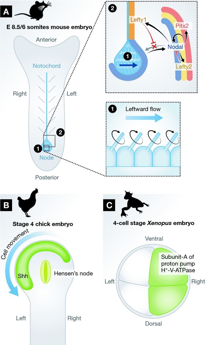
(A) Schematic depiction of a E8.5 mouse embryo. Nodal is expressed around the node. Nodal flow (i) leads to stronger expression of Nodal on the left side (ii) and in the Lateral Plate Mesoderm (LPM) where it positively regulates its own expression by a positive feedback loop. Nodal also activates expression of the homeobox transcription factor Pitx2 and of the TGF-β homologues Lefty2 and Lefty1 in the LPM near the notochord. Lefty1/2 antagonize Nodal diffusion to the right side of the embryo and ultimately shut down Nodal signaling. Pitx2 expression is self-maintained and induces left-sided morphogenesis of the LPM. (B) Schematic depiction of a stage 4 chick embryo’s primitive streak and Hensen’s node. The leftward movement of cells from the right of Hensen’s node induces the asymmetric remodffieling of the node’s morphology as well as asymmetric gene expression patterns (e.g. Shh, green) due to the intermingling of cells with different genetic programs 57,58. (C) Xenopus embryo at the 4-cell stage shows right-sided enrichment in subunit-A of the proton pump H+-V-ATPase, whose activity is necessary for proper lateralization of the animal. Interestingly, this early L/R asymmetric localization appears to be sensitive to actin but not microtubule depolymerization 60.
However, it is not merely the presence of these cilia that is important, but rather their motility. Indeed, inversus viscerum (iv) mutant mice, in which the cilia are present but immotile, show similar randomized lateralization phenotypes 20,21. iv encodes the L/R dynein, another microtubule motor essential for node cilia motility 20. Node cilia rotate clockwise, thereby producing a leftward flow of extra-embryonic fluid, which appears to determine the directionality of embryo lateralization 18,19,22,23. Cilia have been known for some time to be important for lateralization 24, but their role in the production of the Nodal flow was only recently described 18 (Fig 1A). Impairing the flow genetically (with mutant mice) or experimentally (by increasing the viscosity of the medium) leads to L/R patterning defects 25. When the node cilia are missing or immotile, the Nodal flow is abolished and the L/R situs is consequently randomized. Interestingly, the restoration of an artificially generated leftward Nodal flow is sufficient to reinstate normal L/R patterning of mutant mice 25. Conversely, in wild-type mice, superimposition of an artificial rightward Nodal flow is able to override normal patterning and leads to inversion of the axis, demonstrating the importance of the flow in this process 25.
The normal mouse node is thought to comprise between 200 and 300 motile cilia, nevertheless only a couple of them seem to be required for normal lateralization 26. This precision was achieved through thorough analysis of the phenotype of mutant mice, in which ciliogenesis was strongly impaired but that nevertheless retain some cilia at the node. This is for instance the case in mice mutant for the Rfx3 transcription factor necessary for ciliogenesis. The discovery that only two motile cilia—but not one—wherever their position in the node, were sufficient to trigger normal L/R patterning questions the sensitivity of the Nodal flow signal or the existence of a on/off effect of the flow 26. Remarkably, the generation of a small difference or initial bias between the left and right sides by the Nodal flow appears to be sufficient to be turned into robust asymmetry 27. Similar analyses of flow dynamics in various genetic conditions showed that in zebrafish, the flow generated by thirty motile cilia or more reliably predicts the future laterality of the animal 28. Interestingly, the authors revealed distinct sensitivities of different organs to the flow. These observations could account for heterotaxia in conditions in which the flow is compromised but not abolished.
How is the information provided by the Nodal flow implemented for asymmetric morphogenesis, and how does the Nodal-signaling cascade initiate left-sided morphogenesis? Originally detected on both sides of the node, Nodal expression is reinforced on the left side by the Nodal flow. Nodal, a TGF-β family member, diffuses to the LPM surrounding the node where it activates a positive feedback loop inducing its own expression, as well as those of Lefty2 and Pitx2 in the LPM and that of Lefty1 around the midline 29 (Fig 1A). Lefty1 and Lefty2 molecules are monomeric TGF-β family members that compete with Nodal signaling in the extracellular medium. The expression of Lefty1 at the midline antagonizes the Nodal produced on the left side of the embryo LPM, thus preventing the diffusion of Nodal activity to the right side and subsequent ectopic left-sided development 30,31. Consistently, nodal mutants display right-sided characteristics on both sides (right isomerism), whereas both sides of Lefty1 mutants show left-sided characteristics 27,29,30. Downstream of Nodal signaling is the homeodomain-bearing transcription factor Pitx2. Pitx2 expression once activated by Nodal remains expressed in the LPM. Its expression dictates left-sided morphogenesis of the asymmetric organs, thus presaging the development of morphological asymmetries of the body 32–35.
These data show the importance of the flow generated by the node cilia in locking the directionality of the L/R axis. However, cilia rotating around their axis (from their base to their tip) should produce a vortex without any clear directionality and not the laminar flow that is observed experimentally. How can the clockwise rotation of the cilia produce a leftward flow? The answer is twofold. First, the apical surface of the node cells forming the embryonic cavity appears to be convex, and second, their basal body (that anchors the cilium in the cell) is asymmetrically positioned. In the node epithelium, the cilia basal bodies are not positioned in the middle of the apical side but at the posterior end 36,37. These two factors lead to a posterior tilt of the cilia, which in turn leads to an effective stroke toward the left side and an ineffective recovery stroke toward the right side, thereby creating the observed leftward flow 36–38.
How is this coordinated localization of the node cell basal bodies from their initial central apical location to the posterior attained across the node epithelium? A well-known example of the uniform orientation of all cells in the plane of an epithelium is that of PCP. PCP was first described in Drosophila ommatidia and wing bristles, whose coordinated orientation was shown to genetically depend on so-called PCP genes 39. Proper L/R axis establishment is also impaired in mice mutant for the PCP genes dvl and vangl, due to the randomization of the cilia position at the surface of the node pit cells. Thanks to PCP signaling, all node cells have their cilium basal body located similarly at the posterior end of their apical domain and can thus participate in the generation of the coordinated Nodal flow 40–43. Interestingly, the positioning of the cilia basal bodies also depends on actin cytoskeleton remodeling, as the cooperation of the PCP core protein Vangl2 and the actin-severing protein Cofilin1 appears to be important in this process 44. In vangl2;cofilin1 double mutant mice, the basal body fails to migrate posteriorly and remains centrally located leading to L/R patterning randomization 44. Taken together, these data link the generation of the extra-embryonic Nodal flow to the intracellular cell cytoskeleton organization and A/P axis.
Several questions remain, as for example, how does the Nodal flow induce organism lateralization and subsequent asymmetric morphogenesis? How is the Nodal flow sensed? It is now clear that in addition to the node pit cell cilia, a second population of cilia located on the crown cells around the node is crucial for sensing the flow. To date, two not mutually exclusive hypotheses are debated, the first chemical and the other mechanical (for review see 36,45). The former asserts that a morphogen gradient is established by the Nodal flow and sensed by the perinodal crown cells. Nodal Vesicular Parcels are membrane-sheathed vesicles originating from the node cell that are released in an FGF-dependent fashion 46. These Nodal Vesicular Parcels are suggested to be transported by the Nodal flow and to produce a putative gradient of molecules, such as Shh and retinoic acid 18. This hypothesis needs to gather firmer experimental confirmation in order to be corroborated. The latter hypothesis, the mechanical one, claims that the signal carried by the Nodal flow is actually the pressure that is sensed by the sensory cilia of the perinodal crown cells 21.
Whichever the mechanism, it has been shown that the perception of the Nodal flow requires the Ca2+ channel encoded by the Pkd2 and Pkd1l1 genes 47,48. Interestingly, this complex appears to be required solely in the perinodal crown cells for proper L/R establishment. In Pkd2 null-mutant mice, Pkd2 expression was reintroduced by transgenesis in the perinodal crown cells but not in the node pit cells. This localized expression was sufficient to restore normal L/R patterning 49. Consistently, mice with normal Pkd2 expression, in which cilia are absent from node pit cells and only present in the perinodal crown cells, are able to respond to an artificial flow and trigger proper left-sided morphogenesis 19. This suggests that the Pkd2 and Pkd1l1 complex could be responsible for the detection of the Nodal flow and possibly for the resultant Ca2+ signal observed on the left side of the node 47,48,50. However, how this Ca2+ signal impacts on Nodal expression and the subsequent signaling cascade remains to be resolved.
The Nodal flow model is very popular as it provides a comfortable mental frame to link cell polarity to structural chirality and ultimately to organism lateralization, but additional mechanisms could be at play during vertebrate L/R axis establishment. Although no early L/R asymmetry has yet been described in mouse, one study found that blastomere repositioning at the 4- and 8-cell stages affects the stereotypical embryonic axial rotation occurring days later 51. Furthermore, the left–right dynein encoded by the iv locus and known for its role in L/R asymmetry (as mentioned above) has recently been implicated in the process of chromatid segregation 52, thus opening the way for a “chromatid segregation” model hypothesizing a L/R asymmetric imprinting of the chromatin from the zygote first cell division on 53. In addition, recent investigations suggest that a Nodal-independent mechanism, relying on actin polymerization and myosin II activity, could control heart-looping lateralization in zebrafish 54. Other Nodal flow independent mechanisms of L/R patterning in vertebrates and invertebrates are discussed in more detail below.
Ion flux and left–right determination in vertebrates
Several vertebrate species with a node-like structure do not seem to rely on the Nodal flow for their L/R axis determination. In chick for instance, the homologous structure, the Hensen’s node, differs from the mouse node. The mouse or rabbit node is formed of mesodermal pit cells whose motile cilia produce a flow 36,55. In the chick, on the other hand, Hensen’s node cells are endodermal cells with shorter and immotile cilia 56. Interestingly, the chick node itself becomes morphologically asymmetric and adopts a leftward tilt due to cellular rearrangements, cell migration, and interactions with the surrounding tissues (Fig 1B) 57,58. This observation does not seem to be a peculiarity of the chick, or of non-mammalian vertebrates, as it was also reported in the pig embryo 58. Remarkably, these cell migration properties, which precede asymmetric Nodal expression by several hours, directly depend on the L/R program and are downstream of the earlier H+/K+ ATPase activity 58.
A whole body of work has shown the involvement of ion pumps of various kinds in L/R patterning at the earliest stages of development. Initially identified through pharmacological screening for the effect of drugs on lateralization, ion pumps and ion channels such as H+/K+-ATPase, H+-V-ATPase, or Na+/K+-ATPase, were found to possess asymmetric localizations and activities at developmental stages prior to the “Node” and as early as the first cleavages in several vertebrate species (Fig 1C) 59–61. The asymmetric expression of these pumps and channels on one side of the embryo is thought to generate a localized ion flow creating steady differences in pH and transmembrane voltage between left and right sides of the embryo. These pH or electrical gradients are thought to orient lateralization or to mediate the local concentration of small signaling molecules (for review see 14,16). Indeed, when the ion pump or channel activity is missing, the resultant phenotype is often heterotaxia, that is, an uncoordinated L/R axis 59–61. Interestingly, some data indicate that the initial asymmetry of these ion pumps during early development depends on the correct organization of the cell cytoskeleton 60. To our knowledge, no data on whether ion pumps, channels or other mechanisms preceding the Nodal flow stage could be at play in mouse L/R asymmetry establishment are yet available. Taken together, it appears that in several vertebrate species, L/R asymmetry is established at different times of development and via different mechanisms.
Left–right asymmetry determination in non-vertebrate deuterostomes
Several of the actors and mechanisms found in vertebrate L/R determination appear to be conserved in non-vertebrate deuterostomes without Node-like structures, such as the ascidian Ciona intestinalis and Halocynthia roretzi or the echinodermata sea urchin. The C. intestinalis larva possesses two asymmetrically located sensory pigment spots near the brain as well as an asymmetric gut 62. Similarly to the aforementioned vertebrates, Nodal signaling is detected on the left side of C. intestinalis and leads to the expression of the Pitx2 homologue, which in turn directs left-sided morphogenesis 62. Interestingly, H+/K+ ATPase activity also appears to act shortly before Nodal expression in C. intestinalis and its perturbation affects the left-sided expression of the Pitx2 homologue, indicating the requirement for the ion channel in C. intestinalis L/R patterning as well 62. In H. roretzi, another ascidian, Nodal signaling is also detected on the left side of the embryo for L/R morphogenesis. However, in H. roretzi, Nodal expression depends on embryo-wide movements that bring the embryo epidermis and the vitelline membrane in contact. Indeed, a recent study shows that Nodal expression originates from this contact 63. Interestingly, the contact zone is consistently fixed through a cilium-driven stereotypical rotation of the neurula-stage embryo, called the “neurula rotation” 63. These data, once more linking Nodal and ciliary function, suggest that cilia could act in more than one way for L/R determination. Finally, in the sea urchin pluteus larva, the adult rudiment (the progenitor tissue for the future sea urchin) forms on one side of the mesodermal tissues 64,65. Here, Nodal and H+/K+ ATPase activities are also involved in L/R patterning 65,66. But there is a twist to it, as in sea urchin, Nodal is not a left side marker or inducer but is instead found to be expressed on the larval right side, where it prevents left-sided development of the adult rudiment 65,66.
Left–right asymmetry determination in invertebrates
Although they do not all possess asymmetrically positioned organs, most bilaterian animals show some kind of internal L/R asymmetry. Bilateria is a big clade containing the Deuterostomes and Protostomes phyla. All the aforementioned species belong to the Deuterostomes, yet the Protostomes (usually referred to as “invertebrates”) are key to understand the basis of L/R patterning both at the morphological and at the functional level 14,67. Among those, studying three different genera, snails of the Lymnaea genus, the Caenorhabditis elegans nematode, and the Drosophila melanogaster fruit fly, led to some major advances in our understanding of L/R asymmetry, which are discussed below.
Lymnaea snails
In snails, L/R asymmetry can be seen in the asymmetric positioning of organs such as the gonad or renal organ but is most evident in the coiling of their shell, whose direction is firmly controlled. There are snail species with dextral coiling, others with sinistral coiling of their shell. Yet, snails with inverted shell coiling can naturally occur within a strain and prove invaluable to the study of L/R axis determination and patterning. In snails, both nodal and Pitx homologues are asymmetrically expressed during embryogenesis. Their expression is localized to the right side of dextral snails and to the left in sinistral snails and is important for the normal asymmetric production of the shell. Indeed, treatment with a general chemical inhibitor of the TGF-β superfamily (to which Nodal belongs) led to some individuals with non-coiled shells, which could suggest a loss of asymmetry 68. A possible downstream effector of Nodal/Pitx signaling guiding the asymmetric growth of the shell could be the morphogen Dpp, another TGF-β family member. Indeed, Dpp expression appears to predict shell coiling in several species 69.
What controls the asymmetric Nodal/Pitx expression in snails? The exact symmetry-breaking event is unknown, but it appears to happen at the earliest stages of embryo development. At the 8-cell stage, the blastomere arrangement appears chiral. The four micromeres on top have their “axis” slightly shifted to one side compared to the bottom macromeres (Fig 2A). This “spiral” positioning of the blastomeres occurs at the third cleavage and predicts the coiling direction of the shell. It is thus found to the left in sinistral species and to the right in dextral ones 68,70,71. Yet, the situation is strikingly different between variants of a given species, at least for the first stages. Until the 8-cell stage, the situs inversus embryos have all their blastomeres aligned, thus lacking the top micromere tilt of the situs solitus embryos of the same species 70,71. But from the 8-cell stage onwards, an inversed tilt happens and the situs solitus and situs inversus individuals appear to be mirror images. These observations raised the possibility that two distinct mechanisms could be at play to control the dextral and sinistral fates 70. Furthermore, micromanipulations of the blastomere arrangement during the third cleavage (leading to the 8-cell stage) can impose lateralization on the embryos (Fig 2A). Indeed, inversing the normal tilt of the blastomeres in situs solitus embryos or restoring a spiral blastomeric arrangement in situs inversus ones triggers the coiling of the shell of the resulting adults in the direction imposed by the manipulation, as well as Nodal and Pitx asymmetric expression during development 70. These results indicate the crucial importance of the early asymmetric mechanisms at play at the third cleavage stage for L/R axis establishment. Interestingly, treatment of 4-cell stage embryos with the microtubule depolymerizing agent nocodazole does not affect proper L/R development, whereas treatments with actin depolymerizing agents such as latrunculin A or B at the same four-cell stage do impair snail lateralization, indicating the importance of the actin cytoskeleton in this process 71.
Figure 2. Left/right determination in Protostomes.

(A) In snails, L/R asymmetry is manifested in the coiling of the shell. The direction of this coiling depends on the orientation of the first two cell cleavages. The asymmetric spatial arrangement of the blastomeres leads to the spiral orientation of the spindles. Whereas forced inversion at the 2- to 4-cell stage causes only a temporal L/R perturbation, mended at the 4-cell stage, forced inversion at the 4- to 8-cell stage results in a permanent inversion of the L/R axis highlighted by asymmetric Nodal and Pitx expression (green spot). (B) The first clear asymmetric marker in Caenorhabditis elegans is the dextral placement of blastomeres during the 4–6 cell stage transition. The anterior cell and the posterior cell slightly spin so that the mitotic spindle orients rightward, with the result that the midline reorients slightly dextrally. This early asymmetry is propagated later on; one example is the appearance of the functionally asymmetric ASEL/ASER neurons, controlled by the specific expression of lys2 and lys6 genes. (C) Terminalia looping in Drosophila depends on the rotations of two independent rings, the A8a and the A8p, each contributing 180° (white arrowheads on A8a and A8p) to the 360° rotation (blue arrowheads). Although they are in close proximity, the direction of rotation of each of these rings, dextral or sinistral, is independent of each other and only depends on the presence and absence of the dextral determinant MyoID. (D) The gut of the Drosophila embryo is divided in three parts, foregut (red), midgut (blue), and hindgut (green), each displaying a complex L/R asymmetry pattern.
In spite of these indications, the molecular mechanisms regulating snail chirality remain unknown. Genetic experiments have shown that shell chirality depends on a single gene 72,73. Taking advantage of the naturally occurring sinistral individuals of Lymnaea peregra, geneticists performed crossing experiments and found that shell directionality depends on a single locus of the maternal genome 73. Furthermore, injection of dextral egg cytoplasm into sinistral eggs was sufficient to induce normal dextral development, whereas the injection of sinistral egg cytoplasm into dextral eggs had no effect, indicating that the dextral allele is dominant over the sinistral one 73. Interestingly, phylogeny modeling has shown that determination of shell coiling by a single gene is evolutionary conserved 74 and that it could reflect an adaptive prey/predator response to snake asymmetric mandibles 75. However, the exact gene that controls dextral coiling has not yet been identified, despite several attempts 72. And thus, the nature of this maternally inherited and dominant dextral cytoplasmic factor, which is present in the egg and likely acts on the actin cytoskeleton during the first developmental cleavages, remains unknown.
Caenorhabditis elegans
Caenorhabditis elegans is a popular model system, for which the stereotypical developmental fate of each of the one thousand or so cells has been precisely mapped. Caenorhabditis elegans possesses many LR asymmetric features as well as asymmetrically positioned organs, such as the gonad, spermatheca, or vulva (for review on L/R patterning in C. elegans see 76,77). Although the exact symmetry-breaking event during C. elegans development is unknown, the genetic regulation controlling asymmetric morphogenesis has been carefully dissected.
The dextral positioning of blastomeres occurring at the 4- to 6-cell stage transition is the first apparent sign of L/R asymmetry. This process has been heavily used to study early L/R patterning 76,78,79. During the transition from the 4- to 6-cell stage, the anterior and posterior dorsal blastomeres slightly turn to the right, thus orientating the mitotic spindle rightward (Fig 2B). Upon cytokinesis, this asymmetric division leads to the rightward daughter cells to be positioned posteriorly relative to the leftward ones, the whole embryo thus adopting a dextral orientation (Fig 2B). The bias in the direction of the mitotic spindle appears to originate from the earliest stage of embryonic development. The one-cell embryo stereotypically rotates by 120° always in the same direction prior to the first mitosis. This process relies on the organization of the actin cytoskeleton, as depletion of the WAVE-Arp2/3 complex or of the CYK-1 Formin homologue impairs embryo rotation and C. elegans laterality, thus revealing the existence of an actin-based intrinsic chirality 80. This initial chirality seems to be transmitted to the astral microtubules of the spindle, through the cortical G-alpha protein encoded by the gpa-16 gene. Loss of gpa-16 G-alpha protein activity leads to random lateralization of the 6-cell stage blastomere 81. Consistently, disruption of the spindle orientation process similarly results in the randomization of 6-cell blastomere positioning 81,82. These data suggest that these mechanisms are used to orient the mitotic spindle in order to fix consistent L/R development. Among these mechanisms, the non-canonical Wnt signaling pathway has been suggested to act on the cytoskeleton and thereby control blastomere spindle orientation 83,84. From stage 12 onwards, a series of Notch inductions controls L/R patterning 85. Indeed, after the asymmetric blastomere division at the 6-cell stage, a first Notch induction instructs asymmetric L/R patterning 80. Thus, the original L/R asymmetries in spindle orientation are at the basis of later L/R patterning in worms 80,86.
Finally, the C. elegans brain shows two kinds of neuronal L/R asymmetries. First is the stochastic expression of GFP in a reporter line in a set of two neurons that are thus termed “On/Off” 87,88. Through calcium signals between these two neurons, only one of the pair expresses the odorant receptor gene str-2 88. This process rather corresponds to anti-symmetry than to proper stereotyped L/R asymmetry. Second is the stereotyped L/R asymmetry of the neuron pair ASEL/ASER (Fig 2B). Although the ASEL/ASER fate also depends on the 6-cell stage blastomere asymmetry, their future differentiation is determined at the 24-cell stage through two rounds of Notch inductions that leave a L/R mark on the postmitotic neurons 89,90. Recent work identified the nature of the L/R marks and found that a miRNA, encoded by the lsy-6 locus, induced chromatin de-compaction in the neuron committed to the ASEL fate 91,92.
Drosophila melanogaster
In all the model systems reviewed so far, the animal L/R axis appears to be established sequentially from an initial symmetry-breaking event, yet in Drosophila the various L/R organs seem to be able to individually lateralize owing to the existence of L/R organizing centers [93,94 and González-Morales N et al, in preparation]. Furthermore, it is a striking feature of Drosophila that a reset of the lateralization can occur at metamorphosis (for review on L/R patterning in Drosophila see 95). In Drosophila, most L/R research has been performed on the lateralization of two organs at two different times of development: first, the dextral looping of the embryonic hindgut during embryogenesis, and second, the dextral 360° rotation of the male terminalia and the associated coiling of the spermiduct during metamorphosis (Fig 2C and D, 95,96). The dextral orientation of these organs, as well as that of the other Drosophila L/R asymmetric organs, depends on the activity of a single gene: myosin ID (myoID). When myoID activity is missing, Drosophila L/R asymmetry is inverted, thus revealing the activity of an underlying sinistral pathway 94,97. Interestingly, in some of these organs, L/R organizers could be identified in which MyoID activity was exclusively required for normal dextral development of the organ [94 and González-Morales N et al, in preparation]. Using temporally and spatially controlled genetic tools, it was shown that L/R establishment of the embryonic hindgut and terminalia is independent and happens at two distinct developmental times 94,97,98.
Further thorough analysis of myoID expression yielded unanticipated results. Indeed, in the L/R organizer controlling terminalia rotation, MyoID is expressed in two distinct cell rows 94. Interestingly, these two MyoID expression domains each correspond to the two independent rings contributing to the 360° terminalia rotation. Selective depletion of myoID activity in one, the other, or both domains shows that each cell ring contributes 180° to the rotation and that they behave as two genetically independent mini-L/R organizers. Consequently, when myoID activity is present, the ring rotates dextrally by 180° and by 180° sinistrally when myoID activity is missing. These data open startling evolutionary perspectives which could explain the observed diversity in terminalia rotation in diptera, through the appearance and later duplication of a 180° L/R unit 99.
Recently, the Hox transcription factor Abd-B was identified as the upstream regulator of L/R determination in Drosophila (Fig 3A). Abd-B and other Hox genes are key to establish A/P identity 100, nevertheless this new function in L/R patterning appears to be separate. Upon depletion of Abd-B activity in the embryonic hindgut or the male terminalia L/R organizer, loss of myoID expression is observed 93. Nevertheless, unlike myoID loss of function, Abd-B depletion does not result in an inverted asymmetric development of the L/R axis but in the loss of asymmetry leading to a symmetric development of the organs 93. Remarkably, restoring MyoID expression is sufficient to rescue this phenotype indicating that Abd-B controls the expression of the symmetry-breaking factor, the dextral determinant MyoID. Furthermore, Abd-B depletion in a myoID null, and so sinistral, background similarly yields flies developing symmetrically, showing that a genuine sinistral pathway, also under the control of Abd-B, exists (Fig 3A) 93. These data suggest that factors involved in L/R axis establishment might be able to “read” the A/P axis.
Figure 3. Genetic and cellular determination of Drosophila L/R asymmetry.
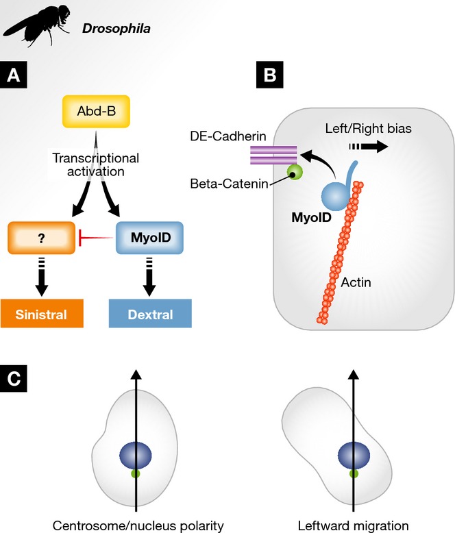
Schematic depiction of genetic and cellular aspects of Drosophila lateralization. (A) The wild-type, or “dextral”, orientation depends on the activity of MyoID (Blue). Dextral determination is dominant over sinistral determination (Red), which only becomes apparent in myoID null flies. Interestingly, Abd-B (Yellow) controls the expression and/or activity of the two opposite pathways. In Abd-B depleted flies, the L/R organs develop symmetrically 93. To date, the putative sinistral counterpart to MyoID is still unknown. (B) In the cells of the L/R organizer, MyoID (blue) binds to cortical actin (red) and needs to associate with the adherens junction components E-cadherin (yellow) and β-catenin (green) at the apical membrane for proper L/R determination 94,109. (C) Several lines of cultured vertebrate cells orient themselves according to their nucleus–centrosome axis (arrow) and are thus able to migrate in a L/R asymmetric manner.
Molecularly, the dextral determinant MyoID is a type I unconventional myosin, a one-headed, monomeric actin-based motor, that is very well conserved in evolution 94,97,101. Type I myosins comprise three domains: an N-terminal single-headed motor domain coupled to a C-terminal tail via an alpha-helical neck 102,103. The motor domain binds actin and hydrolyses ATP. The neck contains a number of IQ domains and binds light chains acting as a lever-arm, thus transmitting the conformational changes that occur in the motor domain after ATP hydrolysis 104,105. Finally, the tail domain is thought to interact with cargos and binds membrane phospholipids through its Pleckstrin Homology domain, a positively charged lipid-binding region 106,107.
How does MyoID act during L/R determination? Interestingly, MyoID activity appears to be required only for a short time to induce a dextral bias 94. To date, the exact mechanism of MyoID action remains unknown, but the actin-binding head domain appears to be central for L/R patterning 98. Additionally, in the cells of the organizer, MyoID requires the adherens junction components β-catenin and E-cadherin as well as a properly organized cortical actin cytoskeleton (Fig 3B) to induce dextral L/R development 94,97,98,108,109. In the epithelium of the embryonic hindgut, MyoID has been shown to cell-autonomously bias cell chirality and induce membrane bending 108. Interestingly, computer simulations showed that mild membrane bending in each cell is sufficient to induce a complete dextral loop of the hindgut 108. MyoID-dependent membrane bending appears to be mediated by E-cadherin, as membranes in E-cadherin null mutants do not bend 108. Taken together, these data suggest that L/R morphogenesis could originate from asymmetric membrane tension generated by a MyoID/E-cadherin complex. Interestingly, unlike in the absence of E-cadherin or β-catenin when no consistent orientation is seen, in the absence of MyoID cell membranes of the hindgut still bend, but this time in the opposite direction 108,109. These observations suggest that the sinistral factor(s), whose activity is only apparent in the absence of MyoID, is also able to induce an orientated cell membrane bias.
Innate cellular chirality
As mentioned above, asymmetric traits are not specific to multicellular structures but can also appear at the single cell level. Indeed, numerous cell types exhibit chiral structures, orientated movements as well as chiral behaviors 110–113. These observations argue that intracellular elements might underlie L/R asymmetry determination. This idea, termed the “intracellular model”, has been around for some time and proposes that the origin of asymmetry in the body plan relies on intracellular structures and in particular the actin cytoskeleton 16. Supporting this model is the fact that in cultured migrating cells, a clear 3D cell polarity can be seen. In addition to the first two axes, rear-front and top-bottom, a third one, drawn from the center of the nucleus to that of the centrosome, demonstrates clear cell chirality and corresponds loosely to the direction followed by these cells during their migration 113. However, when cultured in contact with a repeated pattern, cells consistently migrate with a clear bias toward the left side of this third axis (Fig 3C), strongly suggesting the existence of an intracellular bias present in each individual cell 110,112,113.
The cell chirality depends on the cell cytoskeleton. Disrupting microtubule integrity leads to randomization, revealing the need for an intact microtubule cytoskeleton for this leftward bias 113. Disruption of the actin cytoskeleton instead leads to the “inversion of the cell L/R axis” that is, a rightward bias in cell migration 110. Consistently, the expression of constitutively active GSK3 similarly inverts the cell “L/R axis”. The cells now polarize to the right of the nucleus-to-centrosome axis. These data suggest that GSK3 could act as a link between the unknown original chiral template and the cytoskeleton sensing the spatial cues and orienting cell polarity 113. These data, obtained in vertebrate cells, are reminiscent of the link between the actin cytoskeleton and L/R patterning in Drosophila, C. elegans or the Lymnaea snails, suggesting a conserved mechanism. Furthermore, they also support the existence of a sinistral factor, as cell or organismal orientation can be consistently inverted and not simply randomized. However, a diversity of L/R orientations exists in cultured cells with some cell types having a dextral bias, others a sinistral one and some with no bias at all 111,112. To conclude, cell culture experiments revealed the crucial role of actin dynamics for internal cell chirality and suggest that both dextral and sinistral L/R patterning might originate from intracellular polarity.
Indeed, several pieces of evidence obtained from studies of type I myosins and actin dynamics support the idea that L/R asymmetries can be created de novo from basic cell components 114. Type I myosins, to which Drosophila MyoID belongs, are members of the myosin superfamily of actin-based motors and are found in most eukaryotic cells 115,116. In vertebrates, eight type I myosins (myosin I a–h) are found, whereas only two members exist in Drosophila (myosin ID and IC) 117,118. Recent work, using in vitro binding of murine MyoIc to actin, revealed that MyoIc can asymmetrically guide motility, leading to actin filaments that curl counterclockwise 114. Importantly, this generation of asymmetric motility appears to be a property of MyoIc and not a universal characteristic of myosin I motors, since neither murine MyoIa nor Ib are able to generate a similar asymmetric actin movement 114. Although it is not directly stated, the head domain seems to be responsible for this feature, which is consistent with the fact that, in vivo, the L/R activity of Drosophila MyoID also appears to depend on its head domain 98. The finding that specific myosins can make actin fibers chiral are the earliest described signs of asymmetry somehow related to L/R patterning.
Taken together, it appears that from all the model systems discussed, Nodal flow is rather an exception in L/R axis establishment (Fig 4). Evolutionarily, it could correspond to a refinement that was added to earlier mechanisms happening at the cellular level. The conserved involvement of fundamental cellular elements such as ion channels or cytoskeletal components may point to common ancestral L/R asymmetry mechanisms. Additionally, they allow for the generation of a theoretical model for how, from core molecules at the cellular level, such as the actin cytoskeleton, L/R patterning may be created in metazoans.
Figure 4. L/R asymmetry in metazoa: diversity and convergence.

Common and divergent principles of L/R asymmetry establishment in the model systems discussed in this review (see text for details). Species are aligned along a phylogenetic tree discerning Protostomes (yellow) and Deuterostomes (purple). The mechanisms breaking symmetry (actin-based: red; ion flow: green; cell movement (Cell MVT): orange; cilia-based Nodal flow (Cilia): light blue) are vertically aligned along the developmental time (DVPT TIME) at which they act (early, down; later, up). The direct link between a mechanism and a subsequent one or ultimately to the Nodal-signaling pathway (Nodal, dark blue) is indicated by the color gradient.
Sidebar A: In need of answers —
How is symmetry broken at the cellular level?
What are the mechanisms and molecular elements at the basis of situs inversus phenotypes?
What is, or what are, the origin(s) of L/R asymmetry?
How did L/R asymmetry establishment evolve in metazoa?
Acknowledgments
Work in SN laboratory is supported by CNRS, Inserm, ANR, LABEX SIGNALIFE (Program reference # ANR-11-LABX-0028-01), ARC, FRM and University of Nice. We are grateful to Gaëlle Le Breton for critical reading of this manuscript. We apologize to the colleagues, whose work could not be cited due to space limitation.
Glossary
- A/P
anterior/posterior
- Abd-B
abdominal-B
- ASEL, ASER
left–right asymmetric bilateral sensory neurons in C. elegans
- D/V
dorsal/ventral
- dvl
dishevelled-like
- FGF
fibroblast growth factor
- GSK3
glycogen synthase kinase 3
- Heterotaxia
also situs ambiguus, uncoordinated placing of the internal organs
- Isomerism
situation in which both sides of the body adopt the same fate
- iv
inversus viscerum
- L/R
left–right
- LPM
lateral plate mesoderm
- myoID
myosin ID
- PCP
planar cell polarity
- PH
Pleckstrin Homology
- Pitx2
paired-like homeodomain transcription factor 2
- Pkd1l1
polycystic kidney disease-like 1
- Pkd2
polycystic kidney disease 2
- Shh
sonic hedgehog
- situs inversus
inverted placing of the internal organs
- situs solitus
normal placing of the internal organs
- TGF-β
transforming growth factor beta
- vangl
Van Gogh-like
Conflict of interest
The authors declare that they have no conflict of interest.
References
- Boycott AE, Diver C. On the inheritance of sinistrality in Limnaea peregra. Proc R Soc Lond B, Contain Papers Biol Charact. 1923;95:207–213. [Google Scholar]
- Gurdon JB. Sinistral snails and gentlemen scientists. Cell. 2005;123:751–753. doi: 10.1016/j.cell.2005.11.015. [DOI] [PubMed] [Google Scholar]
- Danos MC, Yost HJ. Linkage of cardiac left-right asymmetry and dorsal-anterior development in Xenopus. Development. 1995;121:1467–1474. doi: 10.1242/dev.121.5.1467. [DOI] [PubMed] [Google Scholar]
- Fujinaga M, Baden JM. A new method for explanting early postimplantation rat embryos for culture. Teratology. 1991;43:95–100. doi: 10.1002/tera.1420430111. [DOI] [PubMed] [Google Scholar]
- Hoyle C, Brown NA, Wolpert L. Development of left/right handedness in the chick heart. Development. 1992;115:1071–1078. doi: 10.1242/dev.115.4.1071. [DOI] [PubMed] [Google Scholar]
- Hummel KP, Chapman DB. Visceral inversion and associated anomalies in the mouse. J Hered. 1959;50:9–13. [Google Scholar]
- Layton WM, Layton MW, Binder M, Kurnit DM, Hanzlik AJ, Van Keuren M, Biddle FG. Expression of the IV (reversed and/or heterotaxic) phenotype in SWV mice. Teratology. 1993;47:595–602. doi: 10.1002/tera.1420470611. [DOI] [PubMed] [Google Scholar]
- Schreiner CM, Scott WJ, Jr, Supp DM, Potter SS. Correlation of forelimb malformation asymmetries with visceral organ situs in the transgenic mouse insertional mutation, legless. Dev Biol. 1993;158:560–562. doi: 10.1006/dbio.1993.1214. [DOI] [PubMed] [Google Scholar]
- Stalsberg H. The origin of heart asymmetry: right and left contributions to the early chick embryo heart. Dev Biol. 1969;19:109–127. doi: 10.1016/0012-1606(69)90051-7. [DOI] [PubMed] [Google Scholar]
- Yokoyama T, Copeland NG, Jenkins NA, Montgomery CA, Elder FF, Overbeek PA. Reversal of left-right asymmetry: a situs inversus mutation. Science. 1993;260:679–682. doi: 10.1126/science.8480178. [DOI] [PubMed] [Google Scholar]
- Yost HJ. Development of the left-right axis in amphibians. Ciba Found Symp. 1991;162:165–176. doi: 10.1002/9780470514160.ch10. discussion 176–181. [DOI] [PubMed] [Google Scholar]
- Levin M, Johnson RL, Stern CD, Kuehn M, Tabin C. A molecular pathway determining left-right asymmetry in chick embryogenesis. Cell. 1995;82:803–814. doi: 10.1016/0092-8674(95)90477-8. [DOI] [PubMed] [Google Scholar]
- Brown NA, Wolpert L. The development of handedness in left/right asymmetry. Development. 1990;109:1–9. doi: 10.1242/dev.109.1.1. [DOI] [PubMed] [Google Scholar]
- Speder P, Petzoldt A, Suzanne M, Noselli S. Strategies to establish left/right asymmetry in vertebrates and invertebrates. Curr Opin Genet Dev. 2007;17:351–358. doi: 10.1016/j.gde.2007.05.008. [DOI] [PubMed] [Google Scholar]
- Hirokawa N, Tanaka Y, Okada Y, Takeda S. Nodal flow and the generation of left-right asymmetry. Cell. 2006;125:33–45. doi: 10.1016/j.cell.2006.03.002. [DOI] [PubMed] [Google Scholar]
- Vandenberg LN, Levin M. A unified model for left-right asymmetry? Comparison and synthesis of molecular models of embryonic laterality. Dev Biol. 2013;379:1–15. doi: 10.1016/j.ydbio.2013.03.021. [DOI] [PMC free article] [PubMed] [Google Scholar]
- Lee JD, Anderson KV. Morphogenesis of the node and notochord: the cellular basis for the establishment and maintenance of left-right asymmetry in the mouse. Dev Dyn. 2008;237:3464–3476. doi: 10.1002/dvdy.21598. [DOI] [PMC free article] [PubMed] [Google Scholar]
- Nonaka S, Tanaka Y, Okada Y, Takeda S, Harada A, Kanai Y, Kido M, Hirokawa N. Randomization of left-right asymmetry due to loss of nodal cilia generating leftward flow of extraembryonic fluid in mice lacking KIF3B motor protein. Cell. 1998;95:829–837. doi: 10.1016/s0092-8674(00)81705-5. [DOI] [PubMed] [Google Scholar]
- Takeda S, Yonekawa Y, Tanaka Y, Okada Y, Nonaka S, Hirokawa N. Left-right asymmetry and kinesin superfamily protein KIF3A: new insights in determination of laterality and mesoderm induction by kif3A−/− mice analysis. J Cell Biol. 1999;145:825–836. doi: 10.1083/jcb.145.4.825. [DOI] [PMC free article] [PubMed] [Google Scholar]
- Supp DM, Witte DP, Potter SS, Brueckner M. Mutation of an axonemal dynein affects left-right asymmetry in inversus viscerum mice. Nature. 1997;389:963–966. doi: 10.1038/40140. [DOI] [PMC free article] [PubMed] [Google Scholar]
- McGrath J, Somlo S, Makova S, Tian X, Brueckner M. Two populations of node monocilia initiate left-right asymmetry in the mouse. Cell. 2003;114:61–73. doi: 10.1016/s0092-8674(03)00511-7. [DOI] [PubMed] [Google Scholar]
- Supp DM, Brueckner M, Kuehn MR, Witte DP, Lowe LA, McGrath J, Corrales J, Potter SS. Targeted deletion of the ATP binding domain of left-right dynein confirms its role in specifying development of left-right asymmetries. Development. 1999;126:5495–5504. doi: 10.1242/dev.126.23.5495. [DOI] [PMC free article] [PubMed] [Google Scholar]
- Okada Y, Nonaka S, Tanaka Y, Saijoh Y, Hamada H, Hirokawa N. Abnormal nodal flow precedes situs inversus in iv and inv mice. Mol Cell. 1999;4:459–468. doi: 10.1016/s1097-2765(00)80197-5. [DOI] [PubMed] [Google Scholar]
- Kartagener H. Bronchiektasien bei situs viscerum inversus. Schweiz Med Wocshenschr. 1935;65:782–784. [Google Scholar]
- Nonaka S, Shiratori H, Saijoh Y, Hamada H. Determination of left-right patterning of the mouse embryo by artificial nodal flow. Nature. 2002;418:96–99. doi: 10.1038/nature00849. [DOI] [PubMed] [Google Scholar]
- Shinohara K, Kawasumi A, Takamatsu A, Yoshiba S, Botilde Y, Motoyama N, Reith W, Durand B, Shiratori H, Hamada H. Two rotating cilia in the node cavity are sufficient to break left-right symmetry in the mouse embryo. Nat Commun. 2012;3:622. doi: 10.1038/ncomms1624. [DOI] [PubMed] [Google Scholar]
- Nakamura T, Mine N, Nakaguchi E, Mochizuki A, Yamamoto M, Yashiro K, Meno C, Hamada H. Generation of robust left-right asymmetry in the mouse embryo requires a self-enhancement and lateral-inhibition system. Dev Cell. 2006;11:495–504. doi: 10.1016/j.devcel.2006.08.002. [DOI] [PubMed] [Google Scholar]
- Sampaio P, Ferreira RR, Guerrero A, Pintado P, Tavares B, Amaro J, Smith AA, Montenegro-Johnson T, Smith DJ, Lopes SS. Left-right organizer flow dynamics: how much cilia activity reliably yields laterality? Dev Cell. 2014;29:716–728. doi: 10.1016/j.devcel.2014.04.030. [DOI] [PubMed] [Google Scholar]
- Brennan J, Norris DP, Robertson EJ. Nodal activity in the node governs left-right asymmetry. Genes Dev. 2002;16:2339–2344. doi: 10.1101/gad.1016202. [DOI] [PMC free article] [PubMed] [Google Scholar]
- Meno C, Shimono A, Saijoh Y, Yashiro K, Mochida K, Ohishi S, Noji S, Kondoh H, Hamada H. Lefty-1 is required for left-right determination as a regulator of lefty-2 and nodal. Cell. 1998;94:287–297. doi: 10.1016/s0092-8674(00)81472-5. [DOI] [PubMed] [Google Scholar]
- Yamamoto M, Mine N, Mochida K, Sakai Y, Saijoh Y, Meno C, Hamada H. Nodal signaling induces the midline barrier by activating nodal expression in the lateral plate. Development. 2003;130:1795–1804. doi: 10.1242/dev.00408. [DOI] [PubMed] [Google Scholar]
- Logan M, Pagan-Westphal SM, Smith DM, Paganessi L, Tabin CJ. The transcription factor Pitx2 mediates situs-specific morphogenesis in response to left-right asymmetric signals. Cell. 1998;94:307–317. doi: 10.1016/s0092-8674(00)81474-9. [DOI] [PubMed] [Google Scholar]
- Piedra ME, Icardo JM, Albajar M, Rodriguez-Rey JC, Ros MA. Pitx2 participates in the late phase of the pathway controlling left-right asymmetry. Cell. 1998;94:319–324. doi: 10.1016/s0092-8674(00)81475-0. [DOI] [PubMed] [Google Scholar]
- Ryan AK, Blumberg B, Rodriguez-Esteban C, Yonei-Tamura S, Tamura K, Tsukui T, de la Peña J, Sabbagh W, Greenwald J, Choe S, et al. Pitx2 determines left-right asymmetry of internal organs in vertebrates. Nature. 1998;394:545–551. doi: 10.1038/29004. [DOI] [PubMed] [Google Scholar]
- Yoshioka H, Meno C, Koshiba K, Sugihara M, Itoh H, Ishimaru Y, Inoue T, Ohuchi H, Semina EV, Murray JC, et al. Pitx2, a bicoid-type homeobox gene, is involved in a lefty-signaling pathway in determination of left-right asymmetry. Cell. 1998;94:299–305. doi: 10.1016/s0092-8674(00)81473-7. [DOI] [PubMed] [Google Scholar]
- Okada Y, Takeda S, Tanaka Y, Izpisua Belmonte JC, Hirokawa N. Mechanism of nodal flow: a conserved symmetry breaking event in left-right axis determination. Cell. 2005;121:633–644. doi: 10.1016/j.cell.2005.04.008. [DOI] [PubMed] [Google Scholar]
- Nonaka S, Yoshiba S, Watanabe D, Ikeuchi S, Goto T, Marshall WF, Hamada H. De novo formation of left-right asymmetry by posterior tilt of nodal cilia. PLoS Biol. 2005;3:e268. doi: 10.1371/journal.pbio.0030268. [DOI] [PMC free article] [PubMed] [Google Scholar]
- Cartwright JH, Piro O, Tuval I. Fluid-dynamical basis of the embryonic development of left-right asymmetry in vertebrates. Proc Natl Acad Sci USA. 2004;101:7234–7239. doi: 10.1073/pnas.0402001101. [DOI] [PMC free article] [PubMed] [Google Scholar]
- Goodrich LV, Strutt D. Principles of planar polarity in animal development. Development. 2011;138:1877–1892. doi: 10.1242/dev.054080. [DOI] [PMC free article] [PubMed] [Google Scholar]
- Antic D, Stubbs JL, Suyama K, Kintner C, Scott MP, Axelrod JD. Planar cell polarity enables posterior localization of nodal cilia and left-right axis determination during mouse and Xenopus embryogenesis. PLoS ONE. 2010;5:e8999. doi: 10.1371/journal.pone.0008999. [DOI] [PMC free article] [PubMed] [Google Scholar]
- Borovina A, Superina S, Voskas D, Ciruna B. Vangl2 directs the posterior tilting and asymmetric localization of motile primary cilia. Nat Cell Biol. 2010;12:407–412. doi: 10.1038/ncb2042. [DOI] [PubMed] [Google Scholar]
- Hashimoto M, Shinohara K, Wang J, Ikeuchi S, Yoshiba S, Meno C, Nonaka S, Takada S, Hatta K, Wynshaw-Boris A, et al. Planar polarization of node cells determines the rotational axis of node cilia. Nat Cell Biol. 2010;12:170–176. doi: 10.1038/ncb2020. [DOI] [PubMed] [Google Scholar]
- Song H, Hu J, Chen W, Elliott G, Andre P, Gao B, Yang Y. Planar cell polarity breaks bilateral symmetry by controlling ciliary positioning. Nature. 2010;466:378–382. doi: 10.1038/nature09129. [DOI] [PMC free article] [PubMed] [Google Scholar]
- Mahaffey JP, Grego-Bessa J, Liem KF, Jr, Anderson KV. Cofilin and Vangl2 cooperate in the initiation of planar cell polarity in the mouse embryo. Development. 2013;140:1262–1271. doi: 10.1242/dev.085316. [DOI] [PMC free article] [PubMed] [Google Scholar]
- Norris DP. Cilia, calcium and the basis of left-right asymmetry. BMC Biol. 2012;10:102. doi: 10.1186/1741-7007-10-102. [DOI] [PMC free article] [PubMed] [Google Scholar]
- Tanaka Y, Okada Y, Hirokawa N. FGF-induced vesicular release of Sonic hedgehog and retinoic acid in leftward nodal flow is critical for left-right determination. Nature. 2005;435:172–177. doi: 10.1038/nature03494. [DOI] [PubMed] [Google Scholar]
- Field S, Riley KL, Grimes DT, Hilton H, Simon M, Powles-Glover N, Siggers P, Bogani D, Greenfield A, Norris DP. Pkd1l1 establishes left-right asymmetry and physically interacts with Pkd2. Development. 2011;138:1131–1142. doi: 10.1242/dev.058149. [DOI] [PMC free article] [PubMed] [Google Scholar]
- Pennekamp P, Karcher C, Fischer A, Schweickert A, Skryabin B, Horst J, Blum M, Dworniczak B. The ion channel polycystin-2 is required for left-right axis determination in mice. Curr Biol. 2002;12:938–943. doi: 10.1016/s0960-9822(02)00869-2. [DOI] [PubMed] [Google Scholar]
- Yoshiba S, Shiratori H, Kuo IY, Kawasumi A, Shinohara K, Nonaka S, Asai Y, Sasaki G, Belo JA, Sasaki H, et al. Cilia at the node of mouse embryos sense fluid flow for left-right determination via Pkd2. Science. 2012;338:226–231. doi: 10.1126/science.1222538. [DOI] [PMC free article] [PubMed] [Google Scholar]
- Kamura K, Kobayashi D, Uehara Y, Koshida S, Iijima N, Kudo A, Yokoyama T, Takeda H. Pkd1l1 complexes with Pkd2 on motile cilia and functions to establish the left-right axis. Development. 2011;138:1121–1129. doi: 10.1242/dev.058271. [DOI] [PubMed] [Google Scholar]
- Gardner RL. Normal bias in the direction of fetal rotation depends on blastomere composition during early cleavage in the mouse. PLoS ONE. 2010;5:e9610. doi: 10.1371/journal.pone.0009610. [DOI] [PMC free article] [PubMed] [Google Scholar]
- Armakolas A, Klar AJ. Left-right dynein motor implicated in selective chromatid segregation in mouse cells. Science. 2007;315:100–101. doi: 10.1126/science.1129429. [DOI] [PubMed] [Google Scholar]
- Sauer S, Klar AJ. Left-right symmetry breaking in mice by left-right dynein may occur via a biased chromatid segregation mechanism, without directly involving the Nodal gene. Front Oncol. 2012;2:166. doi: 10.3389/fonc.2012.00166. [DOI] [PMC free article] [PubMed] [Google Scholar]
- Noel ES, Verhoeven M, Lagendijk AK, Tessadori F, Smith K, Choorapoikayil S, den Hertog J, Bakkers J. A Nodal-independent and tissue-intrinsic mechanism controls heart-looping chirality. Nat Commun. 2013;4:2754. doi: 10.1038/ncomms3754. [DOI] [PubMed] [Google Scholar]
- Feistel K, Blum M. Three types of cilia including a novel 9+4 axoneme on the notochordal plate of the rabbit embryo. Dev Dyn. 2006;235:3348–3358. doi: 10.1002/dvdy.20986. [DOI] [PubMed] [Google Scholar]
- Stephen LA, Johnson EJ, Davis GM, McTeir L, Pinkham J, Jaberi N, Davey MG. The chicken left right organizer has nonmotile cilia which are lost in a stage-dependent manner in the talpid ciliopathy. Genesis. 2014;52:600–613. doi: 10.1002/dvg.22775. [DOI] [PMC free article] [PubMed] [Google Scholar]
- Cui C, Little CD, Rongish BJ. Rotation of organizer tissue contributes to left-right asymmetry. Anat Rec. 2009;292:557–561. doi: 10.1002/ar.20872. [DOI] [PMC free article] [PubMed] [Google Scholar]
- Gros J, Feistel K, Viebahn C, Blum M, Tabin CJ. Cell movements at Hensen’s node establish left/right asymmetric gene expression in the chick. Science. 2009;324:941–944. doi: 10.1126/science.1172478. [DOI] [PMC free article] [PubMed] [Google Scholar]
- Levin M, Thorlin T, Robinson KR, Nogi T, Mercola M. Asymmetries in H+/K+-ATPase and cell membrane potentials comprise a very early step in left-right patterning. Cell. 2002;111:77–89. doi: 10.1016/s0092-8674(02)00939-x. [DOI] [PubMed] [Google Scholar]
- Adams DS, Robinson KR, Fukumoto T, Yuan S, Albertson RC, Yelick P, Kuo L, McSweeney M, Levin M. Early, H+-V-ATPase-dependent proton flux is necessary for consistent left-right patterning of non-mammalian vertebrates. Development. 2006;133:1657–1671. doi: 10.1242/dev.02341. [DOI] [PMC free article] [PubMed] [Google Scholar]
- Aw S, Adams DS, Qiu D, Levin M. H,K-ATPase protein localization and Kir4.1 function reveal concordance of three axes during early determination of left-right asymmetry. Mech Dev. 2008;125:353–372. doi: 10.1016/j.mod.2007.10.011. [DOI] [PMC free article] [PubMed] [Google Scholar]
- Shimeld SM, Levin M. Evidence for the regulation of left-right asymmetry in Ciona intestinalis by ion flux. Dev Dyn. 2006;235:1543–1553. doi: 10.1002/dvdy.20792. [DOI] [PubMed] [Google Scholar]
- Nishide K, Mugitani M, Kumano G, Nishida H. Neurula rotation determines left-right asymmetry in ascidian tadpole larvae. Development. 2012;139:1467–1475. doi: 10.1242/dev.076083. [DOI] [PubMed] [Google Scholar]
- Raff RA, Snoke Smith M. Chapter 7. Axis formation and the rapid evolutionary transformation of larval form. Curr Top Dev Biol. 2009;86:163–190. doi: 10.1016/S0070-2153(09)01007-2. [DOI] [PubMed] [Google Scholar]
- Molina MD, de Croze N, Haillot E, Lepage T. Nodal: master and commander of the dorsal-ventral and left-right axes in the sea urchin embryo. Curr Opin Genet Dev. 2013;23:445–453. doi: 10.1016/j.gde.2013.04.010. [DOI] [PubMed] [Google Scholar]
- Duboc V, Rottinger E, Lapraz F, Besnardeau L, Lepage T. Left-right asymmetry in the sea urchin embryo is regulated by nodal signaling on the right side. Dev Cell. 2005;9:147–158. doi: 10.1016/j.devcel.2005.05.008. [DOI] [PubMed] [Google Scholar]
- Frasnelli E, Vallortigara G, Rogers LJ. Left-right asymmetries of behaviour and nervous system in invertebrates. Neurosci Biobehav Rev. 2012;36:1273–1291. doi: 10.1016/j.neubiorev.2012.02.006. [DOI] [PubMed] [Google Scholar]
- Grande C, Patel NH. Nodal signalling is involved in left-right asymmetry in snails. Nature. 2009;457:1007–1011. doi: 10.1038/nature07603. [DOI] [PMC free article] [PubMed] [Google Scholar]
- Shimizu K, Iijima M, Setiamarga DH, Sarashina I, Kudoh T, Asami T, Gittenberger E, Endo K. Left-right asymmetric expression of dpp in the mantle of gastropods correlates with asymmetric shell coiling. Evodevo. 2013;4:15. doi: 10.1186/2041-9139-4-15. [DOI] [PMC free article] [PubMed] [Google Scholar]
- Kuroda R, Endo B, Abe M, Shimizu M. Chiral blastomere arrangement dictates zygotic left-right asymmetry pathway in snails. Nature. 2009;462:790–794. doi: 10.1038/nature08597. [DOI] [PubMed] [Google Scholar]
- Shibazaki Y, Shimizu M, Kuroda R. Body handedness is directed by genetically determined cytoskeletal dynamics in the early embryo. Curr Biol. 2004;14:1462–1467. doi: 10.1016/j.cub.2004.08.018. [DOI] [PubMed] [Google Scholar]
- Liu MM, Davey JW, Banerjee R, Han J, Yang F, Aboobaker A, Blaxter ML, Davison A. Fine mapping of the pond snail left-right asymmetry (chirality) locus using RAD-Seq and fibre-FISH. PLoS ONE. 2013;8:e71067. doi: 10.1371/journal.pone.0071067. [DOI] [PMC free article] [PubMed] [Google Scholar]
- Freeman G, Lundelius JW. The developmental genetics of dextrality and sinistrality in the gastropod Lymnaea peregra. Wilhelm Roux’s Arc Dev Biol. 1982;191:69–83. doi: 10.1007/BF00848443. [DOI] [PubMed] [Google Scholar]
- Ueshima R, Asami T. Evolution: single-gene speciation by left-right reversal. Nature. 2003;425:679. doi: 10.1038/425679a. [DOI] [PubMed] [Google Scholar]
- Hoso M, Kameda Y, Wu SP, Asami T, Kato M, Hori M. A speciation gene for left-right reversal in snails results in anti-predator adaptation. Nat Commun. 2010;1:133. doi: 10.1038/ncomms1133. [DOI] [PMC free article] [PubMed] [Google Scholar]
- Pohl C. Left-right patterning in the C. elegans embryo: unique mechanisms and common principles. Commun Integr Biol. 2011;4:34–40. doi: 10.4161/cib.4.1.14144. [DOI] [PMC free article] [PubMed] [Google Scholar]
- Wood WB. Handed asymmetry in nematodes. Semin Cell Dev Biol. 1998;9:53–60. doi: 10.1006/scdb.1997.0189. [DOI] [PubMed] [Google Scholar]
- Hutter H, Schnabel R. Establishment of left-right asymmetry in the Caenorhabditis elegans embryo: a multistep process involving a series of inductive events. Development. 1995;121:3417–3424. doi: 10.1242/dev.121.10.3417. [DOI] [PubMed] [Google Scholar]
- Hutter H, Schnabel R. Specification of anterior-posterior differences within the AB lineage in the C. elegans embryo: a polarising induction. Development. 1995;121:1559–1568. doi: 10.1242/dev.121.5.1559. [DOI] [PubMed] [Google Scholar]
- Pohl C, Bao Z. Chiral forces organize left-right patterning in C. elegans by uncoupling midline and anteroposterior axis. Dev Cell. 2010;19:402–412. doi: 10.1016/j.devcel.2010.08.014. [DOI] [PMC free article] [PubMed] [Google Scholar]
- Bergmann DC, Lee M, Robertson B, Tsou MF, Rose LS, Wood WB. Embryonic handedness choice in C. elegans involves the Galpha protein GPA-16. Development. 2003;130:5731–5740. doi: 10.1242/dev.00839. [DOI] [PubMed] [Google Scholar]
- Johnston CA, Afshar K, Snyder JT, Tall GG, Gonczy P, Siderovski DP, Willard FS. Structural determinants underlying the temperature-sensitive nature of a Galpha mutant in asymmetric cell division of Caenorhabditis elegans. J Biol Chem. 2008;283:21550–21558. doi: 10.1074/jbc.M803023200. [DOI] [PMC free article] [PubMed] [Google Scholar]
- Cabello J, Neukomm LJ, Gunesdogan U, Burkart K, Charette SJ, Lochnit G, Hengartner MO, Schnabel R. The Wnt pathway controls cell death engulfment, spindle orientation, and migration through CED-10/Rac. PLoS Biol. 2010;8:e1000297. doi: 10.1371/journal.pbio.1000297. [DOI] [PMC free article] [PubMed] [Google Scholar]
- Bischoff M, Schnabel R. A posterior centre establishes and maintains polarity of the Caenorhabditis elegans embryo by a Wnt-dependent relay mechanism. PLoS Biol. 2006;4:e396. doi: 10.1371/journal.pbio.0040396. [DOI] [PMC free article] [PubMed] [Google Scholar]
- Hermann GJ, Leung B, Priess JR. Left-right asymmetry in C. elegans intestine organogenesis involves a LIN-12/Notch signaling pathway. Development. 2000;127:3429–3440. doi: 10.1242/dev.127.16.3429. [DOI] [PubMed] [Google Scholar]
- Wood WB. Evidence from reversal of handedness in C. elegans embryos for early cell interactions determining cell fates. Nature. 1991;349:536–538. doi: 10.1038/349536a0. [DOI] [PubMed] [Google Scholar]
- Hobert O, Johnston RJ, Jr, Chang S. Left-right asymmetry in the nervous system: the Caenorhabditis elegans model. Nat Rev Neurosci. 2002;3:629–640. doi: 10.1038/nrn897. [DOI] [PubMed] [Google Scholar]
- Troemel ER, Sagasti A, Bargmann CI. Lateral signaling mediated by axon contact and calcium entry regulates asymmetric odorant receptor expression in C. elegans. Cell. 1999;99:387–398. doi: 10.1016/s0092-8674(00)81525-1. [DOI] [PubMed] [Google Scholar]
- Poole RJ, Hobert O. Early embryonic programming of neuronal left/right asymmetry in C. elegans. Curr Biol. 2006;16:2279–2292. doi: 10.1016/j.cub.2006.09.041. [DOI] [PubMed] [Google Scholar]
- Bertrand V, Bisso P, Poole RJ, Hobert O. Notch-dependent induction of left/right asymmetry in C. elegans interneurons and motoneurons. Curr Biol. 2011;21:1225–1231. doi: 10.1016/j.cub.2011.06.016. [DOI] [PMC free article] [PubMed] [Google Scholar]
- Cochella L, Hobert O. Embryonic priming of a miRNA locus predetermines postmitotic neuronal left/right asymmetry in C. elegans. Cell. 2012;151:1229–1242. doi: 10.1016/j.cell.2012.10.049. [DOI] [PMC free article] [PubMed] [Google Scholar]
- Zhang F, O’Meara MM, Hobert O. A left/right asymmetric neuronal differentiation program is controlled by the Caenorhabditis elegans lsy-27 zinc-finger transcription factor. Genetics. 2011;188:753–759. doi: 10.1534/genetics.111.129064. [DOI] [PMC free article] [PubMed] [Google Scholar]
- Coutelis JB, Geminard C, Speder P, Suzanne M, Petzoldt AG, Noselli S. Drosophila left/right asymmetry establishment is controlled by the Hox gene abdominal-B. Dev Cell. 2013;24:89–97. doi: 10.1016/j.devcel.2012.11.013. [DOI] [PubMed] [Google Scholar]
- Speder P, Adam G, Noselli S. Type ID unconventional myosin controls left-right asymmetry in Drosophila. Nature. 2006;440:803–807. doi: 10.1038/nature04623. [DOI] [PubMed] [Google Scholar]
- Geminard C, Gonzalez-Morales N, Coutelis JB, Noselli S. The myosin ID pathway and left-right asymmetry in Drosophila. Genesis. 2014;52:471–480. doi: 10.1002/dvg.22763. [DOI] [PubMed] [Google Scholar]
- Coutelis JB, Petzoldt AG, Speder P, Suzanne M, Noselli S. Left-right asymmetry in Drosophila. Semin Cell Dev Biol. 2008;19:252–262. doi: 10.1016/j.semcdb.2008.01.006. [DOI] [PubMed] [Google Scholar]
- Hozumi S, Maeda R, Taniguchi K, Kanai M, Shirakabe S, Sasamura T, Spéder P, Noselli S, Aigaki T, Murakami R, et al. An unconventional myosin in Drosophila reverses the default handedness in visceral organs. Nature. 2006;440:798–802. doi: 10.1038/nature04625. [DOI] [PubMed] [Google Scholar]
- Hozumi S, Maeda R, Taniguchi-Kanai M, Okumura T, Taniguchi K, Kawakatsu Y, Nakazawa N, Hatori R, Matsuno K. Head region of unconventional myosin I family members is responsible for the organ-specificity of their roles in left-right polarity in Drosophila. Dev Dyn. 2008;237:3528–3537. doi: 10.1002/dvdy.21583. [DOI] [PubMed] [Google Scholar]
- Suzanne M, Petzoldt AG, Speder P, Coutelis JB, Steller H, Noselli S. Coupling of apoptosis and L/R patterning controls stepwise organ looping. Curr Biol. 2010;20:1773–1778. doi: 10.1016/j.cub.2010.08.056. [DOI] [PMC free article] [PubMed] [Google Scholar]
- Maeda RK, Karch F. The bithorax complex of Drosophila an exceptional Hox cluster. Curr Top Dev Biol. 2009;88:1–33. doi: 10.1016/S0070-2153(09)88001-0. [DOI] [PubMed] [Google Scholar]
- Speder P, Noselli S. Left-right asymmetry: class I myosins show the direction. Curr Opin Cell Biol. 2007;19:82–87. doi: 10.1016/j.ceb.2006.12.006. [DOI] [PubMed] [Google Scholar]
- Krendel M, Mooseker MS. Myosins: tails (and heads) of functional diversity. Physiology (Bethesda) 2005;20:239–251. doi: 10.1152/physiol.00014.2005. [DOI] [PubMed] [Google Scholar]
- Nambiar R, McConnell RE, Tyska MJ. Myosin motor function: the ins and outs of actin-based membrane protrusions. Cell Mol Life Sci. 2010;67:1239–1254. doi: 10.1007/s00018-009-0254-5. [DOI] [PMC free article] [PubMed] [Google Scholar]
- Barylko B, Binns DD, Albanesi JP. Regulation of the enzymatic and motor activities of myosin I. Biochim Biophys Acta. 2000;1496:23–35. doi: 10.1016/s0167-4889(00)00006-9. [DOI] [PubMed] [Google Scholar]
- Greenberg MJ, Ostap EM. Regulation and control of myosin-I by the motor and light chain-binding domains. Trends Cell Biol. 2013;23:81–89. doi: 10.1016/j.tcb.2012.10.008. [DOI] [PMC free article] [PubMed] [Google Scholar]
- Hokanson DE, Laakso JM, Lin T, Sept D, Ostap EM. Myo1c binds phosphoinositides through a putative pleckstrin homology domain. Mol Biol Cell. 2006;17:4856–4865. doi: 10.1091/mbc.E06-05-0449. [DOI] [PMC free article] [PubMed] [Google Scholar]
- Hokanson DE, Ostap EM. Myo1c binds tightly and specifically to phosphatidylinositol 4,5-bisphosphate and inositol 1,4,5-trisphosphate. Proc Natl Acad Sci USA. 2006;103:3118–3123. doi: 10.1073/pnas.0505685103. [DOI] [PMC free article] [PubMed] [Google Scholar]
- Taniguchi K, Maeda R, Ando T, Okumura T, Nakazawa N, Hatori R, Nakamura M, Hozumi S, Fujiwara H, Matsuno K. Chirality in planar cell shape contributes to left-right asymmetric epithelial morphogenesis. Science. 2011;333:339–341. doi: 10.1126/science.1200940. [DOI] [PubMed] [Google Scholar]
- Petzoldt AG, Coutelis JB, Geminard C, Speder P, Suzanne M, Cerezo D, Noselli S. DE-Cadherin regulates unconventional Myosin ID and Myosin IC in Drosophila left-right asymmetry establishment. Development. 2012;139:1874–1884. doi: 10.1242/dev.047589. [DOI] [PubMed] [Google Scholar]
- Chen TH, Hsu JJ, Zhao X, Guo C, Wong MN, Huang Y, Li Z, Garfinkel A, Ho CM, Tintut Y, et al. Left-right symmetry breaking in tissue morphogenesis via cytoskeletal mechanics. Circ Res. 2012;110:551–559. doi: 10.1161/CIRCRESAHA.111.255927. [DOI] [PMC free article] [PubMed] [Google Scholar]
- Wan LQ, Ronaldson K, Park M, Taylor G, Zhang Y, Gimble JM, Vunjak-Novakovic G. Micropatterned mammalian cells exhibit phenotype-specific left-right asymmetry. Proc Natl Acad Sci USA. 2011;108:12295–12300. doi: 10.1073/pnas.1103834108. [DOI] [PMC free article] [PubMed] [Google Scholar]
- Wan LQ, Vunjak-Novakovic G. Micropatterning chiral morphogenesis. Commun Integr Biol. 2011;4:745–748. doi: 10.4161/cib.17649. [DOI] [PMC free article] [PubMed] [Google Scholar]
- Xu J, Van Keymeulen A, Wakida NM, Carlton P, Berns MW, Bourne HR. Polarity reveals intrinsic cell chirality. Proc Natl Acad Sci USA. 2007;104:9296–9300. doi: 10.1073/pnas.0703153104. [DOI] [PMC free article] [PubMed] [Google Scholar]
- Pyrpassopoulos S, Feeser EA, Mazerik JN, Tyska MJ, Ostap EM. Membrane-bound myo1c powers asymmetric motility of actin filaments. Curr Biol. 2012;22:1688–1692. doi: 10.1016/j.cub.2012.06.069. [DOI] [PMC free article] [PubMed] [Google Scholar]
- Kim SV, Flavell RA. Myosin I: from yeast to human. Cell Mol Life Sci. 2008;65:2128–2137. doi: 10.1007/s00018-008-7435-5. [DOI] [PMC free article] [PubMed] [Google Scholar]
- Richards TA, Cavalier-Smith T. Myosin domain evolution and the primary divergence of eukaryotes. Nature. 2005;436:1113–1118. doi: 10.1038/nature03949. [DOI] [PubMed] [Google Scholar]
- Berg JS, Powell BC, Cheney RE. A millennial myosin census. Mol Biol Cell. 2001;12:780–794. doi: 10.1091/mbc.12.4.780. [DOI] [PMC free article] [PubMed] [Google Scholar]
- Tzolovsky G, Millo H, Pathirana S, Wood T, Bownes M. Identification and phylogenetic analysis of Drosophila melanogaster myosins. Mol Biol Evol. 2002;19:1041–1052. doi: 10.1093/oxfordjournals.molbev.a004163. [DOI] [PubMed] [Google Scholar]