

Abstract
The subthalamic nucleus (STN) and the zona incerta (ZI) are two major structures of the subthalamus. STN has strong connections between the basal ganglia and related nuclei. ZI has strong connections between brainstem reticular nuclei, sensory nuclei, and nonspecific thalamic nuclei. Both STN and ZI receive heavy projections from a subgroup of layer V neurons in the cerebral cortex. The major goal of this study was to investigate the following two questions on the cortico-subthalamic projections using the lentivirus anterograde tracing method in the rat: 1) whether cortical projections to STN and ZI have independent functional organizations or a global organization encompassing the entire subthalamus as a whole and 2) how the cortical functional zones are represented in the subthalamus. This study revealed that the subthalamus receives heavy projections from the motor and sensory cortices, the cortico-subthalamic projections have a large-scale functional organization that encompasses both STN and two subdivisions of ZI, and the group of cortical axons that originate from a particular area of Cx sequentially innervate and form separate terminal fields in STN and ZI. The terminal zones formed by different cortical functional areas have highly overlapped and fuzzy borders. The somatotopic representations of the sensorimotor cortex in the subthalamus also have extensively overlapped, fuzzy borders. The present study suggests that the layer V neurons in the wide areas of sensorimotor Cx simultaneously control STN and ZI neurons. Together with other known afferent and efferent connections, possible new functionality of the STN and ZI is discussed.
Keywords: cerebral cortex, subthalamus, subthalamic nucleus, zona incerta, AB_10000343, AB_221569, AB_2298772
Introduction
The subthalamic nucleus (STN) and the zona incerta (ZI) are two major structures of the subthalamus, which is also known as the ventral thalamus. STN is a small dish-shaped and very cell dense nucleus located dorsomedial to the cerebral peduncle. ZI is a much larger dish-shaped area than STN and extends across the entire rostrocaudal and mediolateral axis of the subthalamus. At the level of STN, ZI can be subdivided into the dorsal ZI (ZId) and ventral ZI (ZIv) based on cellular composition and efferent and afferent projection sites (Nicolelis et al., 1992; Mitrofanis, 2005).
STN and ZI may have different functional roles. STN receives main inputs from the frontal cortex (Cx), globus pallidus external segment (GPe), thalamus, and the brainstem and projects mainly to basal ganglia nuclei (Kita, 1994). ZI has much wider afferent and efferent projection sites and is considered to be involved in various functions including arousal and attention control and sensory gating by integration of wide area brain activity and control of thalamic and brainstem activity (Ricardo, 1981; Shammah-Lagnado et al., 1987; Mitrofanis, 2005; Bartho et al., 2007; Urbain and Deschenes, 2007), although ZI may also play other roles (Mitrofanis, 2005).
Cx projects heavily to both STN and ZI. A common feature of these projections is that the Cx layer V pyramidal cells projecting to STN and ZI provide extensive collateral projects to other brain areas including the dorsal thalamus, midbrain, pontine nuclei, and the spinal cord (Levesque et al., 1996; Veinante et al., 2000; Kita and Kita, 2012). Some of the Cx neurons project both STN and ZI (Kita and Kita, 2012). However, the Cx areas projecting to ZI are wider than those projecting to STN. Major Cx-STN projections originate from the motor and prefrontal Cx and some from the anterior limbic Cx (Canteras et al., 1990; Degos et al., 2008; Haynes and Haber, 2013). There is no clear data to indicate the existence of projections from the parietal and occipital Cx to STN. ZI receives projections from all the STN projecting areas plus somatosensory and other parietal association Cx (Mitrofanis and Mikuletic, 1999). These overviews lead to an important question; whether Cx projections to STN and ZI have independent functional organizations or a global organization encompassing the subthalamus as a whole.
In the monkey, the prefrontal, the motor, and the limbic Cx form extensively overlapped projection sites in STN (Haynes and Haber, 2013). The projections from the monkey primary and secondary motor Cx to STN also form overlapped projection sites (Nambu et al., 1996). However, it is not known how the functional and sub-functional cortical topography are represented in ZI or the subthalamus as a whole. To investigate these issues and to clarify or confirm some ambiguous Cx projections to the subthalamus described in previous studies using classical tracing methods, we performed anterograde tracing studies using a highly sensitive lentivirus-based expression of GFP (Grinevich et al., 2005).
Methods
Virus preparation and injections
Lentiviruses were produced as previously described by Dittgen et al. (2004) using the FCK(1.3)GW vector containing 1.3 kb recombinant promoter of the mouse calcium/calmodulin-dependent protein kinase II gene and GFP as a reporter gene. The vector backbone is based on a construct FUGW, described in Lois et al. (2002). Lentiviruses were injected into adult male Spraugue Dawley rats (270-320g, Charles River Laboratories, Wilmington, MA, USA) in compliance with the National Institutes of Health Guide for Care and Use of Laboratory Animals and the University of Tennessee Health Science Center Guide for the Use and Care of Laboratory Animals in Research. The rats were anesthetized with an intraperitoneal injection of a mixture of Ketamine (60mg/kg, Pfizer, Inc. New York City, NY, USA) and Xylazine (10mg/kg, Agri Lab. St. Joseph, MO, USA) and placed on a stereotaxic apparatus. For lentivirus injection, each rat received a craniotomy over the intended area, and the tip of the virus-containing glass micropipette (tip diameter <30 μm) glued to the needle of a 10 μl Hamilton syringe was placed approximately 1 mm below the cortical surface. Approximately 0.1μl of viruses (<1×10⁶ infectious particles/μl) for the frontal Cx or 0.2 μl for the parietal and occipital Cx were pressure injected by a pulse motor-driven actuator.
After 3 weeks of survival, the rats were deeply anesthetized with a mixture of Ketamine (100 mg/Kg) and Xylazine (20 mg/Kg) and were perfused through the heart with 10 to 20 ml of isotonic saline followed by 200-300 ml of a fixative (mixture of 4% formaldehyde and 0.2% picric acid in a 0.12 M sodium phosphate buffer, pH 7.4). After perfusion, the brains were removed and postfixed overnight at 4°C and then equilibrated in a 10% followed by a 30% sucrose phosphate buffer (pH 7.4). The brains were cut into 40 μm coronal sections on a freezing microtome.
Immunohistochemistry
Immunostaining for GFP was performed for amplification and transmitted light microscopic visualization of GFP labeled axons and boutons. The sections were rinsed with phosphate buffered (pH 7.6) saline (PBS) and incubated in PBS containing 0.2% Triton X-100 and 0.5% dry milk for 5 hours. Then, the sections were incubated overnight in PBS containing rabbit anti-GFP (Invitrogen, Cat# A-11122, RRID: AB_221569) and 0.5% dry milk, followed by biotinylated anti-rabbit antibody (0.5 μg/ml, BA-1000, Vector Laboratories, Burlingame, CA, USA) for 1.5 hours and avidin-biotin-peroxidase complex (ABC, 1:100, Vector Laboratories, Burlingame, CA, USA) for 1.5 hours. The peroxidase was then visualized by incubating the sections in PBS containing 3,3′-diaminobenzidine (0.05%), NiCl (0.001%), and H2O2 (0.003%). This process stained the GFP-labeled axons a dark-blue color. Some of these sections were further processed for immunostaining for neuron-specific nuclear protein (NeuN) or the calcium binding protein parvalbumin (PV). The sections were incubated overnight in PBS containing anti-NeuN (Millipore, Cat# MAB377, RRID: AB_2298772) or anti-PV monoclonal antibody (Swant, Cat# PV235, RRID: AB_10000343), followed by biotinylated anti-mouse antibody (0.5 μg/ml, BA-2000, Vector Laboratories, Burlingame, CA, USA) for 1.5 hours and avidin-biotin-peroxidase complex (ABC, 1:100, Vector Laboratories, Burlingame, CA, USA) for 1.5 hours. The peroxidase was then visualized by incubating the sections in PBS containing 3,3′-diaminobenzidine (0.05%) and H2O2 (0.003%). The immunostaining for NeuN stained the nuclei neurons a yellow-brown color and stained the cytoplasm a light yellow color. After several rinses, immunostained sections were mounted on gelatin-coated slides, air-dried, dehydrated in graded alcohols to xylene, and coverslipped.
The slides were observed under a microscope (Olympus BX50) with a drawing tube. Five sections with rostrocaudal separations of 160μm (i.e., ones in every 4th consecutively cut sections) covering the rostrocaudal extent of STN were drawn, and the labeled boutons in the subthalamus were plotted. Original drawings were made under an x40 objective, marking one dot for every approximately five boutons. Many landmarks such as blood vessels and fiber bundles were also drawn. For the final computer drawings, each dot represents 10 markings (i.e., approximately 50 boutons) from the original. Microscopic images were acquired by a digital camera (Nikon D70). Images were assembled in Adobe Photoshop (CS4) and Deneva Canvas-X, with adjustments for contrast, brightness, and color balance to obtain optimal visual reproduction of data.
Antibodies
The primary antibodies used in this study are listed in Table 1. The GFP antibody is a rabbit polyclonal raised directly against GFP isolated from Aequorea victoria. It was purified by ion-exchange and has been used previously in Drosophila to label genetically expressed GFP by comparing localization of expression in specimens with and without GFP expression (Kamikouchi et al., 2006). The NeuN monoclonal antibody recognizes NeuN in most central nervous systems of vertebrates (information provided by Millipore). The staining is primarily in the nucleus of neurons (Mullen et al., 1992). The PV monoclonal antibody labeled a subpopulation of neurons in normal brain with high efficiency, but does not stain the brain of parvalbumin knockout mice (manufacturer’s datasheet). It produced a pattern of labeling similar to previous findings in the rat brain (Celio and Heizmann, 1981; Kita and Kita, 2001).
Table 1.
Primary Antibodies Used
| Name, RRID | Immunogen | Manufacturer, species, cat. No. | Dilution |
|---|---|---|---|
| Green fluorescent protein (GFP), AB_221569 Parvalbumin (PV), AB_10000343 Neuronal nuclear antigen (NeuN), AB_2298772 |
Purified full-length native protein Parvalbumin purified from carp muscle Purified cell nuclei from mouse brain |
Invitrogen, rabbit polyclonal, A-11122 Swant, mouse monoclonal, PV235 Millipore, mouse monoclonal, MAB377 |
1:2,000 1:40,000 1:10,000 |
Delineations of STN and ZI
STN contains abundant PV immunoreactive (PV+) fibers and terminals and could be clearly delineated from surrounding structures (Fig. 1). The PV stained sections were also used for subdividing ZI into ZId and ZIv: the former is pale with scattered PV+ neurons, while the latter is darker due to abundant PV+ fibers and terminals and numerous oval-shaped PV+ neurons. The Forel field H2 (H2) is a pale, narrow border area between STN and ZIv and contains small PV negative neurons surrounded by fibers.
Fig. 1.

A: A drawing of a rat coronal section shows subdivisions of the diencephalon. B and C: A parvalbumin (PV) immunostained coronal section showing the main structures in the subthalamus including the subthalamic nucleus (STN), dorsal and ventral subdivision of zona incerta (ZId and ZIv), and the Forel field H2 (H2). VM, ventromedial thalamic nucleus; cp, cerebral peduncle; ml, medial lemniscus.
Results
General features of injection sites and GFP-labeled axons in the subthalamus
The present results were obtained from 2-3 selected injection cases for each of the targeted Cx areas. Either 0.1 or 0.2 μl virus injection in Cx infected pyramidal neurons in an area of 0.5-1.0 mm in diameter. In all the injections, infected neurons were found only in the injection sites. The infection of the neurons was in an all-or none manner, and therefore, clear delineation of the injection sites was possible. The morphological integrity of the neurons observed under the light-microscope suggested that the viruses we used did not damage the infected neurons. Many neurons, probably GABAergic, in the injection sites were not infected (Fig. 2). In the subthalamus, the axons of the infected neurons, even small caliber axons and thin collaterals, expressed abundant GFP. The light microscopic features of the GFP-labeled axons in STN and ZI were the same. The terminals were small to medium and were unevenly distributed along thin axons. Some terminals were also found on short stalks, which were very similar to the previous studies using different anterograde tracers (e.g., Figs. 3 and 8) (Mitrofanis and Mikuletic, 1999; Bartho et al., 2007; Kita and Kita, 2012).
Fig. 2.

A: A low magnification photomicrograph of a coronal section of the lateral agranular cortex showing a lentivirus injection site. The section was doubly immunostained for green fluorescent protein (GFP, dark blue) and neuron specific nuclear protein (NeuN, brown). B: A higher magnification photomicrograph of the area marked in A shows that virus transfected cells are pyramidal and that many other cells stained with NeuN were not transfected.
Fig. 3.

Photomicrographs of a coronal section doubly immunostained for GFP and NeuN showing GFP-labeled axons and boutons in STN and ZI after injection of lentiviruses in the lateral agranular cortex. Thin GFP-labeled axons traverse the subthalamus from the cerebral peduncle (cp) in a dorsomadial direction and form terminal fields in STN, ZIv, and ZId.
Fig. 8.

Photomicrographs of a coronal section doubly immunostained for GFP and NeuN showing labeled axons and boutons in the subthalamus after injection of viruses in S1. The low magnification photomicrograph (A) shows axons leaving the cerebral peduncle (cp), traversing through lateral part of ZI, and innervating the ventral posteromedial thalamic nucleus (VPM). Some of the axons in the ZI emitted very thin collaterals innervating to ZI (B). C and D show fine axons with boutons innervating to STN and ZI, respectively.
Agranular and anterior cingulate Cx
Lentiviruses were injected into different rostrocaudal levels of the lateral or medial agranular Cx (AGl and AGm, respectively). Labeled axons originating from the injection sites descended through the striatum, the pallidum, the internal capsule, and the cerebral peduncle (not shown). Then, thin collateral axons entered from the cerebral peduncle to the subthalamus through its rostral or ventral border and formed en-passant or terminox boutons. Some of the axons innervating STN traveled further in dorsomedial and caudal directions to innervate ZI (Fig. 3).
Figure 4 shows the labeling of large terminal areas in STN, ZIv, and ZId after virus injections into different parts AGl of three rats. In a global view, the labeled terminal fields were in very similar locations, although the labeling density and distributions of heavily labeled patchy areas differed from case to case. In STN, a disk-shaped heavily labeled area expanded from the ventral part of the rostral STN to the dorsal part of the middle STN. The orientation of the disk was parallel to the dorsal border of STN. The ventromedial part of the caudal STN was free of labels or only sparsely labeled by any of the AGl injections. In ZI, virus injections in the rostral part of AGl resulted in the labeling of dense terminal areas in H2, medial parts of caudal ZIv, and medial parts caudal ZId (Figs. 4A and B). The terminal areas in STN and ZI were continuous along the trajectory of axons traversing from the internal capsule in the dorsomedial caudal direction, as mentioned above (Fig. 3). Injections in the caudal part of AGl resulted in only sparse labeling in ZI, although the labeling in STN was dense (Fig. 4C). The labeling density of these labeled areas was heterogeneous with very dense patchy areas to sparse areas (e.g., Fig. 3).
Fig. 4.

A-C: Three examples of lentivirus injection in different rostrocaudal levels of the lateral agranular cortex (AGl). A1-C1: For this and some subsequent figures, the sections showing the virus injection sites in the top row were immunostained for GFP and NeuN. A= is the distance from the interaural line. A2-C2: For each case, boutons in five coronal sections spaced 160μm (i.e, ones in every 4th consecutively cut sections) covering the rostrocaudal extent of the subthalamic nucleus (STN) were plotted. Each red dot represents approximately 50 boutons in 40μm sections. AGm, medial agranular cortex; cp, cerebral peduncle; H2, Forel field H2; ml, medial lemniscus; VM, ventromedial thalamic nucleus; ZId and ZIv, dorsal and ventral subdivision of zona incerta.
Figure 5A-C shows results of virus injections into different rostrocaudal levels of AGm of three rats. In STN, labeled areas diffusely covered a large part of STN except the most lateral part of STN and a medial part of caudal STN. In ZI, the labeled areas diffusely covered a large part of both ZIv and ZId, with heavier labeling in the ventromedial parts than the dorsolateral (Figs. 5A-C). Overall, the heavily labeled locations were very similar to those seen after AGl injections, although AGm injections in general resulted in more diffuse labeling compared to those seen after AGl injections.
Fig. 5.

A-C: Three examples of lentivirus injection in different rostrocaudal levels of the medial agranular Cx (AGm). D and E; Two examples of lentivirus injection in the anterior cingulate Cx (ACg). PrL, prelimbic Cx. Refer to the Fig. 4 legend for other details.
Lentivirus injections into the anterior cingulate Cx (ACg) were performed in four rats. The two rats with the most infected neurons in ACg were selected for analysis (Figs. 5D and E). The other two rats had injection sites extending more into AGm and were not chosen for this study. The GFP-labeled axons from ACg descend through the striatum, the rostrodorsal part of the globus pallidus, and the internal capsule. From the cerebral peduncle, two separate groups of thin axons innervated the subthalamus. One group of thin axons in the internal capsule descended to the cerebral peduncle and then entered STN and formed a dense terminal field in the ventromedial part of middle to caudal STN (Figs. 5D and E). Another large group of medium sized axons entered from the internal capsule to the rostrolateral part of ZId, traversed the ZId and ZIv in the caudomedial direction, and emitted thin collaterals with boutons in these areas. The terminal field with dense patches and surrounding moderate to sparse areas covered most of the ZId and the medial part of ZIv (Figs. 5D and E). The terminal areas in STN were located slightly more medially compared to those labeled after AGm injections. In ZI, labeled terminals were found in the medial 2/3 of ZI and greatly overlapped with the AGl and AGm terminal areas. Because these ACg virus injections infected some AGm neurons, some of the labeling could belong to AGm neurons. The group of axons innervating ZI traveled further medially and dorsally to innervate the ventromedial nucleus, intralaminar nuclei, and posterior thalamic nuclei.
Orbital, and medial limbic Cx
Virus injections into the frontal prelimbic (PrL), the infralimbic (IL), the dorsal peduncular cortex (DP), and the lateral part of the lateral orbital Cx (LO) resulted in sparse to moderate labeling of boutons in the medial part of caudal STN, adjacent H2, and lateral hypothalamus (Figs. 6A, B, and D). Virus injections into the medial part of LO resulted in sparse labeling of boutons in the rostral 2/3 of STN (Fig. 6C). Thin axons innervating these areas entered from the medial part of the cerebral peduncle. ZI contained only occasional axons and very sparse boutons after the orbital and the medial limbic Cx injections (Figs. 6A-D).
Fig. 6.

Results of lentivirus injections into the prelimbic Cx (PrL) (A), infralimbic and dorsal peduncular cortex Cx (IL+ DP) (B), and into the lateral orbital Cx (LO) (C and D). VO, ventral orbital Cx. Refer to the Fig. 4 legend for other details.
Primary somatosensory and parietal Cx
Lentivirus injections were made into the anterior and posterior parts of primary somatosensory Cx (S1) of two rats. GFP-labeled axons from the anterior S1 traveled through the striatum and pallidum and reached the cerebral peduncle. Thin collateral axons emitted in the cerebral peduncle entered the subthalamus through its rostral or ventral border. A moderate density of GFP labeled boutons was observed in the rostrolateral part of STN, the medial parts of caudal ZIv, and the medial parts caudal ZId (Fig. 7A). These labeled locations were very similar to those observed after virus injections into AGl.
Fig. 7.

Results of lentivirus injections into the primary somatosensory Cx (S1) (A and B) and the anterior and posterior parietal Cx (PtA and PtP) (C and D). V2, secondary visual Cx. Refer to the Fig. 4 legend for other details.
For the virus injections into more posterior regions of S1, such as the whisker barrel field, and also for the injections into the parietal Cx described below, most of the thin axons innervating the ZI and STN emerged from the axons that innervated the dorsal thalamus (Levesque et al., 1996). The axons innervating dorsal thalamus branched out from parent axons in the internal capsule and traversed in a dorsomedial direction through the rostrolateral part of ZI and emitted thin secondary collaterals innervating ZI and STN (Fig. 8). Virus injections into the whisker barrel field resulted in the labeling of very heavy terminal fields in the mid-mediolateral caudal 2/3 of ZIv and ZId and moderate density terminal fields in the lateral-most part of STN (Fig. 7B). These labeled areas were located slightly dorsolateral to those AGl projection sites.
Viruses were injected into the anterior and posterior parietal Cx of two rats. Labeled axons originating from the injection sites entered the most dorsal part of the caudal striatum at the level of the internal segment of the pallidum (approximately 6mm from interaural line), traversed a short distance medially through the striatum, and entered into and descended through the internal capsule. Some of the axons in the internal capsule emitted collaterals innervating the dorsal thalamus, and thin collateral axons innervating ZI and STN emerged from those collateral axons, as described above.
Virus injections into the anterior parietal Cx heavily labeled the lateral part of ZIv and moderately labeled the adjacent lateral part of ZId (Fig. 7C). These labeled areas were slightly lateral to the S1 projecting areas. Virus injections into the posterior parietal Cx heavily labeled the ventrolateral part of middle ZIv and moderately labeled the rest of ZIv and the caudal 2/3 of the ZId (Fig. 7D). Virus injections into both anterior and posterior parietal Cx labeled a moderate density of boutons in the narrow lateral part of STN. In all the S1 and parietal injections, the projection sites of STN were laterally continuous to the labeled areas in ZIv.
Visual and auditory Cx injections
Lentivirus injection into the visual Cx approximately 4mm inter-aural and 4mm lateral from the midline and auditory Cx approximately 4mm inter-aural and 7mm lateral from the midline resulted in sparse terminal labeling at the lateral- most part of rostral ZId and ZIv (figures not shown). Axons innervating these areas were collaterals of axons traversing from the subcortical white matter through the caudodorsal striatum and the internal capsule to the dorsal thalamus at the level of STN. No labeled axons were found in STN after visual and auditory Cx injections.
Discussion
The most significant finding of this study is that the Cx-subthalamic projections have a large-scale organization encompassing all subdivisions of the subthalamus (STN, ZIv, and ZId) and that a group of cortical axons originating from a particular area of Cx sequentially innervate and form terminal fields in each subdivision. This study also revealed that the representation of cortical functional zones encompass the subthalamus as a whole, and from the lateral to medial direction, the sensory, motor, and limbic Cx projections form large cloud-like terminal zones with fuzzy borders and extensive overlaps (Fig. 9). The functional implications of these cortical afferents together with other projections are discussed in more detail below.
Fig. 9.
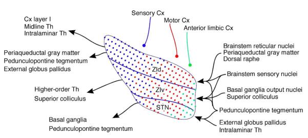
Summary diagram shows that the projections from the sensory, motor, and anterior limbic cortices form large, cloud-like projection zones with fuzzy borders and extensive overlaps that encompass entire subthalamus. The diagram also shows other main afferent and efferent projections to the each subdivision of the subthalamus. Cx, cerebral cortex; Th, thalamic nuclei; STN, subthalamic nucleus; ZId and ZIv, dorsal and ventral subdivision of zona incerta.
Methodological considerations
The most significant advantage of the lentivirus tracing method is that there is absolutely no labeling of fibers of passage. Both high molecular weight (10 k) biotinylated dextran amines (BDA) and Phaseolus vulgaris leucoagglutinin (PHA-L) can be taken up by fibers of passage, although to a lesser degree than low molecular weight BDA and horseradish peroxidase (HRP) (Reiner et al, 2000). The viruses we used appeared to do no damage to neurons at the injection sites as judged by the morphological integrity of the injection sites observed under the light-microscope. The viruses infected neurons in an all-or-none manner, and the somata and axons of all the infected neurons express abundant GFP. Therefore, the extent of the injection site and the projection sites can be clearly identified. In the projection sites, the axons of infected neurons, even small caliber axons and thin collaterals from a long distance away from the injection sites, expressed abundant GFP, and Golgi-like GFP staining characteristics allowed clear visualization of fine axons with boutons. Visualization of thin axons containing small amounts of PHA-L and BDA was often difficult in our experience (Kita and Kitai, 1987; Kita and Kita, 2012).
We know of only three anterograde tracing studies of Cx-subthalamic projections that systematically covered large cortical areas. There were two studies on Cx-STN projections in the monkey (Nambu et al., 1996; Haynes and Haber, 2013) and one on Cx-ZI of the rat (Mitrofanis and Mikuletic, 1999), and all three studies used the BDA method. This is the first study to analyze both STN and ZI using the most advanced method existing today. This study also systematically covered functionally diverse cortical areas.
Functional zones
The large-scale organization of the functional zones found in this study is as follows: The parietal sensory association and somatosensory Cx projecting zones occupy a large part of dorsolateral ZId and ZIv. The locations of the parietal association and the somatosensory Cx projecting zones in ZI were similar to but much larger than those shown using BDA-method (Mitrofanis and Mikuletic, 1999; Mitrofanis, 2005). This study confirmed the observation made by classical HRP and WGA-HRP methods in rats, cats, and monkeys that STN has small somatosensory Cx projecting zones at the dorsolateral-most part of STN, which was located adjacent to the somatosensory zone in ZIv (Carpenter et al., 1981; Canteras et al., 1988; Noda and Oka, 1993), while studies using autoradiographic methods denied the projection (e.g., Afsharpour, 1985). This study revealed that the parietal association Cx also projects to a similar area of STN.
The motor Cx zone includes a large middle part of STN and a large medial 2/3 of the ZI. In general, the locations of the zones were similar to previous reports on STN projections (Afsharpour, 1985; Canteras et al., 1990; Nambu et al., 1996; Haynes and Haber, 2013) and on ZI projections (Mitrofanis and Mikuletic, 1999). However, the areas seen in the present study were much larger than previously implied. We also noted that the STN of primates and rodents have slightly different topographies of the functional zones. The primate has a large dorsolateral prefrontal association Cx that projects to the mid-mediolateral part of STN, and the motor Cx innervates to the lateral part of STN (Parent and Hazrati, 1995; Haynes and Haber, 2013), while in rodents, the area equivalent to the primate dorsolateral prefrontal Cx may be very small or not existent (Vertes, 2004; Preuss, 1995), and projections from the rat motor cortex occupy the mid-mediolateral part of STN.
The ACg has been often considered as a prefrontal structure because of its involvement in various motor related cognitive functions (Harrison and Mair, 1996; Dalley et al., 2004). However, a micro-stimulation study in the rat suggested that the dorsal part of the ACg includes the eye and nose areas of the primary motor Cx (Brecht M, et al 2004). We found that the dorsal part of ACg projects heavily to STN and ZI. The ACg projection areas included the ventromedial part of the caudal half of STN and the medial 2/3 of both ZId and ZIv. These areas greatly overlapped with AGm projecting areas. ACg and AGm projections to other brain sites were also very similar (Gabbott et al., 2005). Mitrofanis and Mikuletic (1999) reported that BDA injections into the caudal part of dorsal ACg densely and diffusely labeled the medial 2/3 of ZId. These observations suggest that the ACg may have dual functions, primarily motor and motor related cognitive, and in the latter, eye and nose movements may play essential roles.
The anteromedial limbic Cx projection zone includes the ventromedial part of STN, adjacent H2, and lateral hypothalamus. No STN projection was seen from the visual Cx. The limbic, orbital, and visual Cx only sparsely project to ZI.
The functional zones described above extensively overlapped with each other. The overlap may be not unique to the rodent STN. The primate prefrontal, premotor, motor, and limbic projections to STN also have extensive overlaps (Nambu et al., 1996; Haynes and Haber, 2013). Also, the extensive overlap in STN may be not unique to cortical projections. Bevan et al (1997) showed that projections from the external segment of globus pallidus and the ventral pallidum, sensorimotor, and limbic structures respectively, to STN converge onto the same neurons. There is currently very little data on the primate Cx-ZI projections, and therefore, it is not possible to compare them at this time. The neurons in the STN and ZI have dendritic arbors extending a long distance in the dorsolateral to ventromedial direction (Kita et al., 1983; Bartho et al. 2007). Both the projection patterns and the morphological characteristics described above suggest that each neuron in the subthalamus may receive inputs from wide areas of Cx.
Somatotopic representations
Previous studies suggested that projections from the primate motor Cx to STN and rat somatosensory Cx projections to ZI have somatotopic representations (Nambu et al., 1996; Nicolelis et al., 1992; Shaw and Mitrofanis, 2002). It was evident in this study that axons originating from different locations in the somatosensory or the motor Cx resulted in the labeling of terminal fields in slightly different but largely overlapping locations in STN and ZI. It was also evident that axons originating from a small area in Cx formed large terminal fields in STN and ZI. These observations suggest that the projections from motor and somatosensory Cx have extensively overlapped, fuzzy somatotopic representations in STN, ZId, and ZIv.
Other afferent and efferent projections of the subthalamus
The STN receives strong afferent projections from GPe, ventral pallidum, intralaminar thalamic nuclei, and PPTg. These projections have extensively overlapped topographical organizations similar to Cx projections (Groenewegen et al., 1993; Maurice et al., 1998; Kita, 2007). The STN sends glutamatergic outputs mainly to other basal ganglia nuclei (Ricardo, 1980; Kita and Kitai, 1987). Thus, STN may be involved in an associative function by integrating motor, prefrontal, and limbic related inputs that are required to perform complex behaviors. This assumption is supported by the observation that STN is more active during complex movement tasks (Lehéricy et al., 2006).
The ZId receives afferent projections from several brainstem nuclei, including the dorsal raphe, periaqueductal grey matter, PPTg, and midbrain and pontine reticular nuclei (Ricardo, 1981; Watanabe and Kawana, 1982; Border et al., 1986; Shammah-Lagnado et al., 1985; Kolmac et al., 1998). The sensory trigeminal nuclear complex and the dorsal column nuclei also project to ZId (Nicolelis et al. 1992). These projections are diffuse and have no clear topography in ZId. Thus, projections from various functional domains converge on ZId (Ricardo, 1981; Shammah-Lagnado et al., 1985 and 1987; Kolmac et al., 1998).
The GABAergic neurons in ZId project to the layer I of a very wide area of Cx in a diffuse and rough topographical manner, to midline and intralaminar thalamic nuclei, to the pretectal area, and to pontine reticular nuclei (Shammah-Lagnado et al., 1987; Lin et al., 1990 and 1997; Nicolelis et al. 1992 and 1995). Known projection sites of glutamatergic neurons in ZId include the globus pallidus, periaqueductal gray, and PPTg (Beitz, 1989; Heise and Mitrofanis, 2004).
The ZIv receives topographically organized projections from the deep layers of the superior colliculus, the sensory trigeminal nuclear complex, and the dorsal column nuclei (Nicolelis et al. 1992; Kolmac et al., 1998). Other afferents known to project to ZIv include the PPTg, the deep cerebellar nuclei, and the basal ganglia output nuclei GPi and SNr. The topographical arrangements of the afferent zones from the brainstem sensory and motor related nuclei roughly match to those Cx afferent zones (see also Roger and Cadusseau, 1985; Shammah-Lagnado et al., 1985).
The ZIv contains mostly GABAergic neurons. These neurons project to the superior colliculus and the higher-order thalamic nuclei such as the posterior and anterior pretectal nuclei and contralateral ZI (Watanabe and Kawana, 1982; Foster et al., 1989; Kolmac et al., 1998; Bartho et al., 2002). These afferent and efferent projections of ZIv appear to show clearer topography than ZId projections. There are heavy mutual intra-incertal connections between ZId and ZIv and also within the each subdivision.
Deep brain stimulation of the motor zone of subthalamus
Deep brain stimulation (DBS) of STN and other brain areas has been used for the treatment of parkinsonisms. DBS sites effective for parkinsonisms include STN and ZI motor zones. Plaha et al. (2006) suggested that DBS of the ZI motor zone might produce more beneficial effects than DBS of STN in some cases. DBS of ZI is also effective for essential tremor and parkinsonian tremor (Xie et al., 2012). We speculate that the DBS of STN and ZI motor zones may exert different effects because these areas have different connections and may have different functional roles as discussed above.
In vivo unit recordings, stimulation of the motor Cx induces a short latency excitation in STN and ZI neurons (Fujimoto and Kita, 1993; Bartho et al., 2007). Thus Cx stimulation may be used to locate effective DBS sites. In order to distinguish STN and ZI motor zones, the short latency response should be combined with additional observations such as unique response and firing patterns of neurons in these nuclei (Bartho et al., 2007; Lavallee et al., 2005). It should also be noted that because of the extensive overlap of different functional zones and extensive local collateral connections, an activation of only motor related circuits by DBS of the subthalamus is not possible.
Functional implications
Based on the present and previous studies, we suggest that the subthalamus integrates and analyzes a broad spectrum of inputs and performs two modes of control. One mode of control is to integrate diverse cortical and brainstem inputs and outputs that control appropriate arousal, attention, sleep-waking cycle, nociceptive behavior, and posture-locomotive responses through the direct and thalamus-mediated projections to the Cx and brainstem, as postulated by several authors (Ricardo, 1981; Steriade et al., 1982; Shammah-Lagnado et al., 1985; Berry et al., 1986; Kolmac et al., 1998; Power and Mitrofanis, 2001; Mitrofanis, 2002 and 2005). The global projection patterns of ZId described above may be well suited for this mode of control (Kolmac et al., 1998; Power and Mitrofanis, 2001). We postulate that integration of coincidentally arriving weak multi-modal inputs from brainstem nuclei or multiple areas of Cx can drive ZI neurons. Several behavioral studies showed that electrical or chemical stimulation of ZI generates locomotor activity, some of which are associated with defense orientation (Mogenson et al., 1985; Milner and Mogenson, 1988; Murer and Pazo, 1993; Supko et al., 1992; Pèrier et al., 2002). For the general sensory function, activation of ZI with glutamate has antinociceptive effects in the rat (Petronilho et al., 2012). Bartho et al. 2007 showed that activity of both ZId and ZIv neurons are synchronous to layer V neurons in the somatosensory cortex. They suggested that the wide spread projections from Cx drive the ZI activity and that the GABAergic efferent signals from ZI neurons may play a critical role in synchronizing thalamocortical and brainstem rhythms. The DBS effects discussed above may be due to a suppression of abnormal oscillations in the motor zones of the subthalamus.
The second mode is to control specific events through more functionally specific input-output channels. Nicolelis et al. (1992) found that individual ZId and ZIv neurons have multi-whisker receptive fields with low-threshold activation to whisker movements. Ma (1990) found that the firing of some ZIv neurons pause prior to the beginning of and during saccades. The pauses were related to the duration of but unrelated to the direction of saccades. These observations suggest that ZI may be involved in the generation of orientative head and eye movements probably through the superior colliculus. We have often tested neurons in the motor zones of ZId and ZIv by electrical stimulation of the motor cortex in rats and monkeys during recording electrode penetrations to STN. We observed that single low intensity stimulation could excite most of the neurons with a very short latency, suggesting direct inputs from a small area of the motor Cx could activate the ZI neurons (Kita, unpublished observations). Urbain and Deschenes (2007) showed that the projection from the motor Cx to ZI gates vibrissal responses in a thalamocortical pathway. For the gating, motor Cx activates GABAergic neurons in the motor zone of ZI, which then inhibits thalamic projecting neurons in the ZIv sensory zone through intra-incertal connections. An opposite direction of control, from the ZI sensory zone to the ZI motor zone through intra-incertal connections, is also possible. Trageser et al. (2006) proposed that PPTg-ZIv-posterior thalamic projections are involved in the behavioral state dependent gating of sensory inputs. These recent studies suggest ZI is involved in the control of specific sensorimotor functions. The present study suggests that the layer V neurons in the wide areas of sensorimotor Cx simultaneously control basal ganglia, sensory gating, arousal, and attention functions respectively through STN, ZIv, and ZId.

Lentivirus anterograde tracing study in the rat revealed that the subthalamus receives heavy projections from the motor and sensory cortices, the cortico-subthalamic projections have a large-scale functional organization that encompasses both the subthalamic nucleus (STN) and two subdivisions of the zona incerta (ZIv and ZId), and the group of cortical axons that originate from a particular area of Cx sequentially innervate and form separate terminal fields in STN, ZIv, and ZId.
141×141mm (72 × 72 DPI)
ACKNOWLEDGMENTS
This work was supported by the National Institute of Neurological Disorders and Stroke Grant NS-57236. We thank B. Chism for technical assistance and R. Kita for editing the manuscript.
Footnotes
CONFLICT OF INTEREST STATEMENT The authors declare no conflicts of interest.
ROLE OF AUTHORS HK designed research. TK and HK performed research and analyzed data. TK, PO, and HK wrote the paper.
References
- Afsharpour S. Topographical projections of the cerebral cortex to the subthalamic nucleus. J Comp Neurol. 1985;236(1):14–28. doi: 10.1002/cne.902360103. [DOI] [PubMed] [Google Scholar]
- Bartho P, Slezia A, Varga V, Bokor H, Pinault D, Buzsaki G, Acsady L. Cortical control of zona incerta. J Neurosci. 2007;27(7):1670–1681. doi: 10.1523/JNEUROSCI.3768-06.2007. [DOI] [PMC free article] [PubMed] [Google Scholar]
- Beitz AJ. Possible origin of glutamatergic projections to the midbrain periaqueductal gray and deep layer of the superior colliculus of the rat. Brain Res Bull. 1989;23(1-2):25–35. doi: 10.1016/0361-9230(89)90159-7. [DOI] [PubMed] [Google Scholar]
- Berry DJ, Ohara PT, Jeffery G, Lieberman AR. Are there connections between the thalamic reticular nucleus and the brainstem reticular formation? J Comp Neurol. 1986;243(3):347–362. doi: 10.1002/cne.902430306. [DOI] [PubMed] [Google Scholar]
- Bevan MD, Clarke NP, Bolam JP. Synaptic integration of functionally diverse pallidal information in the entopeduncular nucleus and subthalamic nucleus in the rat. J Neurosci. 1997;17(1):308–324. doi: 10.1523/JNEUROSCI.17-01-00308.1997. [DOI] [PMC free article] [PubMed] [Google Scholar]
- Border BG, Kosinski RJ, Azizi SA, Mihailoff GA. Certain basilar pontine afferent systems are GABA-ergic: combined HRP and immunocytochemical studies in the rat. Brain Res Bull. 1986;17(2):169–179. doi: 10.1016/0361-9230(86)90113-9. [DOI] [PubMed] [Google Scholar]
- Brecht M, Krauss A, Muhammad S, Sinai-Esfahani L, Bellanca S, Margrie TW. Organization of rat vibrissa motor cortex and adjacent areas according to cytoarchitectonics, microstimulation, and intracellular stimulation of identified cells. J Comp Neurol. 2004;479(4):360–373. doi: 10.1002/cne.20306. [DOI] [PubMed] [Google Scholar]
- Canteras NS, Shammah-Lagnado SJ, Silva BA, Ricardo JA. Somatosensory inputs to the subthalamic nucleus: a combined retrograde and anterograde horseradish peroxidase study in the rat. Brain Res. 1988;458(1):53–64. doi: 10.1016/0006-8993(88)90495-7. [DOI] [PubMed] [Google Scholar]
- Canteras NS, Shammah-Lagnado SJ, Silva BA, Ricardo JA. Afferent connections of the subthalamic nucleus: a combined retrograde and anterograde horseradish peroxidase study in the rat. Brain Res. 1990;513(1):43–59. doi: 10.1016/0006-8993(90)91087-w. [DOI] [PubMed] [Google Scholar]
- Carpenter MB, Carleton SC, Keller JT, Conte P. Connections of the subthalamic nucleus in the monkey. Brain Res. 1981;224(1):1–29. doi: 10.1016/0006-8993(81)91113-6. [DOI] [PubMed] [Google Scholar]
- Celio MR, Heizmann CW. Calcium-binding protein parvalbumin as a neuronal marker. Nature. 1981;293(5830):300–302. doi: 10.1038/293300a0. [DOI] [PubMed] [Google Scholar]
- Dalley JW, Cardinal RN, Robbins TW. Prefrontal executive and cognitive functions in rodents: neural and neurochemical substrates. Neurosci Biobehav Rev. 2004;28(7):771–784. doi: 10.1016/j.neubiorev.2004.09.006. [DOI] [PubMed] [Google Scholar]
- Degos B, Deniau JM, Le Cam J, Mailly P, Maurice N. Evidence for a direct subthalamo cortical loop circuit in the rat. Eur J Neurosci. 2008;27(10):2599–2610. doi: 10.1111/j.1460-9568.2008.06229.x. [DOI] [PubMed] [Google Scholar]
- Deschenes M, Bourassa J, Pinault D. Corticothalamic projections from layer V cells in rat are collaterals of long-range corticofugal axons. Brain Res. 1994;664(1-2):215–219. doi: 10.1016/0006-8993(94)91974-7. [DOI] [PubMed] [Google Scholar]
- Dittgen T, Nimmerjahn A, Komai S, Licznerski P, Waters J, Margrie TW, Helmchen F, Denk W, Brecht M, Osten P. Lentivirus-based genetic manipulations of cortical neurons and their optical and electrophysiological monitoring in vivo. Proc Natl Acad Sci U S A. 2004;101(52):18206–18211. doi: 10.1073/pnas.0407976101. [DOI] [PMC free article] [PubMed] [Google Scholar]
- Feldman SG, Kruger L. An axonal transport study of the ascending projection of medial lemniscal neurons in the rat. J Comp Neurol. 1980;192(3):427–454. doi: 10.1002/cne.901920305. [DOI] [PubMed] [Google Scholar]
- Foster GA, Sizer AR, Rees H, Roberts MH. Afferent projections to the rostral anterior pretectal nucleus of the rat: a possible role in the processing of noxious stimuli. Neuroscience. 1989;29(3):685–694. doi: 10.1016/0306-4522(89)90141-3. [DOI] [PubMed] [Google Scholar]
- Fujimoto K, Kita H. Response characteristics of subthalamic neurons to the stimulation of the sensorimotor cortex in the rat. Brain Res. 1993;609(1-2):185–192. doi: 10.1016/0006-8993(93)90872-k. [DOI] [PubMed] [Google Scholar]
- Gabbott PL, Warner TA, Jays PR, Salway P, Busby SJ. Prefrontal cortex in the rat: projections to subcortical autonomic, motor, and limbic centers. J Comp Neurol. 2005;492(2):145–177. doi: 10.1002/cne.20738. [DOI] [PubMed] [Google Scholar]
- Groenewegen HJ, Berendse HW, Haber SN. Organization of the output of the ventral striatopallidal system in the rat: ventral pallidal efferents. Neuroscience. 1993;57(1):113–142. doi: 10.1016/0306-4522(93)90115-v. [DOI] [PubMed] [Google Scholar]
- Harrison LM, Mair RG. A comparison of the effects of frontal cortical and thalamic lesions on measures of spatial learning and memory in the rat. Behav Brain Res. 1996;75(1-2):195–206. doi: 10.1016/0166-4328(96)00173-8. [DOI] [PubMed] [Google Scholar]
- Haynes WI, Haber SN. The organization of prefrontal subthalamic inputs in primates provides an anatomical substrate for both functional specificity and integration: implications for Basal Ganglia models and deep brain stimulation. J Neurosci. 2013;33(11):4804–4814. doi: 10.1523/JNEUROSCI.4674-12.2013. [DOI] [PMC free article] [PubMed] [Google Scholar]
- Heise CE, Mitrofanis J. Evidence for a glutamatergic projection from the zona incerta to the basal ganglia of rats. J Comp Neurol. 2004;468(4):482–495. doi: 10.1002/cne.10971. [DOI] [PubMed] [Google Scholar]
- Kamikouchi A, Shimada T, Ito K. Comprehensive classification of the auditory sensory projections in the brain of the fruit fly Drosophila melanogaster. J Comp Neurol. 2006;499(3):317–356. doi: 10.1002/cne.21075. [DOI] [PubMed] [Google Scholar]
- Kita H. Globus pallidus external segment. Prog Brain Res. 2007;160:111–133. doi: 10.1016/S0079-6123(06)60007-1. [DOI] [PubMed] [Google Scholar]
- Kita H, Chang HT, Kitai ST. The morphology of intracellularly labeled rat subthalamic neurons: a light microscopic analysis. J Comp Neurol. 1983;215(3):245–257. doi: 10.1002/cne.902150302. [DOI] [PubMed] [Google Scholar]
- Kita H, Kita T. Number, origins, and chemical types of rat pallidostriatal projection neurons. J Comp Neurol. 2001;437(4):438–448. doi: 10.1002/cne.1294. [DOI] [PubMed] [Google Scholar]
- Kita T, Kita H. The subthalamic nucleus is one of multiple innervation sites for long range corticofugal axons: a single-axon tracing study in the rat. J Neurosci. 2012;32(17):5990–5999. doi: 10.1523/JNEUROSCI.5717-11.2012. [DOI] [PMC free article] [PubMed] [Google Scholar]
- Kita H, Kitai ST. Efferent projections of the subthalamic nucleus in the rat: light and electron microscopic analysis with the PHA-L method. J Comp Neurol. 1987;260(3):435–452. doi: 10.1002/cne.902600309. [DOI] [PubMed] [Google Scholar]
- Kolmac CI, Power BD, Mitrofanis J. Patterns of connections between zona incerta and brainstem in rats. J Comp Neurol. 1998;396(4):544–555. doi: 10.1002/(sici)1096-9861(19980713)396:4<544::aid-cne10>3.0.co;2-g. [DOI] [PubMed] [Google Scholar]
- Kolomiets BP, Deniau JM, Mailly P, Menetrey A, Glowinski J, Thierry AM. Segregation and convergence of information flow through the cortico- subthalamic pathways. J Neurosci. 2001;21(15):5764–5772. doi: 10.1523/JNEUROSCI.21-15-05764.2001. [DOI] [PMC free article] [PubMed] [Google Scholar]
- Lavallee P, Urbain N, Dufresne C, Bokor H, Acsady L, Deschenes M. Feedforward inhibitory control of sensory information in higher-order thalamic nuclei. J Neurosci. 2005;25(33):7489–7498. doi: 10.1523/JNEUROSCI.2301-05.2005. [DOI] [PMC free article] [PubMed] [Google Scholar]
- Lehericy S, Bardinet E, Tremblay L, Van de Moortele PF, Pochon JB, Dormont D, Kim DS, Yelnik J, Ugurbil K. Motor control in basal ganglia circuits using fMRI and brain atlas approaches. Cereb Cortex. 2006;16(2):149–161. doi: 10.1093/cercor/bhi089. [DOI] [PubMed] [Google Scholar]
- Levesque M, Gagnon S, Parent A, Deschenes Axonal arborizations of corticostriatal and corticothalamic fibers arising from the second somatosensory area in the rat. Cereb Cortex. 1996;6(6):759–770. doi: 10.1093/cercor/6.6.759. [DOI] [PubMed] [Google Scholar]
- Lin CS, Nicolelis MA, Schneider JS, Chapin JK. A major direct GABAergic pathway from zona incerta to neocortex. Science. 1990;248(4962):1553–1556. doi: 10.1126/science.2360049. [DOI] [PubMed] [Google Scholar]
- Lin RC, Nicolelis MA, Chapin JK. Topographic and laminar organizations of the incertocortical pathway in rats. Neuroscience. 1997;81(3):641–651. doi: 10.1016/s0306-4522(97)00094-8. [DOI] [PubMed] [Google Scholar]
- Lois C, Hong EJ, Pease S, Brown EJ, Baltimore D. Germline transmission and tissue-specific expression of transgenes delivered by lentiviral vectors. Science. 2002;295(5556):868–872. doi: 10.1126/science.1067081. [DOI] [PubMed] [Google Scholar]
- Maurice N, Deniau JM, Menetrey A, Glowinski J, Thierry AM. Prefrontal cortex-basal ganglia circuits in the rat: involvement of ventral pallidum and subthalamic nucleus. Synapse. 1998;29(4):363–370. doi: 10.1002/(SICI)1098-2396(199808)29:4<363::AID-SYN8>3.0.CO;2-3. [DOI] [PubMed] [Google Scholar]
- Milner KL, Mogenson GJ. Electrical and chemical activation of the mesencephalic and subthalamic locomotor regions in freely moving rats. Brain Res. 1988;452(1-2):273–285. doi: 10.1016/0006-8993(88)90031-5. [DOI] [PubMed] [Google Scholar]
- Mitrofanis J. Evidence for an auditory subsector within the zona incerta of rats. Anat Embryol (Berl) 2002;205(5-6):453–462. doi: 10.1007/s00429-002-0268-3. [DOI] [PubMed] [Google Scholar]
- Mitrofanis J. Some certainty for the “zone of uncertainty”? Exploring the function of the zona incerta. Neuroscience. 2005;130(1):1–15. doi: 10.1016/j.neuroscience.2004.08.017. [DOI] [PubMed] [Google Scholar]
- Mitrofanis J, Mikuletic L. Organisation of the cortical projection to the zona incerta of the thalamus. J Comp Neurol. 1999;412(1):173–185. [PubMed] [Google Scholar]
- Mogenson GJ, Swanson LW, Wu M. Evidence that projections from substantia innominata to zona incerta and mesencephalic locomotor region contribute to locomotor activity. Brain Res. 1985;334(1):65–76. doi: 10.1016/0006-8993(85)90568-2. [DOI] [PubMed] [Google Scholar]
- Mullen RJ, Buck CR, Smith AM. NeuN, a neuronal specific nuclear protein in vertebrates. Development. 1992;116(1):201–211. doi: 10.1242/dev.116.1.201. [DOI] [PubMed] [Google Scholar]
- Murer MG, Pazo JH. Circling behaviour induced by activation of GABAA receptors in the subthalamic nucleus. Neuroreport. 1993;4(11):1219–1222. doi: 10.1097/00001756-199309000-00002. [DOI] [PubMed] [Google Scholar]
- Nambu A, Takada M, Inase M, Tokuno H. Dual somatotopical representations in the primate subthalamic nucleus: evidence for ordered but reversed body-map transformations from the primary motor cortex and the supplementary motor area. J Neurosci. 1996;16(8):2671–2683. doi: 10.1523/JNEUROSCI.16-08-02671.1996. [DOI] [PMC free article] [PubMed] [Google Scholar]
- Nicolelis MA, Chapin JK, Lin RC. Somatotopic maps within the zona incerta relay parallel GABAergic somatosensory pathways to the neocortex, superior colliculus, and brainstem. Brain Res. 1992;577(1):134–141. doi: 10.1016/0006-8993(92)90546-l. [DOI] [PubMed] [Google Scholar]
- Nicolelis MA, Chapin JK, Lin RC. Development of direct GABAergic projections from the zona incerta to the somatosensory cortex of the rat. Neuroscience. 1995;65(2):609–631. doi: 10.1016/0306-4522(94)00493-o. [DOI] [PubMed] [Google Scholar]
- Noda T, Oka H. Projections of the anterior coronal gyrus to the subthalamic nucleus in the cat: a combined retrograde and anterograde WGA-HRP study. Brain Res. 1993;605(2):305–308. doi: 10.1016/0006-8993(93)91755-h. [DOI] [PubMed] [Google Scholar]
- Parent A, Hazrati LN. Functional anatomy of the basal ganglia. II. The place of subthalamic nucleus and external pallidum in basal ganglia circuitry. Brain Res Brain Res Rev. 1995;20(1):128–154. doi: 10.1016/0165-0173(94)00008-d. [DOI] [PubMed] [Google Scholar]
- Perier C, Tremblay L, Feger J, Hirsch EC. Behavioral consequences of bicuculline injection in the subthalamic nucleus and the zona incerta in rat. J Neurosci. 2002;22(19):8711–8719. doi: 10.1523/JNEUROSCI.22-19-08711.2002. [DOI] [PMC free article] [PubMed] [Google Scholar]
- Petronilho A, Reis GM, Dias QM, Fais RS, Prado WA. Antinociceptive effect of stimulating the zona incerta with glutamate in rats. Pharmacol Biochem Behav. 2012;101(3):360–368. doi: 10.1016/j.pbb.2012.01.022. [DOI] [PubMed] [Google Scholar]
- Plaha P, Ben-Shlomo Y, Patel NK, Gill SS. Stimulation of the caudal zona incerta is superior to stimulation of the subthalamic nucleus in improving contralateral parkinsonism. Brain. 2006;129(Pt 7):1732–1747. doi: 10.1093/brain/awl127. [DOI] [PubMed] [Google Scholar]
- Power BD, Leamey CA, Mitrofanis J. Evidence for a visual subsector within the zona incerta. Vis Neurosci. 2001;18(2):179–186. doi: 10.1017/s0952523801182027. [DOI] [PubMed] [Google Scholar]
- Power BD, Kolmac CI, Mitrofanis J. Evidence for a large projection from the zona incerta to the dorsal thalamus. J Comp Neurol. 1999;404(4):554–565. [PubMed] [Google Scholar]
- Preuss TM. Do rats have prefrontal cortex? The rose-woolsey-akert program reconsidered. J Cogn Neurosci. 1995;7(1):1–24. doi: 10.1162/jocn.1995.7.1.1. [DOI] [PubMed] [Google Scholar]
- Reiner A, Veenman CL, Medina L, Jiao Y, Del Mar N, Honig MG. Pathway tracing using biotinylated dextran amines. J Neurosci Methods. 2000;103(1):23–37. doi: 10.1016/s0165-0270(00)00293-4. [DOI] [PubMed] [Google Scholar]
- Ricardo JA. Efferent connections of the subthalamic region in the rat. I. The subthalamic nucleus of Luys. Brain Res. 1980;202(2):257–271. doi: 10.1016/0006-8993(80)90140-7. [DOI] [PubMed] [Google Scholar]
- Ricardo JA. Efferent connections of the subthalamic region in the rat. II. The zona incerta. Brain research. 1981;214(1):43–60. doi: 10.1016/0006-8993(81)90437-6. [DOI] [PubMed] [Google Scholar]
- Roger M, Cadusseau J. Afferents to the zona incerta in the rat: a combined retrograde and anterograde study. J Comp Neurol. 1985;241(4):480–492. doi: 10.1002/cne.902410407. [DOI] [PubMed] [Google Scholar]
- Shammah-Lagnado SJ, Negrao N, Ricardo JA. Afferent connections of the zona incerta: a horseradish peroxidase study in the rat. Neuroscience. 1985;15(1):109–134. doi: 10.1016/0306-4522(85)90127-7. [DOI] [PubMed] [Google Scholar]
- Shammah-Lagnado SJ, Negrao N, Silva BA, Ricardo JA. Afferent connections of the nuclei reticularis pontis oralis and caudalis: a horseradish peroxidase study in the rat. Neuroscience. 1987;20(3):961–989. doi: 10.1016/0306-4522(87)90256-9. [DOI] [PubMed] [Google Scholar]
- Shaw V, Mitrofanis J. Anatomical evidence for somatotopic maps in the zona incerta of rats. Anat Embryol (Berl) 2002;206(1-2):119–130. doi: 10.1007/s00429-002-0280-7. [DOI] [PubMed] [Google Scholar]
- Simpson K, Wang Y, Lin RC. Patterns of convergence in rat zona incerta from the trigeminal nuclear complex: light and electron microscopic study. J Comp Neurol. 2008;507(4):1521–1541. doi: 10.1002/cne.21624. [DOI] [PMC free article] [PubMed] [Google Scholar]
- Steriade M, Parent A, Ropert N, Kitsikis A. Zona incerta and lateral hypothalamic afferents to the midbrain reticular core of cat--HRP and electrophysiological study. Brain Res. 1982;238(1):13–28. doi: 10.1016/0006-8993(82)90767-3. [DOI] [PubMed] [Google Scholar]
- Supko DE, Uretsky NJ, Wallace LJ. AMPA/kainic acid glutamate receptor antagonism in the zona incerta dorsal to the subthalamic nucleus inhibits amphetamine-induced stereotypy bur not locomotor activity. Brain Res. 1992;576(1):89–96. doi: 10.1016/0006-8993(92)90612-d. [DOI] [PubMed] [Google Scholar]
- Urbain N, Deschenes M. Motor cortex gates vibrissal responses in a thalamocortical projection pathway. Neuron. 2007;56(4):714–725. doi: 10.1016/j.neuron.2007.10.023. [DOI] [PubMed] [Google Scholar]
- Veinante P, Lavallee P, Deschenes M. Corticothalamic projections from layer 5 of the vibrissal barrel cortex in the rat. J Comp Neurol. 2000;424(2):197–204. doi: 10.1002/1096-9861(20000821)424:2<197::aid-cne1>3.0.co;2-6. [DOI] [PubMed] [Google Scholar]
- Vertes RP. Differential projections of the infralimbic and prelimbic cortex in the rat. Synapse. 2004;51(1):32–58. doi: 10.1002/syn.10279. [DOI] [PubMed] [Google Scholar]
- Watanabe K, Kawana E. The cells of origin of the incertofugal projections to the tectum, thalamus, tegmentum and spinal cord in the rat: a study using the autoradiographic and horseradish peroxidase methods. Neuroscience. 1982;7(10):2389–2406. doi: 10.1016/0306-4522(82)90203-2. [DOI] [PubMed] [Google Scholar]
- Xie T, Bernard J, Warnke P. Post subthalamic area deep brain stimulation for tremors: a mini-review. Transl Neurodegener. 2012;1(1):20. doi: 10.1186/2047-9158-1-20. [DOI] [PMC free article] [PubMed] [Google Scholar]