Abstract
Since its introduction in the early 1990s, the self-expandable metal stent (SEMS) has been increasingly used for the management of malignant colorectal obstruction, not only as a palliative method but also as a preoperative treatment in surgical candidates. However, more recently, concerns have been raised over stent complication rates. Early complications include pain, perforation, and rectal bleeding, and late complications include stent migration and stent obstruction. With the increasing use of SEMS for treatment, physicians need to be more aware of complications occurring after the placement of these stents. This review covers the technical considerations and management of complications after colonic stenting.
Keywords: Colon, Stents, Complications
INTRODUCTION
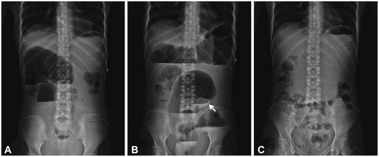
Malignant colorectal obstruction can occur as a result of primary or other metastatic cancers. Acute colorectal obstruction may be observed in 8% to 25% of patients with colorectal cancer, and it often leads to emergency surgical decompression.1 Since its introduction in the early 1990s, the self-expandable metal stent (SEMS) has been increasingly used for the management of malignant colorectal obstruction, not only as a palliative method but also as a preoperative treatment in surgical candidates.2 For palliative purposes, where surgical management might be considered unethical, stenting is likely to be a useful option, as it avoids a colostomy and improves the quality of life in many patients. In patients with inoperable tumors, the placement of SEMS offers a potentially effective palliation without the need for a permanent colostomy (Fig. 1). Several studies have assessed the effectiveness of SEMS, and it has been reported that the procedure reduces the hospital stay, medical costs, rates of stoma operations, and patient morbidity and mortality.3
Fig. 1.
(A) Abdominal radiograph of a 48-year-old woman with metastatic colonic carcinoma and local invasion who presented with acute colonic obstruction. Palliative decompression was offered because of her poor general condition and high surgical risk. (B) Radiograph obtained immediately after stent deployment showing partial expansion of the stent (arrow), with adequate flow of contrast material. (C) Abdominal radiograph obtained 24 hours after stent deployment showing decompression of the colonic obstruction; adequate position of the stent is noted.
However, minor complications related to colon stent placement, such as mild to moderate rectal bleeding, transient anorectal pain, and temporary incontinence, are common. More severe life-threatening complications have been described, such as perforation or death.4,5 The study of Maruthachalam et al.6 showed that endoscopic stent insertion increased the levels of carcinoembryonic antigen and/or cytokeratin 20 mRNA expression in the peripheral circulation of patients with colorectal cancer. In theory, SEMS insertion is an endoscopic procedure that could have deleterious effects on both tumor development and metastasis, and thus the effect of SEMS on the long-term outcome of those patients whose disease is potentially curable is still unclear. Zhang et al.7 compared the effects and results of stenting as a bridge method with those of emergency surgery. The use of a stent as a bridge method for left-sided colon cancer reduces the colostomy rate and postoperative complications. The mortality and long-term survival rates were not different.7 Concerning the risk factors associated with complications, complete colorectal obstruction and the use of covered stents contribute to the complications.8 The degree of occlusion has been reported to be one of the important risk factors, especially because a total obstruction may be associated with friable and damaged tissue.4 This review covers the technical considerations and management of complications after colonic stenting.
TECHNICAL CONSIDERATIONS
Fluoroscopy vs. colonoscopy
Methods of colonic stenting include fluoroscopy alone, colonoscopy alone, and a combination of fluoroscopy and colonoscopy. In the rectum and distal sigmoid colon, fluoroscopy alone or colonoscopy alone could be performed. However, in more proximal lesions, the combined approach is better. Patients with proximal lesions usually have marked colonic angulations. Colonoscopy can help in visualizing the approach the guidewire, and fluoroscopy is useful for documenting the length of an obstruction and for the diagnosis of perforation. The combined approach also reduces radiation exposure and is considered a priority treatment in most cases.
Stricture position and lesion
Although stent placements in the right colon are technically challenging, both right- and left-sided obstructions can be overcome. Surgical options are also considered for these lesions.9,10 Clearly, it may be a challenge to maneuver the colonoscope properly to reach the right side of the colon. A proximal location is reported to be associated with procedure failure.11 Other studies have reported that a distal location is more associated with a higher frequency of complications due to the bending load.4 Another study did not find any significant differences in terms of the obstruction site or malignant origins.8 The discrepancy between studies may be attributed to a true variation in the study design and baseline populations.
Most stent insertions can be performed in elective to emergency surgery. No difference in success rates and complications has been found between elective and emergency surgery. However, a close evaluation of the cancer is important because undetected synchronous lesions or an extrinsic compression mass can cause failure in adequately expanding the stent.12,13 Extracolonic malignancy has been considered a risk factor for the higher rate of complications because of its possible relation to carcinomatosis or immobilized bowel.14 The less friable luminal mucosa of an extracolonic malignant obstruction can be related with stent migration, which negatively affects the long-term clinical outcomes. Furthermore, colon stenting can also treat a benign obstructive lesion. Athreya et al.5 reviewed three cases of benign obstructive lesions, two of which were obstructions due to diverticulitis and one was due to ischemic change.
COMPLICATIONS
Mortality
Colonic stenting is considered a relatively safe procedure, and its mortality rate is roughly 1%.5 Another review carried out by Khot et al.15 showed similar results. In other studies, the mortality rate is 2.7%; it is safer than the surgical procedure, which has a mortality rate of 15% to 20%.16,17 In the review by Khot et al.,15 three patients died; two were found to have perforation, and decompression could not be achieved in the other. In the report by Dharmadhikari and Nice,18 two patients died; one death was due to perforation with comorbidities and the other was due to a sudden cardiac arrest.
Perforation
The perforation rate is about 4.8% in several meta-analysis studies.15,19 Incomplete initial stent expansion needs additional procedures such as balloon dilation. However, additional balloon dilation is not recommended because it could cause perforation. In addition, the peristaltic movements of the colon after decompression may result in more stent expansion. Khot et al.15 reported a perforation rate of 10% in a balloon-dilated group and 2% in a non-balloon-dilated group of patients. Fortunately, modern stents can achieve suitable expansion without additional dilation, even in tight strictures. Perforation can also occur in areas of tumor necrosis or during manipulation with angiographic catheters and guidewires in the presence of a diverticulum.20 The use of soft-tipped guidewires may help reduce perforation. Athreya et al.5 found four perforations among 87 patients; one patient had a perforation after balloon dilatation, and two patients was discovered a perforation during autopsy. The reasons for the perforation are as follows: one is that the sharp edge of the stent could perforate the proximally thinned bowel wall; another reason is that tumor ingrowth after colonic stenting could obstruct the lesion and perforate the more proximal site. If a patient complains of severe abdominal pain, perforation should be suspected, especially if the pain occurred immediately or shortly after the procedure.
Inadequate decompression
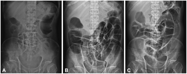
If adequate bowel decompression is not achieved, malpositioning of the stent or incomplete expansion should be suspected.21 Initial stent dilatation might usually be performed insufficiently on the first day, and physicians should pay attention to the gradual expansion of the stent during the next couple of days.18 Persistent colonic obstruction can also be related to undetected synchronic carcinomas that can occur in up to 5% of patients.12 Our patient presented with acute colonic obstruction with advanced gastric cancer, seeding metastasis, and segmental rectal wall thickening. Although palliative decompression was done, the symptom was not resolved because of the seeding metastasis (Fig. 2). Occasionally, rectal fecal impaction and retained stool may cause persistent obstruction.22
Fig. 2.
(A) Abdominal radiograph of an 82-year-old patient who presented with acute colonic obstruction with advanced gastric cancer, seeding metastasis, and segmental rectal wall thickening. (B) Palliative decompression was offered by means of rectal stenting. (C) However, the symptom was not resolved because of the seeding metastasis.
Migration
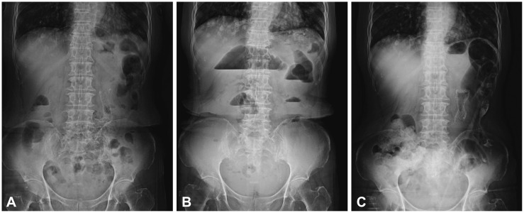
Several studies reported that the rate of stent migration is 4% to 10%.5,15,23 This most frequently occurs within the first 24 hours; however, migration at 3 and 7 days after the procedure has been reported by Canon et al.21 Stent migration occurs because of technical factors such as inadequate expansion. In addition, the smaller the stent diameter, the more the colon is angulated; also, the more insufficient the length is to allow stent flaring, the easier could stent migration occur. Furthermore, migration more frequently occurs after radiation therapy or chemotherapy, and in patients with a benign etiology.15 Although all of these reasons cause stent loosening, the lumen expands further and stent loosening has a benign outcome. Dharmadhikari and Nice18 reported that a stool softener might prevent migration because fecal impaction can attribute to migration. What is most interesting is that most patients remain asymptomatic even with stent migration, and further intervention is useless. However, some patients may complain about rectal irritation or symptoms of reobstruction, such as pain and constipation. Our patient had signs of reobstruction such as abdominal pain and nausea. She had a palliative procedure, and the stent migrated after 2 months. Restenting was done and the symptoms were relieved (Fig. 3).
Fig. 3.
(A) Abdominal radiograph of a female patient with signs of reobstruction such as abdominal pain and nausea. She underwent a palliative procedure and her symptoms were relieved. (B) However, the stent migrated after 2 months. She again presented symptoms of reobstruction. (C) Restenting was done and the symptoms were relieved.
Obstruction after a successful decompression
Obstruction after a successful decompression is sometimes a problem in palliative cases, and the most common cause of obstruction is tumor overgrowth in about 30%.24 To reduce tumor overgrowth, covered stents are recommended and overlap the edge of stent with non-diseased sections about 2 cm.18 In this complication, a second stent can be considered an option. In the study by Athreya et al.,5 two of 102 patients had obstruction after a successful decompression due to tumor overgrowth. One patient had palliative colostomy. The other patient had underlying diseases and metastatic disease; therefore, any kind of further treatments were impossible.5 Other causes of recurrent obstruction are stent migration and fecal impaction. Dehydration and medication can be associated with fecal impaction, and it can be treated by hydration or stopping medication.18
Bleeding
There are a few reported cases of bleeding.20 Mild to moderate low gastrointestinal bleeding has been reported in about 0% to 5%. In most cases, treatment was not required.24
Pain
The most common complication after colonic stenting is abdominal and rectal pain. However, specific treatments are not necessary.18 In the study by Choi et al.,8 one of 152 patients with rectal cancer experienced tenesmus, which was managed conservatively. Although the lesion was not within 5 cm from the anal verge, mucosal irritation caused by the distal end of the stent might have caused the tenesmus.8 Stenting in the very low rectum may cause severe tenesmus; therefore, it should be avoided.
CONCLUSIONS
Colonic stenting is a safe and effective method to resolve symptoms of obstruction. It shows good outcomes with technical and clinical success. Colonic stenting can also overcome obstruction caused by benign and malignant lesions. In patients with comorbidities, who are unfit for surgery, colonic stenting is more useful. Complications after stenting are more frequent in cases of complete obstruction or dilatation of a proximal lesion before stenting. Stenting may be dangerous and should be avoided in such cases. In a previous study, 13% of patients showed a complication within 30 days, and the late complication rate was 26.7%.10 The longer survival time of these patients might be the reason for the higher chances of stent-related complications. Although colonic stenting has a few fatal complications, the most frequent and severe complications may be successfully resolved with simple methods, such as repeat stenting or medications. Of course, awareness about the critical timing of seeking the aid of radiology and surgical teams is important, especially in fatal complications. Finally, colonic stenting could improve prognosis and outcome.
Footnotes
The authors have no financial conflicts of interest.
References
- 1.Keymling M. Colorectal stenting. Endoscopy. 2003;35:234–238. doi: 10.1055/s-2003-37265. [DOI] [PubMed] [Google Scholar]
- 2.Trompetas V. Emergency management of malignant acute left-sided colonic obstruction. Ann R Coll Surg Engl. 2008;90:181–186. doi: 10.1308/003588408X285757. [DOI] [PMC free article] [PubMed] [Google Scholar]
- 3.Helfer KS. Auditory and auditory-visual perception of clear and conversational speech. J Speech Lang Hear Res. 1997;40:432–443. doi: 10.1044/jslhr.4002.432. [DOI] [PubMed] [Google Scholar]
- 4.Small AJ, Coelho-Prabhu N, Baron TH. Endoscopic placement of self-expandable metal stents for malignant colonic obstruction: long-term outcomes and complication factors. Gastrointest Endosc. 2010;71:560–572. doi: 10.1016/j.gie.2009.10.012. [DOI] [PubMed] [Google Scholar]
- 5.Athreya S, Moss J, Urquhart G, Edwards R, Downie A, Poon FW. Colorectal stenting for colonic obstruction: the indications, complications, effectiveness and outcome: 5 year review. Eur J Radiol. 2006;60:91–94. doi: 10.1016/j.ejrad.2006.05.017. [DOI] [PubMed] [Google Scholar]
- 6.Maruthachalam K, Lash GE, Shenton BK, Horgan AF. Tumour cell dissemination following endoscopic stent insertion. Br J Surg. 2007;94:1151–1154. doi: 10.1002/bjs.5790. [DOI] [PubMed] [Google Scholar]
- 7.Zhang Y, Shi J, Shi B, Song CY, Xie WF, Chen YX. Self-expanding metallic stent as a bridge to surgery versus emergency surgery for obstructive colorectal cancer: a meta-analysis. Surg Endosc. 2012;26:110–119. doi: 10.1007/s00464-011-1835-6. [DOI] [PubMed] [Google Scholar]
- 8.Choi JH, Lee YJ, Kim ES, et al. Covered self-expandable metal stents are more associated with complications in the management of malignant colorectal obstruction. Surg Endosc. 2013;27:3220–3227. doi: 10.1007/s00464-013-2897-4. [DOI] [PubMed] [Google Scholar]
- 9.Binkert CA, Ledermann H, Jost R, Saurenmann P, Decurtins M, Zollikofer CL. Acute colonic obstruction: clinical aspects and cost-effectiveness of preoperative and palliative treatment with self-expanding metallic stents: a preliminary report. Radiology. 1998;206:199–204. doi: 10.1148/radiology.206.1.9423673. [DOI] [PubMed] [Google Scholar]
- 10.Abbott S, Eglinton TW, Ma Y, Stevenson C, Robertson GM, Frizelle FA. Predictors of outcome in palliative colonic stent placement for malignant obstruction. Br J Surg. 2014;101:121–126. doi: 10.1002/bjs.9340. [DOI] [PubMed] [Google Scholar]
- 11.Jung MK, Park SY, Jeon SW, et al. Factors associated with the long-term outcome of a self-expandable colon stent used for palliation of malignant colorectal obstruction. Surg Endosc. 2010;24:525–530. doi: 10.1007/s00464-009-0604-2. [DOI] [PubMed] [Google Scholar]
- 12.Arenas RB, Fichera A, Mhoon D, Michelassi F. Incidence and therapeutic implications of synchronous colonic pathology in colorectal adenocarcinoma. Surgery. 1997;122:706–709. doi: 10.1016/s0039-6060(97)90077-5. [DOI] [PubMed] [Google Scholar]
- 13.Miyayama S, Matsui O, Kifune K, et al. Malignant colonic obstruction due to extrinsic tumor: palliative treatment with a self-expanding nitinol stent. AJR Am J Roentgenol. 2000;175:1631–1637. doi: 10.2214/ajr.175.6.1751631. [DOI] [PubMed] [Google Scholar]
- 14.Keswani RN, Azar RR, Edmundowicz SA, et al. Stenting for malignant colonic obstruction: a comparison of efficacy and complications in colonic versus extracolonic malignancy. Gastrointest Endosc. 2009;69(3 Pt 2):675–680. doi: 10.1016/j.gie.2008.09.009. [DOI] [PubMed] [Google Scholar]
- 15.Khot UP, Lang AW, Murali K, Parker MC. Systematic review of the efficacy and safety of colorectal stents. Br J Surg. 2002;89:1096–1102. doi: 10.1046/j.1365-2168.2002.02148.x. [DOI] [PubMed] [Google Scholar]
- 16.Tekkis PP, Kinsman R, Thompson MR, Stamatakis JD Association of Coloproctology of Great Britain, Ireland. The Association of Coloproctology of Great Britain and Ireland study of large bowel obstruction caused by colorectal cancer. Ann Surg. 2004;240:76–81. doi: 10.1097/01.sla.0000130723.81866.75. [DOI] [PMC free article] [PubMed] [Google Scholar]
- 17.Leitman IM, Sullivan JD, Brams D, DeCosse JJ. Multivariate analysis of morbidity and mortality from the initial surgical management of obstructing carcinoma of the colon. Surg Gynecol Obstet. 1992;174:513–518. [PubMed] [Google Scholar]
- 18.Dharmadhikari R, Nice C. Complications of colonic stenting: a pictorial review. Abdom Imaging. 2008;33:278–284. doi: 10.1007/s00261-007-9240-2. [DOI] [PubMed] [Google Scholar]
- 19.Sebastian S, Johnston S, Geoghegan T, Torreggiani W, Buckley M. Pooled analysis of the efficacy and safety of self-expanding metal stenting in malignant colorectal obstruction. Am J Gastroenterol. 2004;99:2051–2057. doi: 10.1111/j.1572-0241.2004.40017.x. [DOI] [PubMed] [Google Scholar]
- 20.Mainar A, Tejero E, Maynar M, Ferral H, Castañeda-Zúñiga W. Colorectal obstruction: treatment with metallic stents. Radiology. 1996;198:761–764. doi: 10.1148/radiology.198.3.8628867. [DOI] [PubMed] [Google Scholar]
- 21.Canon CL, Baron TH, Morgan DE, Dean PA, Koehler RE. Treatment of colonic obstruction with expandable metal stents: radiologic features. AJR Am J Roentgenol. 1997;168:199–205. doi: 10.2214/ajr.168.1.8976946. [DOI] [PubMed] [Google Scholar]
- 22.Lopera JE, Ferral H, Wholey M, Maynar M, Castañeda-Zúñiga WR. Treatment of colonic obstructions with metallic stents: indications, technique, and complications. AJR Am J Roentgenol. 1997;169:1285–1290. doi: 10.2214/ajr.169.5.9353443. [DOI] [PubMed] [Google Scholar]
- 23.Clark JS, Buchanan GN, Khawaja AR, et al. Use of the Bard Memotherm self-expanding metal stent in the palliation of colonic obstruction. Abdom Imaging. 2003;28:518–524. doi: 10.1007/s00261-002-0059-6. [DOI] [PubMed] [Google Scholar]
- 24.Suzuki N, Saunders BP, Thomas-Gibson S, Akle C, Marshall M, Halligan S. Colorectal stenting for malignant and benign disease: outcomes in colorectal stenting. Dis Colon Rectum. 2004;47:1201–1207. doi: 10.1007/s10350-004-0556-5. [DOI] [PubMed] [Google Scholar]


