Abstract
Background and Purpose
The aim of this study was to elucidate the role of therapy-related cardiotoxicity in multiple sclerosis (MS) patients treated with mitoxantrone and to identify potential predictors for individual risk assessment.
Methods
Within a multicenter retrospective cohort design, cardiac side effects attributed to mitoxantrone were analyzed in 639 MS patients at 2 MS centers in Germany. Demographic, disease, treatment, and follow-up data were collected from hospital records. Patients regularly received cardiac monitoring during the treatment phase.
Results
None of the patients developed symptomatic congestive heart failure. However, the frequency of patients experiencing cardiac dysfunction of milder forms after mitoxantrone therapy was 4.1% (26 patients) among all patients. Analyses of the risk for cardiotoxicity revealed that cumulative dose exposure was the only statistically relevant risk factor associated with cardiac dysfunction.
Conclusions
The number of patients developing subclinical cardiac dysfunction below the maximum recommended cumulative dose is higher than was initially assumed. Interestingly, a subgroup of patients was identified who experienced cardiac dysfunction shortly after initiation of mitoxantrone and who received a low cumulative dose. Therefore, each administration of mitoxantrone should include monitoring of cardiac function to enhance the treatment safety for patients and to allow for early detection of any side effects, especially in potential high-risk subgroups (as determined genetically).
Keywords: multiple sclerosis, mitoxantrone, cardiotoxicity, dose dependency
Introduction
Mitoxantrone (MX) is an anthracenedione derivative that intercalates into DNA, inhibits topoisomerase II, and causes strand breaks that can result in chromosomal translocations.1 The results of a pivotal placebo-controlled, double-blinded, randomized, multicenter MX trial by the Mitoxantrone in Multiple Sclerosis Study Group (MIMS) about 10 years ago opened up a treatment option for patients suffering from highly active forms of relapsing-remitting multiple sclerosis (MS) (RRMS) and secondary progressive MS (SPMS).2 MX has therefore been used as an escalating immunotherapy in MS. Due to its approval and its promising results concerning disability and relapses among MS patients, as well as there being few alternative strategies available for SPMS, the number of patients receiving MX has increased in the last decade.
However, the MX lifetime dosage is limited by its potential toxicity, and in particular its cardiac and hematologic adverse reactions. In oncologic indications, MX-associated congestive heart failure (CHF) was reported in 2.6-6% of patients who received MX doses of up to 140 mg/m2 body surface area (BSA).3,4 The cardiotoxicity of the anthracycline family is considered to be dose-dependent and to result in myocardial damage that reduces the left ventricular ejection fraction (LVEF). In rare but potentially severe cases, cardiotoxicity results in CHF and other heart dysfunctions such as electrocardiogram (ECG) changes, arrhythmias, and clinical heart failure.5 Data on MX-related cardiotoxicity in MS are sparse, with the incident rate of symptomatic heart failure ranging between 0.2% and 2.0% for symptomatic heart failure.6,7,8,9,10,11 We have reported previously the occurrence of LVEF reduction in 4 out of 18 prospectively assessed patients at a very early stage of MX treatment.12 At present, the risk factors contributing to MX-related cardiotoxicity are not completely understood. This study was therefore performed to assess cardiac dysfunction subsequent to MX treatment in a large German cohort of MS patients, and to identify potential clinical predictors for individual risk assessment.
Methods
Within a multicenter retrospective cohort study performed at two MS centers (Departments of Neurology at the University Medical Centers at Mainz and Bochum) between 1995 and 2010, we collected data for 639 German MS patients treated with at least one infusion of MX, and analyzed the cardiac side effects attributed to MX. Patients diagnosed with worsening RRMS, SPMS, or primary progressive MS according to the current diagnostic criteria regularly received intravenous MX infusions every 3 months at a dose of 12 mg/m2 BSA per infusion, based on the MIMS protocol. Individualized dose adaptation by leukocyte nadirs was commonly performed. Patients were evaluated medically before treatment initiation, including an assessment of their scores on the Expanded Disability Status Scale (EDSS).
No patients experienced cardiac dysfunction before MX treatment. All new cardiac adverse events during treatment-including reduction of resting LVEF measured using transthoracic echocardiogram (<50% or an absolute reduction of ≥10% from the baseline), diastolic and systolic contraction abnormalities, and newly emerging arrhythmias that could not be attributed to any other origin-were regarded as therapy-related cardiac dysfunction. Both the clinical diagnosis and transthoracic echocardiography (TTE) were evaluated by a cardiologist. Assessments of LVEF and CHF by clinical examination, and ECG and TTE were performed at baseline and then annually after discontinuation or completion of the treatment, as recommended by the guidelines for the diagnosis and treatment of neurological diseases by the German Society of Neurology. During the treatment phase, the frequency of cardiac check-ups varied slightly between the patients: a TTE check-up was performed before each infusion in patients who had received MX in recent years, and biannually in patients who had received MX less recently. This slight discrepancy was due to changes associated with continuous reevaluations of guidelines concerning the monitoring of patients under MX treatment over the course of our relatively long observational period (1995-2012). The treatment was stopped when any type of cardiac dysfunction as described above was diagnosed.
Mean values and standard deviations are given for the descriptive analyses, in addition to the median with the 25th and 75th percentiles for the cumulative MX dose. Differences between two groups were assessed using the nonparametric two-tailed Mann-Whitney U test. Fisher's exact test was used to evaluate the effects of gender and disease course.
Logistic regression modeling was used to determine the significance of the effects of potential contributing factors (cumulative dose, age at initial diagnosis, EDSS score, gender, and disease course) on cardiotoxicity risk. The odds ratios are presented with 95% confidence intervals.
Since this was an explorative study, no adjustments for multiple testing were performed. The statistical tests were conducted for illustrative purposes rather than for hypothesis testing. Probability values are given for descriptive reasons only, and should be interpreted with caution and in connection with effect estimates.
All statistical analyses were performed using SPSS version 15.0 (SPSS Inc., Chicago, IL, USA), and the local ethics committees at the two involved centers approved the final protocol.
Results
We identified 639 MX-treated MS patients (400 females, 239 males) in the databases of the 2 university medical centers. Their mean age was 36.1 years (range, 21-74 years), and 102 had RRMS, 441 had SPMS, and 96 had primary progressive multiple sclerosis. Patients received a mean cumulative MX dose of 65.8 mg/m2 BSA, and 19.4% received more than 100 mg/m2 BSA (maximum, 184 mg/m2 BSA). The median cumulative dose was 60.0 mg/m2 BSA (25th percentile, 36.0 mg/m2; 75th percentile, 90.0 mg/m2). The mean baseline EDSS score was 5.6 (range, 2.0-8.5). The demographics and characteristics of the patients in this cohort are presented in Table 1.
Table 1.
Descriptive statistics of the study cohort
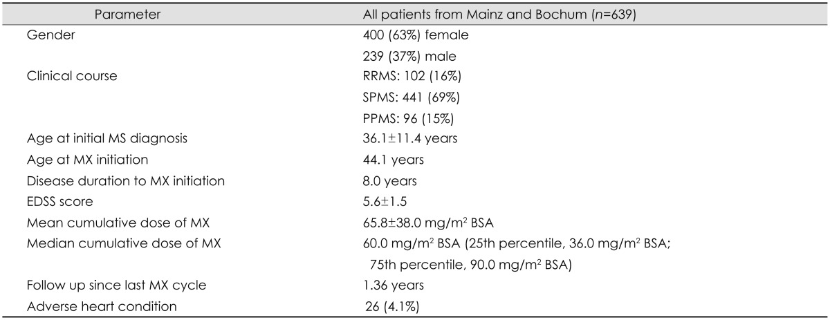
Data are mean±SD or (%) values.
BSA: body surface area, EDSS: Expanded Disability Status Scale, MS: multiple sclerosis, MX: mitoxantrone, PPMS: primary progressive multiple sclerosis, RRMS: relapsing-remitting multiple sclerosis, SPMS: secondary progressive multiple sclerosis.
Subclinical cardiac dysfunction was recorded for 26 of the 639 patients (4.1%) during the observation period. Among these, eight patients (1.25%) had an LVEF that had decreased below 50%, and nine patients (1.41%) exhibited an absolute LVEF reduction of ≥10% from the baseline. Nine additional cardiac-related adverse events were observed: arrhythmia (four patients, 0.63%), systolic dysfunction (three patients, 0.47%), diastolic relaxation dysfunction (one patient, 0.15%), and pericardial effusion (one patient, 0.15%). None of the patients in our cohort developed symptomatic CHF. The cumulative dose in patients with cardiac dysfunction varied between 12 and 184 mg/m2 BSA (mean, 84.2 mg/m2 BSA). Interestingly, delayed cardiac dysfunction occurred in one patient 6 months after treatment completion (reported hypokinesis and systolic dysfunction). The characteristics of patients with cardiac dysfunction are summarized in Table 2.
Table 2.
Characteristics of patients with cardiac dysfunction attributed to MX treatment
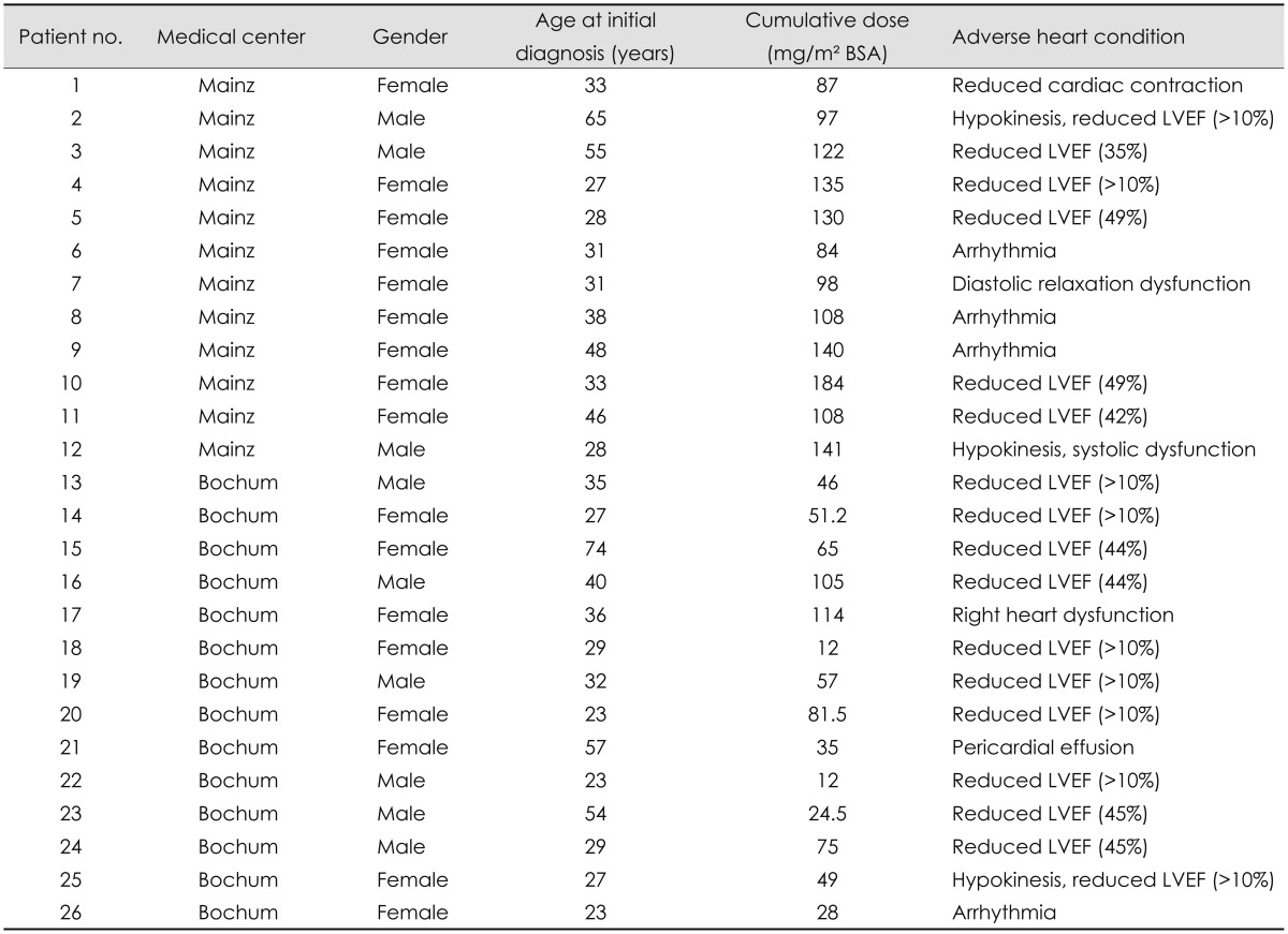
BSA: body surface area, LVEF: left ventricular ejection fraction, MX: mitoxantrone.
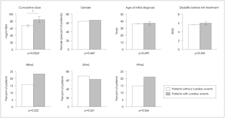
We examined the potential relationships between cardiotoxicity and clinical or demographic risk factors in our cohort. The mean cumulative dose was significantly higher in patients with cardiac dysfunction (n=26, 84.2 mg/m2 BSA) than in those without cardiac events (n=613, 64.9 mg/m2 BSA, p=0.0262) (Fig. 1). There was a statistically significant dose dependency when the cumulative dose was analyzed as a dichotomous covariate. Among patients with cardiac events, 15 (57.2%) received doses of >80 mg/m2 BSA, compared to 11 patients (42.3%) who received ≤80 mg/m2 BSA (p=0.0064). The incidence of cardiac dysfunction was 8.19% in patients receiving a cumulative dose of >80 mg/m2 BSA compared to 2.75% in patients receiving a cumulative dose of ≤80 mg/m2 BSA.
Fig. 1.
Analyses of potential relationships between the occurrence of cardiac events and patient characteristics. The cumulative dose was significantly higher in the patients group with cardiac events. No significant difference was observed between gender, age at initial diagnosis, disability before MX treatment or disease course, and the incidence of cardiac events. Data are mean and SEM values; *p<0.05. BSA: body surface area, EDSS: Expanded Disability Status Scale, MX: mitoxantrone, PPMS: primary progressive multiple sclerosis, RRMS: relapsing-remitting multiple sclerosis, SPMS: secondary progressive multiple sclerosis.
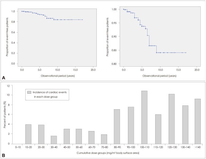
Furthermore, stepwise backward and forward logistic regression revealed that cumulative-dose exposure is the only statistically relevant risk factor associated with the risk of cardiac dysfunction (p=0.003). We concluded that the odds of cardiac dysfunction were 1.019 times higher for every mg/m2 BSA increase in MX cumulative dose, and that the 95% confidence interval for the odds ratio was 1.006-1.032 (Table 3). Fig. 2A illustrates the proportion of patients without cardiac dysfunction over the observational period in years as a Kaplan-Meier curve. Fig. 2B demonstrates the relationship between the incidence of cardiac dysfunction and cumulative dose. Interestingly, albeit at a lower incidence, cardiac dysfunction seems to occur shortly after initiation of MX and when receiving a low cumulative dose (10-30 mg/m2 BSA) in susceptible individuals.
Table 3.
Results of the logistic regression analysis
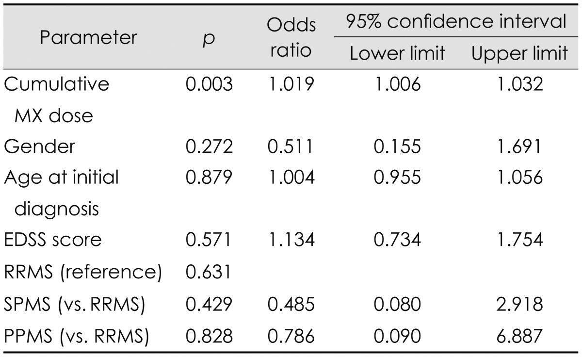
EDSS: Expanded Disability Status Scale, MX: mitoxantrone, PPMS: primary progressive multiple sclerosis, RRMS: relapsing-remitting multiple sclerosis, SPMS: secondary progressive multiple sclerosis.
Fig. 2.
Patients with cardiac events. A: Event-free curves (Kaplan-Meier curves) indicating the proportion of patients without cardiac dysfunction over the observation period (in years). B: The incidence of cardiac events by cumulative dose shown as the percentage of patients.
We did not identify a statistically significant association between gender, age at initial diagnosis, disability (EDSS score) or disease course, and the occurrence of cardiac events (Table 3, Fig. 1).
Seventeen of the 26 patients with cardiac dysfunction attended a clinical follow-up at least once following the initial detection of a cardiac event. We observed a transient cardiac dysfunction within 6 of these patients and persistent dysfunction in the other 11. The remaining nine patients either were lost to follow-up or could not be evaluated after discontinuation because the end of the observational period was less than 1 year after the last MX cycle.
Discussion
The benefit of MX as an escalation therapy for worsening RRMS and SPMS has been demonstrated in clinical trials.2,13 The restriction of a cumulative lifetime dosage of MX accounts for potential risks of treatment. Dose dependency is widely accepted, and particularly in terms of cardiotoxicity, although there are reports of patients with cardiac failure following low-dose treatment.12,14 The mechanisms of MX-associated cardiotoxicity are not completely understood, but mediation of myocardial tissue damage via interactions with cellular iron metabolism leading to formation of reactive oxygen has been suggested.15
The overall incidence of cardiac dysfunction in our study cohort (4.1%), as measured by cardiac echo and/or ECG, was higher than that found in the initial phase II and phase III MX clinical trials,2,16 but comparable to the range of other postmarketing studies. In a prospective French study, 4.9% of 802 patients presented with decreased LVEF.8 In the prospective Registry to Evaluate Novantrone Effects in Worsening Multiple Sclerosis study involving 509 patients, a reduction of LVEF was observed in 5.3% during the treatment phase and 5.6% during the follow-up phase.7
A retrospective analysis of 1,378 MS patients reported a reduction of LVEF in 1.8% of patients receiving cumulative doses of <100 mg/m2 BSA versus 5% of those receiving doses of >100 mg/m2 BSA.6 In our cohort the incidence of cardiac dysfunction was significantly higher in patients receiving a cumulative dose of >80 mg/m2 BSA (8.19%) than in those receiving a cumulative dose of >80 mg/m2 BSA (2.75%). Interestingly, albeit at a lower incidence, we observed cardiac dysfunction shortly after initiation of MX in the low-cumulative-dose groups, indicating that there may be a subgroup of susceptible individuals. This is in line with our previously reported reduction of LVEF in 4 out of 18 prospectively assessed MS patients very early in the MX treatment course.12,14
The reasons for early cardiac dysfunction in our cohort remain unclear, and so we are currently evaluating potential pharmacogenetic markers. None of our patients had a history of cardiac disease. Speculation has arisen as to whether early cardiotoxicity provides evidence of a dose-independent cross-reaction with the myocardium. The observed early change in LVEF might be effectively clinically compensated during first-treatment courses, although a subclinical decline in cardiac dysfunction or a temporary reduction of the LVEF might be appear transiently.
With regard to the parameters assessed herein, we did not identify a statistically significant association between gender, age at initial diagnosis, disease duration, disability (EDSS score), or disease course, and the occurrence of cardiac events. As reported previously, none of these indicators can be regarded as prognostic factors for MX-associated cardiac dysfunction.6,10 This highlights the need to identify pharmacogenetic biomarkers that allow individualized risk stratification. In this context, it has recently been reported that the bioactivation of MX mediated through CYP2E1 metabolism, which occurs locally in the cardiomyoblasts, plays a significant role in the cellular damage promoted by MX.17 Thus, cytochrome P450 polymorphisms might contribute to individual susceptibility to MX-induced cardiotoxicity. Furthermore, the relevance of ABC-transporter gene polymorphisms as potential pharmacogenetic markers for MX response and its cardiotoxicity in MS has been demonstrated.18 These potential pharmacogenetic risk factors are currently being investigated in prospective studies.19,20
Despite our large cohort, the present data must be interpreted with caution due to the use of a retrospective approach over different observation periods. In addition, the mean follow-up period was too short to comprehensively detect delayed MX-related cardiotoxicity, some of which might have developed after treatment discontinuation. However, two recent prospective studies have demonstrated a cardiotoxicity incidence similar to that reported here.7,8
Notwithstanding the above-mentioned limitation, the data reported here demonstrate that MX therapy in MS patients is associated with cardiotoxicity that is dose-dependent, but can occur very early in susceptible individuals. Since reliable markers for predicting the individual susceptibility to MX-induced cardiotoxicity are not yet available, cautious reevaluation of treatment indications should be performed regularly during the treatment course, and physicians should use the lowest possible cumulative dose that achieves the desired clinical effect. Studies have demonstrated a concomitant cardioprotective effect of dexrazoxane in combination with MX, but thus far this has not influenced the treatment recommendation.21,22 Nonetheless, treatment recommendations have narrowed the indications for MX treatment from the beginning for SPMS, progressive relapsing MS, or worsening RRMS, since the initial trials have been restricted to this cohort of patients.
We emphasize the importance of vigilant monitoring of patients for cardiac complications by assessment of cardiac signs and symptoms by history, physical examination, ECG, and TTE before the administration of each dose of MX, and then annually for up to 5 years following the cessation of treatment conducted according to the recommendations of the United States Food and Drug Administration published in 2008.23
Acknowledgements
This study was supported financially by the Johannes Gutenberg University of Mainz [JGU MAIFOR grant and a grant from the Inneruniversitäre Forschungsförderung (Stufe I) to F.L. and by the Competence Network for Multiple Sclerosis funded by the Federal Ministry of Education and Research.
We thank Darragh O'Neill for editing the manuscript as a native English speaker, and Eva Fucik and Michael Baier for supporting the collection of clinical data.
Footnotes
The authors have no financial conflicts of interest.
References
- 1.Neuhaus O, Kieseier BC, Hartung HP. Mechanisms of mitoxantrone in multiple sclerosis--what is known? J Neurol Sci. 2004;223:25–27. doi: 10.1016/j.jns.2004.04.015. [DOI] [PubMed] [Google Scholar]
- 2.Hartung HP, Gonsette R, König N, Kwiecinski H, Guseo A, Morrissey SP, et al. Mitoxantrone in progressive multiple sclerosis: a placebo-controlled, double-blind, randomised, multicentre trial. Lancet. 2002;360:2018–2025. doi: 10.1016/S0140-6736(02)12023-X. [DOI] [PubMed] [Google Scholar]
- 3.Arlin ZA, Silver R, Cassileth P, Armentrout S, Gams R, Daghestani A, et al. Phase I-II trial of mitoxantrone in acute leukemia. Cancer Treat Rep. 1985;69:61–64. [PubMed] [Google Scholar]
- 4.Mather FJ, Simon RM, Clark GM, Von Hoff DD. Cardiotoxicity in patients treated with mitoxantrone: Southwest Oncology Group phase II studies. Cancer Treat Rep. 1987;71:609–613. [PubMed] [Google Scholar]
- 5.Praga C, Beretta G, Vigo PL, Lenaz GR, Pollini C, Bonadonna G, et al. Adriamycin cardiotoxicity: a survey of 1273 patients. Cancer Treat Rep. 1979;63:827–834. [PubMed] [Google Scholar]
- 6.Ghalie RG, Mauch E, Edan G, Hartung HP, Gonsette RE, Eisenmann S, et al. A study of therapy-related acute leukaemia after mitoxantrone therapy for multiple sclerosis. Mult Scler. 2002;8:441–445. doi: 10.1191/1352458502ms836oa. [DOI] [PubMed] [Google Scholar]
- 7.Rivera VM, Jeffery DR, Weinstock-Guttman B, Bock D, Dangond F. Results from the 5-year, phase IV RENEW (Registry to Evaluate Novantrone Effects in Worsening Multiple Sclerosis) study. BMC Neurol. 2013;13:80. doi: 10.1186/1471-2377-13-80. [DOI] [PMC free article] [PubMed] [Google Scholar]
- 8.Le Page E, Leray E, Edan G French Mitoxantrone Safety Group. Long-term safety profile of mitoxantrone in a French cohort of 802 multiple sclerosis patients: a 5-year prospective study. Mult Scler. 2011;17:867–875. doi: 10.1177/1352458511398371. [DOI] [PubMed] [Google Scholar]
- 9.Goffette S, van Pesch V, Vanoverschelde JL, Morandini E, Sindic CJ. Severe delayed heart failure in three multiple sclerosis patients previously treated with mitoxantrone. J Neurol. 2005;252:1217–1222. doi: 10.1007/s00415-005-0839-3. [DOI] [PubMed] [Google Scholar]
- 10.Zingler VC, Nabauer M, Jahn K, Gross A, Hohlfeld R, Brandt T, et al. Assessment of potential cardiotoxic side effects of mitoxantrone in patients with multiple sclerosis. Eur Neurol. 2005;54:28–33. doi: 10.1159/000087242. [DOI] [PubMed] [Google Scholar]
- 11.Kingwell E, Koch M, Leung B, Isserow S, Geddes J, Rieckmann P, et al. Cardiotoxicity and other adverse events associated with mitoxantrone treatment for MS. Neurology. 2010;74:1822–1826. doi: 10.1212/WNL.0b013e3181e0f7e6. [DOI] [PMC free article] [PubMed] [Google Scholar]
- 12.Paul F, Dörr J, Würfel J, Vogel HP, Zipp F. Early mitoxantrone-induced cardiotoxicity in secondary progressive multiple sclerosis. J Neurol Neurosurg Psychiatry. 2007;78:198–200. doi: 10.1136/jnnp.2006.091033. [DOI] [PMC free article] [PubMed] [Google Scholar]
- 13.Edan G, Miller D, Clanet M, Confavreux C, Lyon-Caen O, Lubetzki C, et al. Therapeutic effect of mitoxantrone combined with methylprednisolone in multiple sclerosis: a randomised multicentre study of active disease using MRI and clinical criteria. J Neurol Neurosurg Psychiatry. 1997;62:112–118. doi: 10.1136/jnnp.62.2.112. [DOI] [PMC free article] [PubMed] [Google Scholar]
- 14.Dörr J, Bitsch A, Schmailzl KJ, Chan A, von Ahsen N, Hummel M, et al. Severe cardiac failure in a patient with multiple sclerosis following low-dose mitoxantrone treatment. Neurology. 2009;73:991–993. doi: 10.1212/WNL.0b013e3181b878f6. [DOI] [PubMed] [Google Scholar]
- 15.Xu X, Persson HL, Richardson DR. Molecular pharmacology of the interaction of anthracyclines with iron. Mol Pharmacol. 2005;68:261–271. doi: 10.1124/mol.105.013383. [DOI] [PubMed] [Google Scholar]
- 16.Millefiorini E, Gasperini C, Pozzilli C, D'Andrea F, Bastianello S, Trojano M, et al. Randomized placebo-controlled trial of mitoxantrone in relapsing-remitting multiple sclerosis: 24-month clinical and MRI outcome. J Neurol. 1997;244:153–159. doi: 10.1007/s004150050066. [DOI] [PubMed] [Google Scholar]
- 17.Rossato LG, Costa VM, de Pinho PG, Arbo MD, de Freitas V, Vilain L, et al. The metabolic profile of mitoxantrone and its relation with mitoxantrone-induced cardiotoxicity. Arch Toxicol. 2013;87:1809–1820. doi: 10.1007/s00204-013-1040-6. [DOI] [PubMed] [Google Scholar]
- 18.Cotte S, von Ahsen N, Kruse N, Huber B, Winkelmann A, Zettl UK, et al. ABC-transporter gene-polymorphisms are potential pharmacogenetic markers for mitoxantrone response in multiple sclerosis. Brain. 2009;132(Pt 9):2517–2530. doi: 10.1093/brain/awp164. [DOI] [PubMed] [Google Scholar]
- 19.Hasan SK, Buttari F, Ottone T, Voso MT, Hohaus S, Marasco E, et al. Risk of acute promyelocytic leukemia in multiple sclerosis: coding variants of DNA repair genes. Neurology. 2011;76:1059–1065. doi: 10.1212/WNL.0b013e318211c3c8. [DOI] [PubMed] [Google Scholar]
- 20.Chan A, Lo-Coco F. Mitoxantrone-related acute leukemia in MS: an open or closed book? Neurology. 2013;80:1529–1533. doi: 10.1212/WNL.0b013e31828cf891. [DOI] [PubMed] [Google Scholar]
- 21.Bernitsas E, Wei W, Mikol DD. Suppression of mitoxantrone cardiotoxicity in multiple sclerosis patients by dexrazoxane. Ann Neurol. 2006;59:206–209. doi: 10.1002/ana.20747. [DOI] [PubMed] [Google Scholar]
- 22.Weilbach FX, Chan A, Toyka KV, Gold R. The cardioprotector dexrazoxane augments therapeutic efficacy of mitoxantrone in experimental autoimmune encephalomyelitis. Clin Exp Immunol. 2004;135:49–55. doi: 10.1111/j.1365-2249.2004.02344.x. [DOI] [PMC free article] [PubMed] [Google Scholar]
- 23.U.S. Food and Drug Administration [Internet] Mitoxantrone Hydrochloride (marketed as Novantrone and generics) - Healthcare Professional Sheet text version. Rockville, MD: U.S. Food and Drug Administration; [cited 2014 Jan 1]. Available from: http://www.fda.gov/drugs/drugsafety/postmarketdrugsafetyinformationforpatientsandproviders/ucm126445.htm. [Google Scholar]


