Abstract
Differences in the pattern and quantity of high molecular weight isoforms of L1 neural cell adhesion molecule were found between premature and term newborns compared to children and adults. These patterns were disrupted in two patients with neurologic disease.
Keywords: L1 neural cell adhesion molecule, Cerebrospinal fluid, Corticospinal tract, Spasticity, Ectodomain shedding, Proteolysis
Introduction
Neuronal cell adhesion molecules, such as L1, play a role in nervous system development via axon elongation, targeting, and fasciculation as well as nerve regeneration (Bateman et al., 1996; Nayeem et al., 1999; Ramanathan et al., 1996; Rathjen and Schachner, 1984). High molecular weight (HMW) bands identified with L1 antibodies are detected in human tissues (Fogel et al., 2003). These different isoforms may be L1 with immature posttranslational modifications (Faissner et al., 1985; Michelson et al., 2002), or the result of extracellular proteolysis of L1(Gutwein et al., 2003). These proteolytic fragments may regulate L1 expression and function (Fogel et al., 2003; Michelson et al., 2002; Nayeem et al., 1999). We hypothesized that, in human CSF, there are developmental changes in both the content and the concentration of the HMW isoforms of L1.
Methods
Sixteen CSF samples discarded from the bacteriology laboratory at the University Hospitals of Cleveland were obtained after Institutional Review Board approval. Samples obtained were from six premature newborns (P), 30 to 34 weeks gestational age, who had their CSF collected within the first 24 h of life; four term newborns (T) who had CSF collected either in the first 24 h of life or at 2 months of age; three children (C) aged 10 months old to 5 years old; and adults (A) aged 28, 53, and 55 years. All of the neonates (P and T) had CSF collected to rule out meningitis and/or sepsis. CSF had been collected from the children and adults for various reasons including seizures, intracranial mass, and metastatic cancer evaluations. There were two additional samples of CSF obtained from patients with known central nervous system disease, a 2-month-old female infant with congenital hydrocephalus (HC) and an adult with amyotrophic lateral sclerosis (ALS).
All samples were handled identically. Samples were stored at 4°C for 1 week after lumbar puncture, then frozen at −80°C until analysis by SDS-PAGE and Western blot. Length of storage at −80°C had no effect on banding pattern or density. Immunoblots were developed using primary antibodies of rabbit polyclonal antibodies to both full-length human L1 (L1FL) (Wong et al., 1995) and L1 cytoplasmic domain (L1CD) (Schaefer et al., 1999) and mouse monoclonal antibody to a human L1-Fc chimera containing the extracellular domain of L1 (L1ED) (all antibodies kindly provided by Dr. Vance Lemmon). Secondary antibodies were goat anti-rabbit or goat anti-mouse conjugated to horseradish peroxidase obtained from Jackson Laboratories and developed using epichemiluminescence. Only immunoblots probed initially with L1FL were quantified using scanning densitometry and NIH Image. The molecular weight of each band was determined by comparison to migration lengths of proteins with known molecular weights using NIH Image. Immunoblots using secondary antibodies alone showed no HMW immunoreactivity in any CSF (data not shown).
Statistical analysis
The analysis for determining variability with age of each band quantity used 34 immunoblots of CSF from 16 patients developed initially with L1FL. Measurements were made under 4 different experimental conditions, and therefore, there were 1 to 4 observations per patient. For the purpose of analysis, subjects were grouped into 4 age categories: P, T, C, and A. A mixed effects linear model approach was used to test the main effect of age group on band density on log 10 scale. This method allows for within-patient correlation and corrects for differences in results due to experimental conditions. Results are reported as the P value for the corrected age group effect. Analyses were done using Proc Mixed in SAS v. 8.2 (The SAS Institute, Carey, NC) with a restricted maximum likelihood parameter estimation.
Results
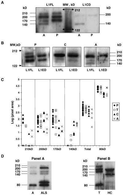
The immunoblots of CSF from preterm and term newborns show four HMW bands at 210, 200, 170 and 80 kDa. The immunoblots with L1FL using CSF from children and adults have five HMW bands at 210, 200, 170, 140 and 80 kDa. The 140-kDa band is not detected in any immunoblot of preterm or term newborn CSF. Representative blots are shown of preterm neonates (Figs. 1A and B), term neonate (Fig. 1D), child (Fig. 1B) and adults (Figs. 1A, B, D) using L1FL. The 80-kDa band is shown in an adult and term newborn (Fig. 1D). Blots prepared with L1CD show 200-, 140- and 80-kDa bands in the adult CSF and 200 and 80-kDa bands in preterm CSF (Fig. 1A). This result indicates that the 200-, 140- and 80-kDa HMW forms contain at least one epitope on the cytoplasmic domain of L1. L1ED, a monoclonal antibody to the extracellular domain of L1, detects HMW bands at 200 and 170 kDa in preterm newborn, child and adult CSF (Fig. 1B), indicating that these HMW forms contain a similar epitope on the L1 extracellular domain.
Fig. 1.
(A) Comparison of immunoreactivity of the high molecular weight bands of L1 between antibodies to full-length L1 versus antibodies to the cytoplasmic domain of L1. Two samples of CSF, one from a 53-year-old adult (A) and one from a 34-week premature infant (P) were run on SDS-PAGE and transferred to a PDVF membrane. The membrane was divided and half immunoblotted with polyclonal antibody to full-length L1 (L1FL) and the other half with polyclonal antibody to the cytoplasmic domain of L1 (L1CD). Following completion of the immunoblotting procedure, the two halves were reunited and exposed to X-ray film. Molecular weight markers are indicated by double headed arrows, estimated molecular weights of the HMW are indicated by lines. (B) Comparison of immunoreactivity of the high molecular weight bands of L1 between antibodies to full-length L1 versus antibodies to the extracellular domain of L1. Three samples of CSF, one from a 34-week premature infant (Premature), one from a 10-month-old child (Child), and one from a 53-year-old adult (Adult), were run on SDS-PAGE and immunoblotted with polyclonal antibody to full-length L1 (L1FL). The membranes were stripped, and reblotted with monoclonal antibody to the extracellular domain of L1 (L1ED). As can be seen, the 210-kDa band does not appear on the blots with L1ED. In addition, L1ED does not recognize the 140-kDa HMW band. Molecular weight markers are indicated by arrowheads, estimated molecular weights of the HMW are indicated by lines. (C) Quantification of band density for the HMW bands of L1. Band density was measured as described. Values were converted to log values to allow comparison across blots. Total is log value of the sum of the pixel density of the 210-, 200-, 170- and 140-kDa bands. (D) Comparison of HMW band pattern of patients with primary CNS disease compared to typical newborn and adult patterns. Immunoblots of CSF samples from a normal adult (A) and an adult with amyotrophic lateral sclerosis (ALS) are shown in panel A, and a normal newborn (N) and a 2-month-old female infant with congenital hydrocephalus (HC) in panel B. Immunoblots were prepared with L1FL as the primary antibody. The 80-kDa band can be seen in both the adult and newborn without primary CNS disease (A, N). However, a novel band is present above the 80-kDa band in the patient with ALS and the 80-kDa band is absent in the patient with HC. In addition, the patient with HC has several differences in the HMW banding pattern from both the normal newborn (N) and adult (A) patterns.
Immunoblots initially blotted with L1FL were used to determine changes in quantity of the HMW of L1 with age. Results are shown in Fig. 1C. The 210-kDa band density monotonically decreases with increasing age across age groups (P = 0.03). The 170-kDa band density also monotonically decreases with increasing age across age groups (P < 0.001). This indicates that there is more 210 kDa and 170 kDa L1 in the CSF of the younger subjects. The amounts of both the 200- and 80-kDa form are inconsistent across age groups. The 140-kDa form monotonically increases with increasing age across age groups (P < 0.001). However, the sum of the 210-, 200-, 170-, and 140-kDa band densities monotonically decreases with increasing age across age groups (P = 0.02).
As shown in Fig. 1D panel A, the adult with ALS (ALS) has an additional band at 100 kDa. The infant with HC (HC) has no bands at 210, 170 or 80 kDa, prominent bands at both 200 and 140 kDa, and an aberrant band with a molecular weight between 200 and 170 kDa as compared to a term newborn (T).
Discussion
This study produced two novel findings: (1) there is an age-related decrease in the 210- and 170-kDa isoforms of L1 in an equal volume of human CSF with a greater amount being found in preterm and term newborn infants when compared with children and adults, and (2) the 140-kDa isoform appears only in the CSF of children and adults. It is not surprising that differences in L1 isoforms in CSF are found between preterm and term neonates compared to children and adults since, in the younger age groups, this is the time of peak brain and neural pathway development. The identity of the HMW isoforms of L1 in CSF may provide clues to the regulation and promotion of corticospinal tract (CST) development.
To further characterize these HMW bands, we used two different antibodies to determine if the cytoplasmic domain or the extracellular domain was present in each of the four HMW bands. We interpret these findings as follows: the 200-kDa form is full-length fully glycosylated L1 as described in lysates from whole brain (Faissner et al., 1985; Michelson et al., 2002; Poltorak et al., 1995; Vawter et al., 1998). The protein in this band is immunoreactive with both antibodies to L1CD as well as L1ED, implying the full-length molecule is represented. The 210-kDa form may also be full-length L1, but with immature glycosylation. The immaturity of the glycosylation may interfere with the immunoreactivity with L1CD and L1ED, since both these antibodies were raised against proteins produced either as recombinants in bacteria (Schaefer et al., 1999) or from transfected COS7 cells (Fransen et al., 1998) and thus expected to have different posttranslational modifications. These full-length transmembrane forms of L1 may be found in CSF as part of membrane vesicles which are released in response to different signals (Gutwein et al., 2003). The 200-kDa protein does not decrease with age. Hence, this form of L1 secreted into vesicles does not appear to be more important during development of the CST.
However, the 170-kDa band does decrease markedly with age. This HMW form of L1 is immunoreactive with L1ED and not L1CD, implying that this is the ADAM10 cleaved extracellular domain (Gutwein et al., 2003) as described previously (Poltorak et al., 1995). Its prominence in preterm and term newborn CSF may indicate a critical role in the development of the CST. This soluble form of L1 has been implicated in cell migration (Gutwein et al., 2000, 2003; Mechtersheimer et al., 2001). The extracellular domain of L1 has been shown to be sufficient for mediating neurite outgrowth (Bearer et al., 1999; Fransen et al., 1998) suggesting that the 170-kDa soluble form may also enhance axon elongation. Thus, the soluble form of L1 may be important in CST development and regeneration. It is interesting to note that mutations that result in unregulated shedding of extracellular fragments result in more severe disease than point mutations in the extracellular domain or deletions of the cytoplasmic domain (Fransen et al., 1996; Yamasaki et al., 1997).
Surprisingly, the 140-kDa form reacts with L1CD but not with antibodies to the extracellular domain. This form may be the same as that described by Poltorak et al. (1995) and assumed to be the plasmin cleavage product of L1. However, that form would lack the cytoplasmic domain. Other possibilities include L1 without glycosylation (Michelson et al., 2002) or a proteolytic fragment containing the cytoplasmic domain cleaved by a protease present in adult CSF.
Although only two cases of primary CNS disease were available, the prominent disruption of L1 banding patterns in these cases suggest that further studies of L1 expression in ALS and HC would be of value.
Acknowledgments
We thank Kevin Buck, Michelle Stamm and Rashmi Phanindra for their technical help. Supported by a grant from the National Institute on Alcohol Abuse and Alcoholism/NIH (R01 AA11839)(CFB). Cynthia F. Bearer had full access to all of the data in the study and takes responsibility for the integrity of the data and the accuracy of the data analysis.
References
- Bateman A, Jouet M, MacFarlane J, Du J-S, Kenwrick S, Chothia C. Outline structure of the human L1 cell adhesion molecule and the sites where mutations cause neurological disorders. EMBO J. 1996;15:6050–6059. [PMC free article] [PubMed] [Google Scholar]
- Bearer CF, Swick AR, O’Riordan MA, Cheng G. Ethanol inhibits L1-mediated neurite outgrowth in postnatal rat cerebellar granule cells. J. Biol. Chem. 1999;274:13264–13270. doi: 10.1074/jbc.274.19.13264. [DOI] [PMC free article] [PubMed] [Google Scholar]
- Faissner A, Teplow D, Kubler D, Keilhauer G, Kinzel V, Schachner M. Biosynthesis and membrane topography of the neural cell adhesion molecule L1. EMBO J. 1985;4:3105–3113. doi: 10.1002/j.1460-2075.1985.tb04052.x. [DOI] [PMC free article] [PubMed] [Google Scholar]
- Fogel M, Gutwein P, Mechtersheimer S, Riedle S, Stoeck A, Smirnov A, Edler L, Ben-Arie A, Huszar M, Altevogt P. L1 expression as a predictor of progression and survival in patients with uterine and ovarian carcinomas. Lancet. 2003;362:869–875. doi: 10.1016/S0140-6736(03)14342-5. [DOI] [PubMed] [Google Scholar]
- Fransen E, Vits L, Van Camp G, Willams PJ. The clinical spectrum of mutations in L1, a neuronal cell adhesion molecule. Am. J. Med. Genet. 1996;64:73–77. doi: 10.1002/(SICI)1096-8628(19960712)64:1<73::AID-AJMG11>3.0.CO;2-P. [DOI] [PubMed] [Google Scholar]
- Fransen E, D’Hooge R, Van Camp G, Verhoye M, Sijbers J, Reyniers E, Soriano P, Kamiguchi H, Willemsen R, Koekkoek SKE, De Zeeuw CI, De Deyn PP, Van der Linden A, Lemmon V, Kooy RF, Willems PJ. L1 knockout mice show dilated ventricles, vermis hypoplasia and impaired exploration patterns. Hum. Mol. Genet. 1998;7:999–1009. doi: 10.1093/hmg/7.6.999. [DOI] [PubMed] [Google Scholar]
- Gutwein P, Oleszewski M, Mechtersheimer S, Agmon-Levin N, Krauss K, Altevogt P. Role of src kinases in the ADAM-mediated release of L1 adhesion molecule from human tumor cells. J. Biol. Chem. 2000;275:15490–15497. doi: 10.1074/jbc.275.20.15490. [DOI] [PubMed] [Google Scholar]
- Gutwein P, Mechtersheimer S, Riedle S, Stoeck A, Gast D, Joumaa S, Zentgraf H, Fogel M, Altevogt P. ADAM10-mediated cleavage of L1 adhesion molecule at the cell surface and in released membrane vesicles. FASEB J. 2003;17:292–294. doi: 10.1096/fj.02-0430fje. [DOI] [PubMed] [Google Scholar]
- Mechtersheimer S, Gutwein P, Agmon-Levin N, Stoeck A, Oleszewski M, Riedle S, Postina R, Fahrenholz F, Fogel M, Lemmon V, Altevogt P. Ectodomain shedding of L1 adhesion molecule promotes cell migration by autocrine binding to integrins. J. Cell Biol. 2001;155:661–673. doi: 10.1083/jcb.200101099. [DOI] [PMC free article] [PubMed] [Google Scholar]
- Michelson P, Hartwig C, Schachner M, Gal A, Veske A, Finckh U. Missense mutations in the extracellular domain of the human neural cell adhesion molecule L1 reduce neurite outgrowth of murine cerebellar neurons. Hum. Mutat. 2002;20:481–482. doi: 10.1002/humu.9096. [DOI] [PubMed] [Google Scholar]
- Nayeem N, Silletti S, Yang X-M, Lemmon VP, Reisfeld WB, Stallcup WB, Montgomery AMP. A potential role for the plasmin(ogen) system in the posttranslational cleavage of the neural cell adhesion molecule L1. J. Cell Sci. 1999;112:4739–4749. doi: 10.1242/jcs.112.24.4739. [DOI] [PubMed] [Google Scholar]
- Poltorak M, Khoja I, Hemperly JJ, Williams JR, El-Mallakh R, Freed WJ. Disturbances in cell recognition molecules (N-CAM and L1 antigen) in the CSF of patients with schizophrenia. Exp. Neurol. 1995;131:266–272. doi: 10.1016/0014-4886(95)90048-9. [DOI] [PubMed] [Google Scholar]
- Ramanathan R, Wilkemeyer M, Mittal B, Perides G, Charness M. Alcohol inhibits cell–cell adhesion mediated by human L1. J. Cell Biol. 1996;133:381–390. doi: 10.1083/jcb.133.2.381. [DOI] [PMC free article] [PubMed] [Google Scholar]
- Rathjen FG, Schachner MEJ. Immunocytological and biochemical characterization of a new neuronal cell surface component (L1 antigen) which is involved in cell adhesion. EMBO J. 1984;3:1–10. doi: 10.1002/j.1460-2075.1984.tb01753.x. [DOI] [PMC free article] [PubMed] [Google Scholar]
- Schaefer AW, Kamiguchi H, Wong EV, Beach CM, Landreth G, Lemmon V. Activation of the MAPK signal cascade by the neural cell adhesion molecule L1 requires L1 internalization. J. Biol. Chem. 1999;274:37965–37967. doi: 10.1074/jbc.274.53.37965. [DOI] [PubMed] [Google Scholar]
- Vawter MP, Cannon-Spoor EC, Hemperly JJ, Hyde TM, VanderPutten DM, Kleinman JE, Freed WJ. Abnormal expression of cell recognition molecules in schizophrenia. Exp. Neurol. 1998;149:424–432. doi: 10.1006/exnr.1997.6721. [DOI] [PubMed] [Google Scholar]
- Wong E, Cheng G, Payne H, Lemmon V. The cytoplasmic domain of the cell adhesion molecule L1 is not required for homophilic adhesion. Neurosci. Lett. 1995;200:155–158. doi: 10.1016/0304-3940(95)12100-i. [DOI] [PubMed] [Google Scholar]
- Yamasaki M, Thompson P, Lemmon V. CRASH syndrome: mutations in the L1CAM correlate with severity of the disease. Neuropediatrics. 1997;28:175–178. doi: 10.1055/s-2007-973696. [DOI] [PMC free article] [PubMed] [Google Scholar]


