Paraneoplastic cerebellar degeneration (PCD) is a rare condition that often heralds an underlying tumor. The syndrome is often associated with small cell carcinoma of the lung, breast cancer, gynecologic cancers, and Hodgkin disease. Treatment options are not well defined and improvement is often limited. Diagnosis can often be aided by detection of antibodies to various cerebellar proteins, including Yo, voltage-gated calcium channels (VGCC), and the Delta/Notch-like epidermal growth factor–related receptor (the target antigen of the Tr immune response). Current therapeutic goals are aimed at suppression or removal of immune-mediated activity, resection of primary tumor, and symptomatic treatment. We report a case of PCD with VGCC autoantibodies and noncutaneous metastatic Merkel cell carcinoma.
Case report.
A healthy 50-year-old Caucasian man developed acute ataxia followed by vertigo the next day. He was treated for benign paroxysmal positional vertigo and then labyrinthitis, without relief. His ataxia and vertigo progressed and he was admitted 1 week later. His basic serologies and MRI/magnetic resonance angiogram were unremarkable. Two days after admission, he developed dysarthria. His CSF demonstrated leukocytosis (white blood cell count 134: lymphs 86%) with normal protein and glucose. On transfer to our hospital, his neurologic examination demonstrated pure cerebellar dysfunction with preservation of strength, sensation, and reflexes. CT chest/abdomen/pelvis was unremarkable. He recalled an upper respiratory infection 2 weeks prior. He was diagnosed with postinfectious cerebellitis and given 3 days of IV methylprednisolone with mild improvement.
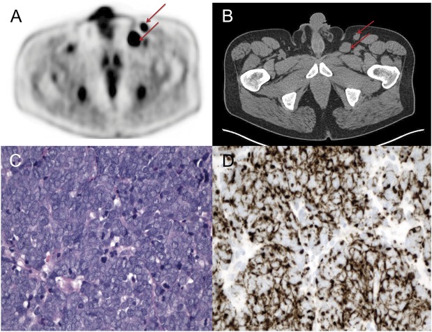
At 2-month follow-up, his ataxia was severe. Repeat MRI brain was unremarkable, and repeat lumbar puncture showed resolution of the leukocytosis. However, a paraneoplastic panel (Athena Diagnostics, Worcester, MA) from his CSF showed elevated levels of P/Q-type VGCC antibodies to 220 pmol/L. Antibodies to Hu, Yo, Ri, and CAR were negative. Electrophysiologic studies did not show evidence of Lambert-Eaton myasthenic syndrome (LEMS). He was started on high-dose oral steroids. A PET scan revealed hypermetabolic lesions in the descending colon and left inguinal lymph nodes (figure, A and B). Meanwhile, his function and neurologic examination worsened so he was started on IV immunoglobulin (IVIg) (0.4 g/kg for 5 days). Colonoscopy demonstrated an adenomatous polyp and high-grade dysplasia in the descending colon. Excisional biopsy of his left inguinal node revealed high-grade neuroendocrine carcinoma with tumor cells expressing Pan-CK, CK20 (perinuclear dot-like), chromogranin, CD56, and Ki-67, consistent with Merkel cell carcinoma (figure, C and D). Subsequent lymphadenectomy demonstrated metastatic carcinoma involving 1 of 14 nodes without extracapsular extension. He was diagnosed with Merkel cell carcinoma stage IIIA (T0N1aM0) with unknown primary. His dermatologic examination was unremarkable.
Figure. Four months after symptom onset.
(A) Two fluorodeoxyglucose positive left inguinal lymph nodes. (B) CT pelvis showing 2 enlarged inguinal lymph nodes: posterior node is 24 × 19 mm, anterior node is 16 × 13 mm. (C) Inguinal lymphadenectomy showing high-grade neuroendocrine carcinoma (hematoxylin & eosin staining, 400× magnification). (D) Immunohistochemical staining for CK20 showing perinuclear “dot-like” staining, characteristic of Merkel cell carcinoma (400× magnification).
After tumor resection, he had 3 monthly IVIg treatments; however, his symptoms persisted. A 3-month repeat CT chest/abdomen/pelvis and colonoscopy did not show recurrent disease. Monthly rituximab (375 mg/m2) was added to IVIg. After 2 doses, he progressively improved; he required a walker to ambulate, had mild dysarthria, and had no upper extremity ataxia. His peripheral B-cell count was 0 so no further rituximab was given.
Discussion.
We describe a case of PCD associated with the VGCC autoimmune response and a noncutaneous metastatic Merkel cell tumor with unknown primary. Merkel cell carcinoma is a rare and aggressive tumor of neuroendocrine cells that typically presents as a cutaneous lesion in sun-exposed areas of the body in older Caucasian men.
The association between Merkel cell carcinoma and PCD was reported in 2005, although the specific antibody had not yet been identified.1 Two cases of Merkel cell carcinoma associated with LEMS have been described.2 LEMS is associated with antibodies to Cav2.1 VGCC. It is plausible that Merkel cell tumors may provoke the Cav2.1 antibody response, since Cav2.1 is expressed by Merkel cells.3 A minority of patients with LEMS have coexisting cerebellar ataxia, and a small group may have cerebellar symptoms without a neuromuscular disorder.4 VGCC antibodies may be found in 11%–40% of patients with otherwise unexplained subacute cerebellar degeneration.5,6 In PCD with VGCC antibodies, an underlying tumor, typically small cell carcinoma of the lung, is detected in approximately one-third of patients.7 VGCC antibodies inhibit VGCC currents in neurons or transfected cells, alter cerebellar synaptic transmission, and can cause cerebellar dysfunction in an animal model.8 VGCC autoantibodies are therefore probably pathogenic, supporting the use of therapies that may decrease antibody production, such as rituximab.
This case highlights several important diagnostic and therapeutic considerations for patients with PCD. Careful evaluation for malignancy is necessary in patients with otherwise unexplained cerebellar symptoms. VGCC antibodies should prompt vigilance for lung cancer. Small cell lung cancer is the most common type of neuroendocrine tumor associated with paraneoplastic disorders; however, other neuroendocrine tumors can also trigger autoimmunity, so cancer screening should not be confined to the chest.9 Prompt immunotherapy and cancer therapy should be considered. However, responses to therapy are often incomplete. Merkel cell tumors, although rare, may be particularly associated with VGCC antibodies, which may be associated with LEMS and/or PCD.
Footnotes
Author contributions: Dr. Zhang: responsible for drafting the manuscript for content and revising the manuscript. Dr. Emery: responsible for selecting and formatting pathology images for the figure. Dr. Lancaster: responsible for providing critical revisions of the manuscript, significant intellectual content, and final review of the manuscript.
Study funding: No targeted funding reported.
Disclosure: C. Zhang and L. Emery report no disclosures. E. Lancaster has received research support from Talecris Inc and Lundbeck Inc. Go to Neurology.org/nn for full disclosures. The Article Processing Charge was paid by the authors.
References
- 1.Balegno S, Ceroni M, Corato M, et al. Antibodies to cerebellar nerve fibres in two patients with paraneoplastic cerebellar ataxia. Anticancer Res 2005;25:3211–3214 [PubMed] [Google Scholar]
- 2.Eggers SDZ, Salomao DR, Dinapoli RP, Vernino S. Paraneoplastic and metastatic neurologic complications of Merkel cell carcinoma. Mayo Clinic Proc 2001;76:327–330 [DOI] [PubMed] [Google Scholar]
- 3.Piskorowski R, Haeberle H, Panditrao MV, Lumpkin EA. Voltage-activated ion channels and Ca2+-induced Ca2+ release shape Ca2+ signaling in Merkel cells. Pflugers Arch 2008;457:197–209 [DOI] [PMC free article] [PubMed] [Google Scholar]
- 4.Clouston PD, Saper CB, Arbizu T, et al. Paraneoplastic cerebellar degeneration. III. Cerebellar degeneration, cancer, and the Lambert–Eaton myasthenic syndrome. Neurology 1992;42:1944–1950 [DOI] [PubMed] [Google Scholar]
- 5.Burk K, Wick M, Roth G, Decker P, Voltz R. Antineuronal antibodies in sporadic late-onset cerebellar ataxia. J Neurol 2010;257:59–62 [DOI] [PubMed] [Google Scholar]
- 6.Lennon VA, Kryzer TJ, Griesmann GE, et al. Calcium-channel antibodies in the Lambert-Eaton syndrome and other paraneoplastic syndromes. N Engl J Med 1995;332:1467–1474 [DOI] [PubMed] [Google Scholar]
- 7.Greenlee JE. Treatment of paraneoplastic cerebellar degeneration. Curr Treat Options Neurol 2013;15:185–200 [DOI] [PMC free article] [PubMed] [Google Scholar]
- 8.Liao YJ, Safa P, Chen YR, Sobel RA, Boyden ES, Tsien RW. Anti-Ca2+ channel antibody attenuates Ca2+ currents and mimics cerebellar ataxia in vivo. Proc Natl Acad Sci U S A 2008;105:2705–2710 [DOI] [PMC free article] [PubMed] [Google Scholar]
- 9.Brammer JE, Lulla P, Lynch GR. Retrospective review of extra-pulmonary small cell carcinoma and prognostic factors. Int J Clin Oncol Epub 2013 Oct 12 [DOI] [PubMed]


