Summary
We describe three cases of incidentally found lesions in the dural venous sinuses on magnetic resonance imaging, that other had erroneously considered pathological entities. We made the diagnosis of giant arachnoid granulations. The differential diagnosis with thrombosis or intrasinusal tumoral lesions was easily made on the basis of three typical radiological features of the granulations: the hyperintensity of the lesions on FLAIR, a blood vessel within the lesion and bone erosion.
Keywords: giant arachnoid granulations, CT, MR
Introduction
Arachnoid granulations (AG) are extensions of the arachnoid membrane into the dural venous sinuses, serving to drain the cerebrospinal fluid (CSF) from the subarachnoid space into the venous system. In some people, these extensions grow to become “giant” AG 1-4. We describe three new cases of giant AG, two in the transverse sinus and one in the superior sagittal sinus.
Case 1
A 50-year old woman underwent MRI of the brain at another institution because of pain and numbness in the left V2 division of the trigeminal nerve.
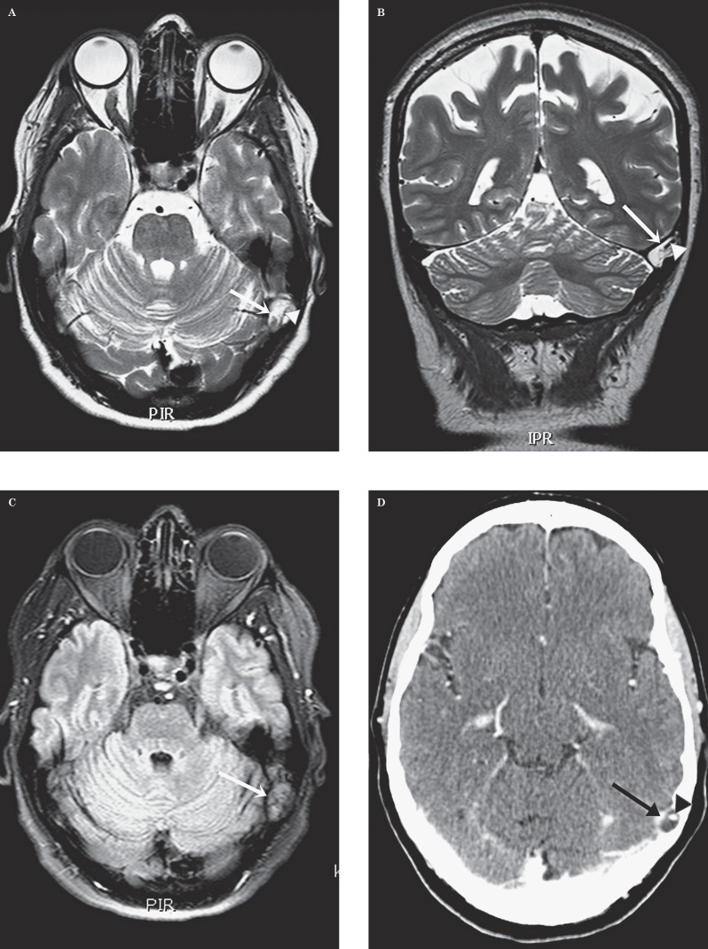
On this MR scan, no lesion was found to explain the trigeminal neuralgia. In the lateral aspect of the left lateral sinus an ovalar lesion was found partially obstructing the lumen of the sinus. The lesion was hyperintense on the axial and coronal T2-weighted sequences (Figure 1A,B) with evidence of a central strongly hypointense tubular structure, probably representing a flow void in a vascular structure. The lesion was also hyperintense on the axial CISS and gradient-echo sequence. On the FLAIR sequence (Figure 1C), there was no attenuation and the lesion remained hyperintense. There was no diffusion restriction in the lesion. Intravenous contrast was not administered.
Figure 1.
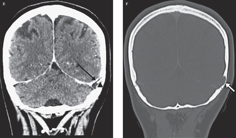
Patient 1. Giant arachnoid granulation in the left transverse sinus. Transverse (A) and coronal (B) T2-weighted images. An ovalar hyperintense structure (arrow) can be seen in the left transverse sinus at the transition to the sigmoid sinus. Intralesional flow void (arrowhead). C) Transverse FLAIR. There is attenuation of the content of this “lesion” (arrow) that remains hyperintense. Transverse (D) and coronal (E) enhanced CT. The “lesion” (arrow) is hypodense with a central enhancing vascular structure (arrowhead). E) Coronal CT with bone windows. Note the scalloping of the overlying bone (arrow).
Since this lesion was read as a tumour, the patient was referred to the neurosurgery department at our hospital for a second opinion. When studying the MR together with the neurosurgical staff, we suggested the diagnosis of a giant AG and proposed a contrast-enhanced CT for confirmation.
CT showed a hypodense (density of +/- 20 Hounsfield units) non-enhancing nodular lesion in the left transverse sinus, in the lateral third, at the level of the transition to the sigmoid sinus (Figure 1D,E). There was scalloping of the adjacent temporal bone (Figure 1E). A small enhancing vein was visible in the superior part of the lesion (Figure 1D,E). These findings confirmed the diagnosis of a giant AG. The patient was reassured she had no tumour and did not require further treatment.
Case 2
A 36-year-old man had a known history of right parietal haemorrhages since his youth with several repetitive episodes. During a new recent haemorrhage, on imaging at another institution a lesion was found in the right transverse sinus, interpreted as a thrombosis and patient was put on anticoagulation.
The patient was referred to our institution for a second opinion and the images were shown to us.
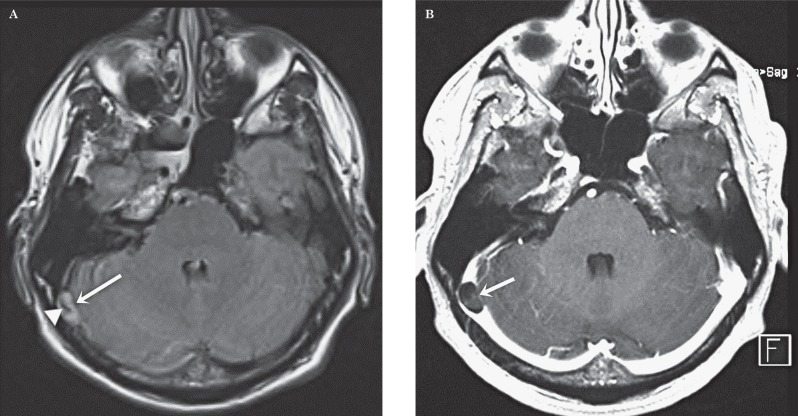
On MRI an ovalar structure was found in the right transverse sinus. This structure was hyperintense on T2-weighted images and the FLAIR sequence and was hypointense on T1. On FLAIR a rounded intralesional area of flow void was seen (Figure 2A).
There was no enhancement after contrast (Figure 2B). There was erosion of the inner table of the skull, as confirmed by subsequent CT. Diagnosis of a giant AG was made. The real cause of the haemorrhages is still under investigation.
Figure 2.
Patient 2. Giant arachnoid granulation in the right transverse sinus. A) Transverse FLAIR. Note the hyperintense “lesion” (arrow) in the left transverse sinus at the transition to the sigmoid sinus. Central flow void pointing to a vascular structure (arrowhead). B) Transverse gadolinium-enhanced image. The hypointense ovalar lesion (arrow) in the transverse sinus is confirmed. The vascular structure less well visible due to volume-averaging.
Case 3
A 12-year-old boy complained of three episodes of sudden headache accompanied by blurred vision, diplopia and neck pain.
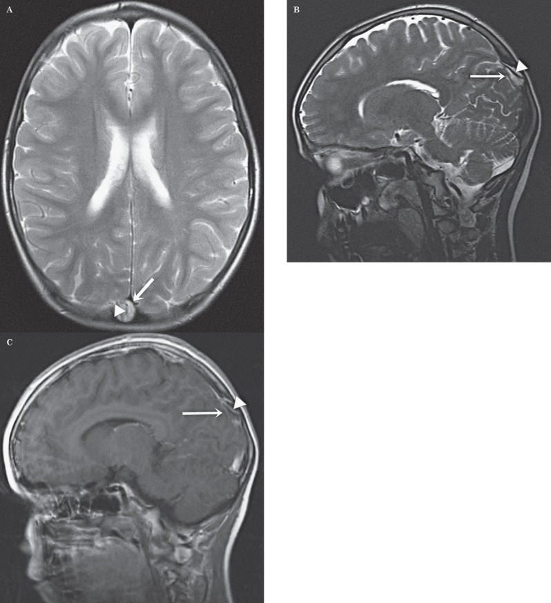
There was no history of previous headaches. Clinical neurological examination was normal. MRI of the brain was performed to rule out a Chiari malformation. The cerebellar tonsils had a normal location. In the superior sagittal sinus a large lesion was present with T2 hyperintensity and with a central linear area of flow void (Figure 3A,B). This structure enhanced strongly whereas the rest of the lesion did not enhance at all (Figure 3C). Again a tumoral lesion was suspected but we made the diagnosis of a giant AG. Since the headache was not continuous and given the absence of papillary oedema or other signs of intracranial hypertension, it was considered that there was no relation between the attacks of headache and the AG.
Figure 3.
Patient 3. Giant arachnoid granulation in the superior sagittal sinus. Transverse (A) and sagittal (B) T2-weighted image. Huge ovalar hyperintense structure (arrow) within the superior sagittal sinus in the under third. Intralesional vascular flow-void (arrowhead). C) Sagittal gadolinium-enhanced image. Confirmation of the granulation (arrow) and the vascular structure (arrowhead).
Discussion
Arachnoid granulations (AG), first described by Antonio Pacchioni in 1705, are cerebrospinal fluid-filled protrusions extending from the subarachnoid space into the venous sinuses through apertures in the dura 2,4,5. They represent distended arachnoid villi and are macroscopically visible 1.
The prevalence of AG increases with age 6. AG normally measure a few millimetres. In some cases they grow to fill and dilate the dural sinuses even with expansion of the inner table of the skull and are then called “giant AG” 1. There is no consensus on the definition of “giant” in the literature. Sometimes a large AG is called “giant” when larger than 1 cm 4, but Kan et al. referred to AG as “giant” when they fill the lumen of a dural sinus, causing local dilatation or filling defects 1. Most commonly AG are found in the transverse sinuses, particularly within the middle and lateral portions of the sinus, with a slight left predominance 6. The second most common location is the superior sagittal sinus, but they can be found everywhere in the dural venous sinuses 2,4. There is no difference in sex distribution 6.
Mostly, giant AG are discovered as incidental findings without any relation to the patient’s symptoms as in our three cases. In some cases, however, they fill and dilate the dural sinuses, causing symptoms of increased intracranial pressure based on venous hypertension secondary to partial sinus occlusion. They usually present with symptoms of headache 1,7,8. Endovascular treatment with stents might be necessary in case of invalidating symptoms 9.
The imaging characteristics of normal and giant AG are well-known and can be considered typical 4,5. On skull radiography, they can be visible as radiolucent zones or they can cause impressions on the inner table of the calvaria 4. The CT density of granulations in the literature varies from hypodense to isodense with the brain parenchyma 4,6. On MR imaging, the arachnoid granulations are iso- to hypointense relative to brain parenchyma on T1-weighted images. They all show a hyperintense signal on T2-weighted images 4,6. The fluid in the granulations is mostly not attenuated on a FLAIR sequence, but remains hyperintense, most likely due to pulsation artifacts from the adjacent sinus and differing CSF flow characteristics within the AG 2. This was the case in all three of our patients. In the series reported by Trimble et al. 4, approximately 80% of giant arachnoid granulations contain CSF-incongruent fluid on at least one MR image and nearly half contain fluid that does not parallel CSF on at least two sequences. FLAIR is the most reliable technique, differing in signal intensity with CSF in 100% of cases 4.
On angiographic studies such as CT angiography, MR angiography or catheter angiography, AG appear as ovoid filling defects in the dural venous sinuses in the venous phase 2,5. Vascular structures presumed to be veins can be seen as focal linear contrast enhancement on enhanced CT or MR or as linear flow voids on non-enhanced MR as seen in two of our patients. It seems that larger granulations are more likely to contain these internal veins 2,4,6. Non-vascular soft tissue has also been reported in giant AG, and was variously interpreted as stromal collagenous tissue, hypertrophic arachnoid mesangial cell proliferation, or invaginating brain tissue 4. AG can be mistaken for pathologic processes in the dural venous sinuses. It is important to distinguish these benign giant AG from more serious dural venous sinus pathology such as thrombosis and neoplasia to avoid unnecessary invasive procedures, because of their infrequency and large size. The differential diagnosis with thrombosis can be made because thrombosis usually involves an entire segment of sinus or multiple sinuses, and can extend into cortical veins, while AG produce focal, well-defined defects 7,8. It has to be mentioned that fresh thrombus is hyperdense on CT and hyperintense on T1-weighted MR, whereas AG have other imaging characteristics 8. The differential diagnosis with tumour can be made because of the shape, the lack of contrast enhancement and the lack of diffusion restriction 1,3,4.
References
- 1.Kan P, Stevens EA, Couldwell WT. Incidental giant arachnoid granulation. Am J Neuroradiol. 2006;27:1491–1492. [PMC free article] [PubMed] [Google Scholar]
- 2.Leach JL, Meyer K, Jones BV, et al. A large arachnoid granulations involving the dorsal superior sagittal sinus: findings on MR imaging and MR venography. Am J Neuroradiol. 2008;29:1335–1339. doi: 10.3174/ajnr.A1093. doi: 10.3174/ajnr.A1093. [DOI] [PMC free article] [PubMed] [Google Scholar]
- 3.Mamourian AC, Towfighi J. MR of giant arachnoid granulation, a normal variant presenting as a mass within the dural venous sinus. Am J Neuroradiol. 1995;16(4 Suppl):901–904. [PMC free article] [PubMed] [Google Scholar]
- 4.Trimble CR, Harnsberger HR, Castillo M, et al. "Giant" arachnoid granulations just like CSF? Not. Am J Neuroradiol. 2010;31:1724–1728. doi: 10.3174/ajnr.A2157. doi: 10.3174/ajnr.A2157. [DOI] [PMC free article] [PubMed] [Google Scholar]
- 5.Roche J, Warner D. Arachnoid granulations in the transverse and sigmoid sinuses: CT, MR, and MR angiographic appearance of a normal anatomic variation. Am J Neuroradiol. 1996;17(4):677–683. [PMC free article] [PubMed] [Google Scholar]
- 6.Leach JL, Jones BV, Tomsick TA, et al. Normal appearance of arachnoid granulations on contrast-enhanced CT and MR of the brain: differentiation from dural sinus disease. Am J Neuroradiol. 1996;17:1523–1532. [PMC free article] [PubMed] [Google Scholar]
- 7.Chin SC, Chen CY, Lee CC, et al. Giant arachnoid granulation mimicking dural sinus thrombosis in a boy with headache: MRI. Neuroradiology. 1998;40(3):181–183. doi: 10.1007/s002340050564. doi: 10.1007/s002340050564. [DOI] [PubMed] [Google Scholar]
- 8.Choi HJ, Cho CW, Kim YS, et al. Giant arachnoid granulation misdiagnosed as transverse sinus thrombosis. J Korean Neurosurg Soc. 2008;43:48–50. doi: 10.3340/jkns.2008.43.1.48. doi: 10.3340/jkns.2008.43.1.48. [DOI] [PMC free article] [PubMed] [Google Scholar]
- 9.Zheng H, Zhou M, Zhao B, et al. Pseudotumor cerebri syndrome and giant arachnoid granulation: treatment with venous sinus stenting. J Vasc Interv Radiol. 2010;21:927–929. doi: 10.1016/j.jvir.2010.02.018. doi: 10.1016/j.jvir.2010.02.018. [DOI] [PubMed] [Google Scholar]


