We read with great interest the study of Alves et al. 1 demonstrating that the qualitative evaluation of CBV and MTT maps obtained with computed tomography perfusion (CTP) imaging may overestimate the true ischemic penumbra in ischemic stroke. The authors conclude that CBV abnormalities are reliable markers for irreversible ischemia. They demonstrate that CBV may, rarely, overestimate the ischemic “core” and that MTT tends to overestimate the extent of final infarct areas, as these values do not differentiate the true “at risk” penumbra from benign oligemia. The authors also highlight some important methodological considerations related to the use of perfusion analysis software. Software applications from different vendors do not generate equivalent quantitative perfusion results. Caution should thus be exercised when interpreting quantitative/qualitative CTP measures, because these values may vary considerably, depending on the post-processing software. The major mathematical technique utilized for calculation of perfusion parameters is the deconvolution approach. Deconvolution can be performed using several methods. The classic deconvolution method is termed “standard singular value decomposition” (sSVD). This technique is robust, and its results are independent of the underlying vascular anatomy (i.e. intra/extracranial stenoses and/or leptomeningel collaterals). However, sSVD is sensitive to delays in the arrival of the contrast agent. When such delays occur, MTT and CBV are overestimated and CBF is underestimated. The use of abnormal map values may lead to overestimation of the ischemic penumbra by including non-ischemic brain regions into delayed contrast agent arrival calculations. To overcome these difficulties, new deconvolution methods have been developed to minimize the effects of bolus delay and dispersion, including delay-corrected deconvolution (dSVD) and block-circulant deconvolution (bSVD) algorithms. As a general rule, the use of a delay-insensitive deconvolution method is recommended when imaging stroke patients, a population in whom arterial stenoses are common.
The role of computed tomography angiography (CTA) in the interpretation of the core and the ischemic penumbra was not discussed in the Alves et al. article. Several groups have suggested that CTA source images (CTA-SI), like DWI MRI, may sensitively delineate tissue destined to infarct in spite of successful recanalization. The superior accuracy of CTA-SI in identifying infarct “core” compared with unenhanced CT has been unequivocally established in multiple studies 2,3. Theoretical modeling indicates that CTA-SI obtained using early generation protocols generates predominantly blood volume-weighted rather than blood flow-weighted images. Due to the relatively slow acquisition, images acquired with these protocols reflect an approximate steady state of contrast in the brain arteries and parenchyma. However, newer, faster MDCT CTA-SI protocols, such as those used with 64-slice scanners, with injection rates up to 7 mL/s and short preparation delay times, change the temporal shape of the “time-density” infusion curve, eliminating the near-steady state during the timing of the CTA-SI acquisition.
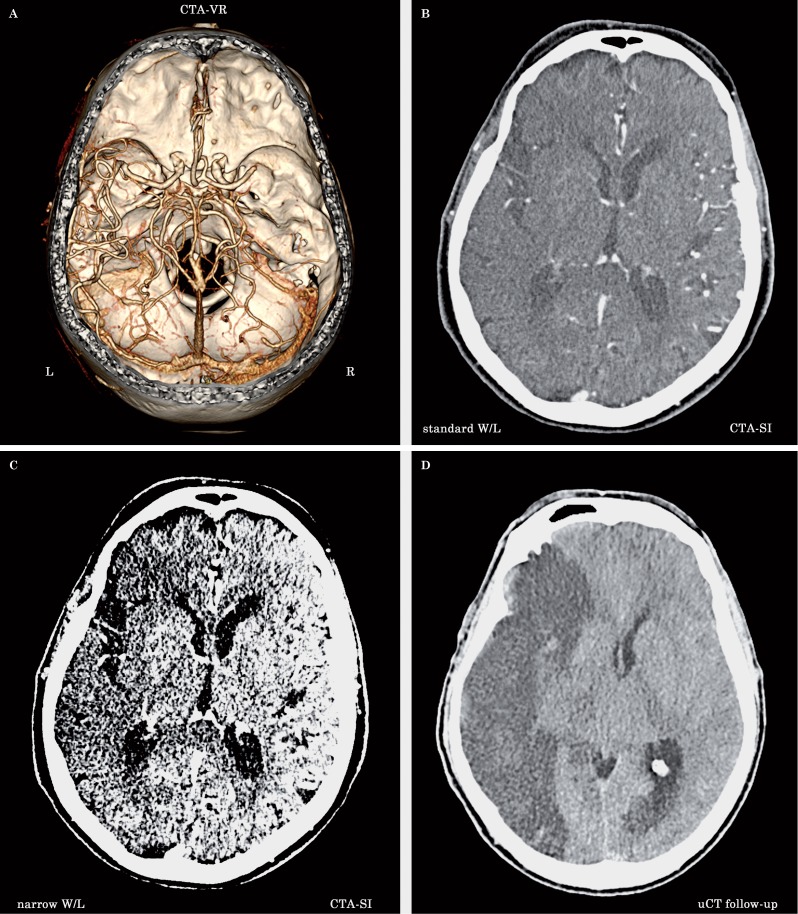
Hence, using current generation CTA protocols on faster state-of-the art MDCT scanners, the CTA-SI maps typically achieve more flow- rather than volume-weighted images. With early-generation CTA protocols, in the absence of early recanalization, CTA-SI typically defines minimal final infarct size and, hence, like DWI MRI, can be used to identify an “infarct core” in the acute setting (Figure 1 A-D). CTA-SI subtraction maps, obtained by coregistration and subtraction of the unenhanced head CT from the CTA-SI, result in quantitative blood volume maps of the entire brain and are particularly appealing for clinical use because, unlike quantitative first-pass CT perfusion maps, they provide whole brain coverage. Thus, radiologists should interpret CTP maps in conjunction with CTA-SI data. Finally, it is again worth underscoring that with the newer, more flow-weighted CTA protocols, not every acute CTA-SI hypodense ischemic lesion is destined to infarct; a substantial portion of a CTA-SI hypodense lesion may reflect “at-risk” ischemic penumbra, or even benign oligemia 3. Moreover, Kamalian et al. 4 reported that CBF maps optimally correlate with admission diffusion-weighted imaging. The accuracy of CBF versus CBV in determining the infarct core is supported by the fact that calculation of CBV is typically more sensitive to time-density curve truncation than is CBF. It is critical for radiologists who utilize PCT for stroke imaging to be familiar with the postprocessing software implemented at their institution and to analyze comprehensive datasets from advanced stroke imaging (CT, CTA, CTP and relative source image). The optimal method to delineate the core from the ischemic penumbra using CTP maps has not yet been established, and further studies should be performed to better understand the hemodynamic phenomena that occur in the acute phases of ischemic stroke.
Figure 1.
A 78-year-old man with ischemic stroke of unknown onset presented to the emergency department with left hemiplegia. A) Volume rendering CTA (cranio-caudal view) shows occlusion of the right cerebral artery in the M1-M2 segment. B) CTA-SI at level of the basal nuclei with standard window width and center level (W/L) (286/66) is also illustrated. The CTA protocol acquisition occurs at an approximate steady state of contrast in the brain arteries and parenchyma so that the image is predominantly blood volume-weighted. Image review should be performed with narrow window width and center level display settings to maximize gray-white matter differentiation, thereby improving the detection of subtle, hypodense ischemic regions. C) With the same CTA-SI viewed with narrow W/L (38/48), it is possible to better appreciate the hypodense area of ischemic origin in the right cerebral hemisphere attributable to infarct "core" (irreversibly damaged tissue). D) A follow-up unenhanced CT scan clearly delineates the extent of ischemia, corresponding to the region identified with the CTA-SI in Figure 1C. Accurate inspection of CTA-SI with narrow windows enables definition of the ischemic core with the additional advantage of whole brain coverage.
References
- 1.Alves JE, Carneiro A, Xavier J. Reliability of CT perfusion in the evaluation of the ischaemic penumbra. Neuroradiol J. 2014;27(1):91–95. doi: 10.15274/NRJ-2014-10010. doi: 10.15274/NRJ-2014-10010. [DOI] [PMC free article] [PubMed] [Google Scholar]
- 2.Schramm P, Schellinger PD, Fiebach JB, et al. Comparison of CT and CT angiography source images with diffusion-weighted imaging in patients with acute stroke within 6 hours after onset. Stroke. 2002;33(10):2426–2432. doi: 10.1161/01.str.0000032244.03134.37. doi: 10.1161/01.STR.0000032244.03134.37. [DOI] [PubMed] [Google Scholar]
- 3.Lin K, Rapalino O, Law M, et al. Accuracy of the Alberta Stroke Program Early CT Score during the first 3 hours of middle cerebral artery stroke: comparison of noncontrast CT, CT angiography source images, and CT perfusion. Am J Neuroradiol. 2008;9(5):931–936. doi: 10.3174/ajnr.A0975. doi: 10.3174/ajnr.A0975. [DOI] [PMC free article] [PubMed] [Google Scholar]
- 4.Kamalian S, Kamalian S, Maas MB, et al. CT cerebral blood flow maps optimally correlate with admission diffusion-weighted imaging in acute stroke but thresholds vary by postprocessing platform. Stroke. 2011;42(7):1923–1928. doi: 10.1161/STROKEAHA.110.610618. doi: 10.1161/STROKEAHA.110.610618. [DOI] [PMC free article] [PubMed] [Google Scholar]


