. 2014 Oct 14;8:471. doi: 10.3332/ecancer.2014.471

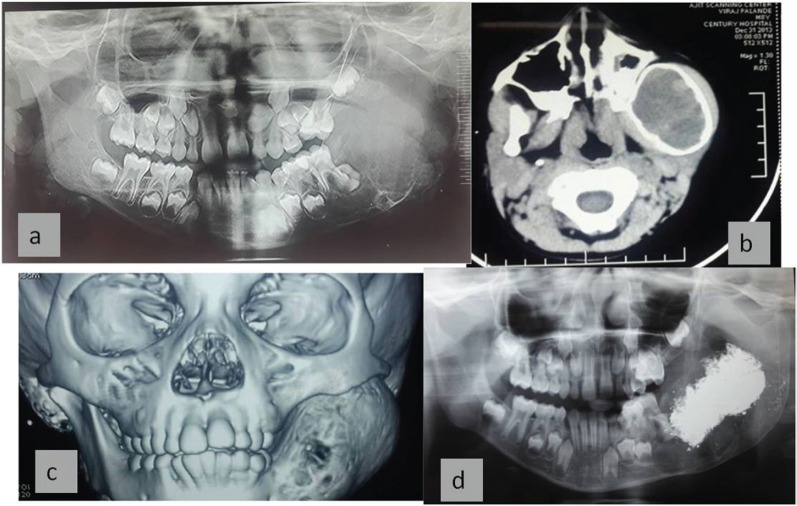
Figure 3. X-ray and CT scan show (a) large multilocular mixed radiolucent radiopaque lesion involving left side of mandible extending from mandibular, second deciduous molar. Superiorly it extends up to condylar and coronoid processes. Inferior margin showed multiple septae with thinning of the cortical plates, Anterior displacement of the tooth bud 37 was noted. (b) The CT sections show large severely expansile lesion in the left ramus of the mandible extending up to the cortical plates. (c) 3D CT shows expansion and thinning of cortical plates localised areas with perforation of cortical plates noted. (d) shows postoperative follow-up after two months with bony healing inferiorly.

Figure 3.