Abstract
Background
End-stage chronic respiratory diseases (CRD) have systemic consequences, such as weight loss and susceptibility to infection. However the mechanisms of such dysfunctions are as yet poorly explained. We hypothesized that the genes putatively involved in these mechanisms would emerge from a systematic analysis of blood mRNA profiles from pre-transplant patients with cystic fibrosis (CF), pulmonary hypertension (PAH), and chronic obstructive pulmonary disease (COPD).
Methods
Whole blood was first collected from 13 patients with PAH, 23 patients with CF, and 28 Healthy Controls (HC). Microarray results were validated by quantitative PCR on a second and independent group (7PAH, 9CF, and 11HC). Twelve pre-transplant COPD patients were added to validate the common signature shared by patients with CRD for all causes. To further clarify a role for hypoxia in the candidate gene dysregulation, peripheral blood mononuclear cells from HC were analysed for their mRNA profile under hypoxia.
Results
Unsupervised hierarchical clustering allowed the identification of 3 gene signatures related to CRD. One was common to CF and PAH, another specific to CF, and the final one was specific to PAH. With the common signature, we validated T-Cell Factor 7 (TCF-7) and Interleukin 7 Receptor (IL-7R), two genes related to T lymphocyte activation, as being under-expressed. We showed a strong impact of the hypoxia on modulation of TCF-7 and IL-7R expression in PBMCs from HC under hypoxia or PBMCs from CRD. In addition, we identified and validated genes upregulated in PAH or CF, including Lectin Galactoside-binding Soluble 3 and Toll Like Receptor 4, respectively.
Conclusions
Systematic analysis of blood cell transcriptome in CRD patients identified common and specific signatures relevant to the systemic pathologies. TCF-7 and IL-7R were downregulated whatever the cause of CRD and this could play a role in the higher susceptibility to infection of these patients.
Introduction
Dependence on oxygen supplementation is an end-stage condition of several chronic respiratory diseases (CRD). In France, almost 150,000 patients receive long-term oxygen therapy, with a median survival of 1 to 4 years depending on the underlying cause [1]. Chronic Obstructive Pulmonary Disease (COPD), Cystic Fibrosis (CF) and Pulmonary Arterial Hypertension (PAH) have this end-stage supplementation in common despite distinct pathophysiologies and treatments. COPD results from damage to airways and lung parenchyma [2]; CF is caused by a mutation in the Cystic Fibrosis Transmembrane conductance Regulator gene (CFTR), affecting bronchial epithelium mucus production leading to lung impairment and infection [3]; PAH is a condition involving a remodelling of pulmonary vessels causing right heart failure [4], [5]. Chronic tissue hypoxia resulting from these diseases induces peripheral damage including weight loss and metabolism dysfunction directly impacting the patient outcome [6], [7]. Using high-throughput approaches in genomics, transcriptomics or proteomics, previous studies have identified biological signatures relevant for these diseases, characterized notably by immunological abnormalities [8]–[10]. However, the mechanisms of the systemic consequences of CRD are still poorly understood. Little attention has been paid to date to the impact of CRD on blood cells, which may carry disease-specific information due to direct or indirect modifications. We hypothesized that CRD-induced metabolic changes could impact blood cell gene expression, and that this compartment offers a means to detect genes implicated in unexplored CRD-related changes.
To test this hypothesis, a systematic analysis of blood mRNA profiles was performed in CRD patients awaiting lung transplant. Microarray analysis was performed on a first group (microarray cohort) composed of PAH, CF and Healthy Controls (HC). We distinguished a common transcriptomic signature related to CRD and specific signatures for each disease. These patterns were validated in an external and independent cohort of HC and patients with CF, PAH and COPD (validation cohort).
Methods
Study population
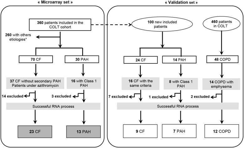
The study protocol was approved by the Comité de Protection des Personnes Ouest 1-Tours (reference number: 2009-A00036-51), and written informed consent was obtained from all subjects. Blood samples were collected from pre-transplant patients included in a multicentric longitudinal cohort, intituled Cohort of Lung Transplantation (COLT). This cohort consists in monitoring patients during 5 years following lung transplantation in order to detect predictive factors of chronic lung allograft dysfunction. We took advantage of the biocollection to study blood of patients with CRD before transplantation. The strategy of selecting the included samples is described in Figure 1. To increase the chance of detecting a signature of CRD that is independent of the primary disease, we first focused on 2 diseases with highly contrasted pathophysiology, CF and Class 1 PAH. Secondarily, to validate this common signature as being present in any CRD, a supplementary group of COPD patients was included. Patients were selected among the cohort so that each group was as homogeneous as possible regarding the form of the disease (CF documented as exempt of secondary PAH, Class 1 PAH, COPD with documented emphysema) and treatment (CF with azithromycin). Some samples were secondarily excluded due to unsatisfying mRNA quality. The “microarray cohort” was therefore composed of 13 patients diagnosed with PAH and 23 with CF (Table 1A). Twenty-eight samples from healthy controls (HC) collected by the French Blood Establishment were used in the microarray analysis (Table 1A). In order to overcome the age difference between CF and PAH, we selected a HC population mixed in age: 46.43% of HC were matched with CF (born after 1970) and 53.57% with PAH patients (born before 1960).
Figure 1. Strategy for selecting patients from the COLT (COhort of Lung Transplantation).
*Emphysema, Sarcoïdosis, Lymphangiomatosis, Secondary PAH, histiocytosis, fibrosis, bronchiectasis and COPD (Chronic Obstructive Pulmonary Disease). CF: Cystic Fibrosis; PAH: Pulmonary Arterial hypertension; COPD: Chronic Obstructive Pulmonary Disease. Patients were excluded during the RNA process following specific qualities criteria.
Table 1. Summary of clinical data of patients included in the microarray (A) and in the validation (B) cohorts.
| Patients Group | Age | Gender (Male/All) | Smoking status (Yes/No) | BMI (Kg/m2) | FEV1 (%) | PaO2 (kPa) | PAPm (mmHg) | |
| A. | Patients included in the microarray cohort | |||||||
| CF n = 23 | 11/23 | 0/23* | ||||||
| mean ± SD | 24±7 | 18.4±2.25 | 24.7±8.50 | 8.1±0.74 | ||||
| min | 16 | 14.5 | 14 | 7 | ||||
| max | 41 | 22.4 | 42 | 9.6 | ||||
| PAH n = 13 | 5/13 | 0/13φ | ||||||
| mean ± SD | 41±15.3 | 22.3±6.10 | 74.9±30.05 | 7.9±1.66 | 66.7±17.17 | |||
| min | 16 | 16.3 | 36 | 5.9 | 53 | |||
| max | 61 | 34 | 122 | 10 | 110 | |||
| HC n = 28 | 9/28 | |||||||
| mean ± SD | 42.5±14.20 | |||||||
| min | 21 | |||||||
| max | 68 | |||||||
| B. | Patients included in the validation cohort | |||||||
| CF n = 9 | 5/9 | 0/9 | ||||||
| mean ± SD | 26±6.3 | 18±1.20 | 24±5.43 | 7.2±1.58 | ||||
| min | 15 | 16 | 15 | 4.9 | ||||
| max | 36 | 19 | 35 | 8.4 | ||||
| PAH n = 7 | 0/7 | 0/7£ | ||||||
| mean ± SD | 34±18.5 | 24±7.81 | 80±15.06 | 9.1±0.22 | 64±23.39 | |||
| min | 15 | 20 | 30 | 8.9 | 40 | |||
| max | 67 | 27 | 95 | 11.2 | 100 | |||
| COPD n = 12 | 8/12 | 0/12§ | ||||||
| mean ± SD | 58±2.6 | 21.2±4.53 | 18.1±5.40 | 7.84±1.9 | ||||
| min | 54 | 16 | 10 | 3.8 | ||||
| max | 61 | 28.6 | 32 | 10.17 | ||||
| HC n = 11 | 6/11 | |||||||
| mean ± SD | 41.5±13.47 | |||||||
| min | 23 | |||||||
| max | 58 | |||||||
PAH = pulmonary arterial hypertension; CF = cystic fibrosis; HC = healthy controls; COPD = chronic obstructive pulmonary disease; BMI = Body Mass Index; FEV1 = Forced Expiratory Volume in 1 second; PaO2 = arterial oxygen tension; PAPm = mean pulmonary artery pressure (mmHg);
*2/23 are wean smokers; φ2/13 are wean smokers; £1/7 is a wean smoker; §12/12 are wean smokers.
The relevance of candidate genes from the microarrays analysis was confirmed by quantitative polymerase chain reaction (q-PCR) performed on a second group of patients referred to as the “validation cohort”. This group was composed of 7 PAH and 9 CF patients newly included in COLT since the first selection, 12 COPD patients selected among the whole COLT population, and 11 HC (Table 1B). The same criteria were applied as in the “microarray” set. In this validation cohort again, we matched HC according to the patients age: 54.54% were born before 1960 and 45.46% after 1970.
RNA Isolation
Samples were collected in PAXgene tubes (PreAnalytix, Qiagen), and stored at −80°C. Total RNA was extracted using the PAXgene Blood RNA System kit with an on-column DNase digestion protocol. Quality and quantity of total RNA were determined using a 2100 Bioanalyzer (Agilent Technologies Incorporation) and a NanoDrop ND-1000 spectrophotometer (NanoDrop Technologies). Microarray and qPCR analyses were performed on RNA with 260/280 and 260/230 OD ratios above 1.8 and a RNA integrity number (RIN) above 7.
Gene expression microarray analysis
RNA samples were prepared and hybridized on Agilent Human Gene Expression 8×60 K microarrays (Agilent Technologies, part number: G4851A). In order to avoid any bias due to blood populations differences between groups, the Lowess (locally weighted scatterplot smoothing) normalization procedure was applied on all the microarrays together [11]. Thereby spots with half of the samples exhibiting signals less than the mean of all median signals were removed (threshold: 90.83). 30,146 probes were kept out of 58,717. Raw microarray data were deposited in the Gene Expression Omnibus (GEO) database (accession number GSE38267). Unsupervised hierarchical clustering was performed with Cluster (v3.0) and TreeView software using uncentred Pearson correlation with a median-centred gene dataset. To select the genes participating to the same biological process, we selected 5 clusters (A–E), based on a combined approach: selected genes were clustered together and exhibited a between-group t-test p-value below 1% [12]. The biological significance for each cluster was determined using GOminer software. Thus over-represented GO ontology (GO) categories within the list of genes were identified by comparison with the others genes expressed on the microarrays (i.e. the 17,163 expressed genes). In addition, Ingenuity Pathway Analysis (IPA) (Ingenuity Systems Inc.) was used to construct network pathways.
Sample classification methods from gene expression data
Prediction Analysis of Microarray (PAM) was performed using R v2.13.0 software with a pamr package to identify minimum gene sets that differentiated patient groups. Additional hierarchical clustering was performed with MultiExperiment Viewer software [13] using the uncentered Pearson correlation as a similarity metric and average linkage clustering. Principal component analysis (PCA) and receiver operating curve (ROC) were performed using R v2.13.0 software with ade4 [14] or pROC package, respectively.
Quantitative PCR (qPCR) for microarray validation
Microarray results were validated by qPCR with a new set of independent samples. Complementary DNA was synthesized starting from 500 ng of RNA using an Omniscript kit (Qiagen). Real-time quantitative PCR was performed on a ViiA7 Fast Real-Time PCR System (Applied Biosystems) using commercially available primers: HPRT1 (Hs99999909_m1), β2M (Hs00984230_m1), ACTB (Hs99999903_m1), TCF-7 (Hs00175273_m1), CD6 (Hs00198752_m1), IL-7R (Hs00233682_m1), LGALS3 (Hs00173587_m1), MDK (Hs00171064_m1) TLR4 (Hs00152939_m1), NLRC4 (Hs00802666_m1) and TLR8 (Hs00607866_mH). Samples were analysed in duplicate and the geometric mean of quantification cycle values (Cq) for HPRT1, β2M and ACTB was used to normalize cDNA amounts. Relative expression between a sample and a reference was calculated according to the 2−ΔΔCq method [15].
Cellular culture under hypoxic and normoxic conditions
PBMCs from HC cultured in 24-well plates with 1 mL of RPMI 1640 media supplemented 10% FBS, 200 mg/mL penicillin, 200 U/mL streptomycin, 4 mM L-glutamine were placed either in a hypoxia incubator, created by displacing O2 (2% O2) with infusion of N2 (93%), or a normoxic incubator (21% O2) for 12 h at 37°C. RNA was extracted using a Macherey Nagel kit according to the supplier’s recommendations. Complementary DNA was synthetized from 250 ng using a superscript III kit (invitrogen) and qPCR was performed to study TCF-7 and IL-7R expression. Finally, median fluorescence intensity was measured for CD127 (also called IL-7R) protein on CD3+CD4+ T cells by flow cytometry using the following antibodies (1/100e): CD3-PE-Cy7, CD4-PercP-Cy 5.5 and CD127-PE (BD, Biosciences). A Viability dye (BD Horizon V450, 1/1000e) may be used to exclude dead cells from analysis (LSR II BD Biosciences and FlowJo software).
Analysis of lymphocyte flow cytometry profile in PBMCs of patients in CRDs
PBMC from 7 CF, 8 PAH and 6 COPD patients and 6 HC were rapidly thawed by placing cryovials at 37°C. Cells were washed, resuspended in supplemented RPMI 1640. 3.106 cells were stained with CD3-PE-Cy7, CD4-PercP-Cy 5.5, CD127-PE, BD Horizon V450 (BD, Biosciences). Results were generated by flow cytometry (LSR II BD Biosciences and FlowJo software).
Statistics
Regarding microarray analysis, the selection of genes of interest is based on a combined approach including a t-test with a p-value inferior to 0.01 and a clustering selection. This approach is based on the assumption that genes participating to same biological functions are clustered together as demonstrated by Alizadeh et al. [16]. A test called SP Calc was used for calculating sample size and power in our microarray study [17]. Our analysis exhibited a reasonable statistical power superior to 75% despite the small sample size. qPCR and Flow cytometry results are given as mean ±standard error of the mean. The non-parametric Kruskal Wallis tests with Dunn’s ad-hoc pairwise comparisons and Mann and Whitney test were applied using GraphPad Prism, v4. Differences between groups were defined as significant when the p-value was <0.05.
Results
Clinical Parameters
Table 1A shows the characteristics of the patients whose blood samples were used for the microarray process. The mean ages of PAH patients and HC were similar (41±15.3 years vs 42.5±14.20, mean ± SD, p≥0.05). However, CF patients were significantly younger than HC and PAH (24±7 vs 42.5±14.20 for HC, p≤0.01, and 41±15.3 for PAH, p≤0.05). In contrast, CF and PAH were comparable in mean body mass index (18.4±2.25 vs 22.3±6.10) and PaO2 (8.1±0.74 and 7.9±1.66 kPa) measured without supplementary oxygen. PAH patients were selected in line with the Group 1 of the Pulmonary Hypertension World Health Organisation (WHO) clinical classification system and displayed a mean pulmonary artery pressure (mPAP) of 66.7±17.17 mmHg. Table 1B gives the characteristics of the subjects whose blood samples served to confirm the microarray data by qPCR (validation cohort). Age of CF, PAH, COPD and HC did not significantly differ (26±6.3 (CF), 34±18.5 (PAH), 58±2.6 (COPD) and 41.5±13.47 (HC)). The means of PaO2 values did not differ significantly between groups of patients. Finally, Figure S1 shows significant differences in the microarray cohort between CF and PAH concerning total count (in Giga/L) of leukocytes, neutrophils and eosinophils (Figure S1A). However, these results do not influence the microarray analysis, normalized on the number of blood cells among each population. Noteworthy, no significant difference was found regarding proportions of leukocyte subpopulations between CF and PAH (Figure S1B). We observed no modification across all blood populations between COPD, PAH and CF in the validation cohort (Figure S1C, D).
Overall gene expression profiles
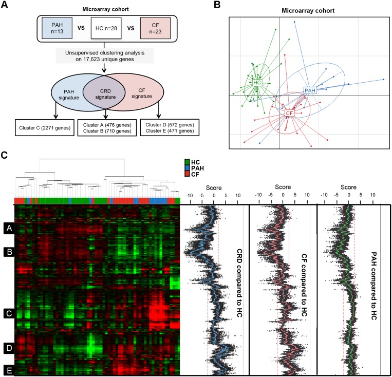
Gene expression microarrays were performed using total RNA from peripheral whole blood from 23 CF, 13 PAH and 28 HC (Figure 2A). Using the expression values of 17,163 unique genes, the principal component analysis (PCA) graph based on the first 2 components displayed a clear separation between HC and the CRD patients, whereas there was less distinction between CF and PAH (Figure 2B). In addition, a similar segregation was observed in the sample dendrogram of the unsupervised hierarchical clustering (Figure 2C). We then used a gene clustering approach to select signatures associated with CRD, CF and PAH, assuming that genes clustering together participate in a common function [18]. Based on unsupervised hierarchical clustering and an associated student t-test (p-value<0.01), we identified two clusters of under-expressed genes (clusters A (476 genes) and B (710 genes)) in both CF and PAH groups compared to HC (Figure 2A, C). In addition, compared to HC one cluster was associated with PAH (cluster C = 2,271 genes) and two with CF (clusters D and E, composed of 572 and 471 genes, respectively) (Figure 2A, C). These latter 3 clusters are composed of over-expressed genes relative to HC. In order to investigate the biological significance of these 5 clusters, GOminer analysis was performed to annotate all genes in each cluster. The ingenuity pathway analysis was used to identify key functional pathways.
Figure 2. Unsupervised hierarchical clustering analysis.
A) Tree analysis. Clustering analysis based on the 30,146 probes corresponding to 17,163 unique genes expressed in PAH, CF patients and Healthy Controls (HC). 3 signatures were found: 1 common between CF and PAH (named CRD signature), 1 specific to CF and 1 to PAH; B) Principal Component Analysis (PCA) displayed a clear separation between HC and patients with CRD, whereas CF and PAH patients were less distinct; C) 5 groups of genes (or clusters) were selected, A to E, based on a combined approach: selected genes were clustered together and exhibited a t-test p-value below 1% between the CRD group (PAH+CF), CF or PAH versus HC. Green represents relatively low expression, and red indicates relatively high expression.
Identification and validation of genes associated with both CRDs
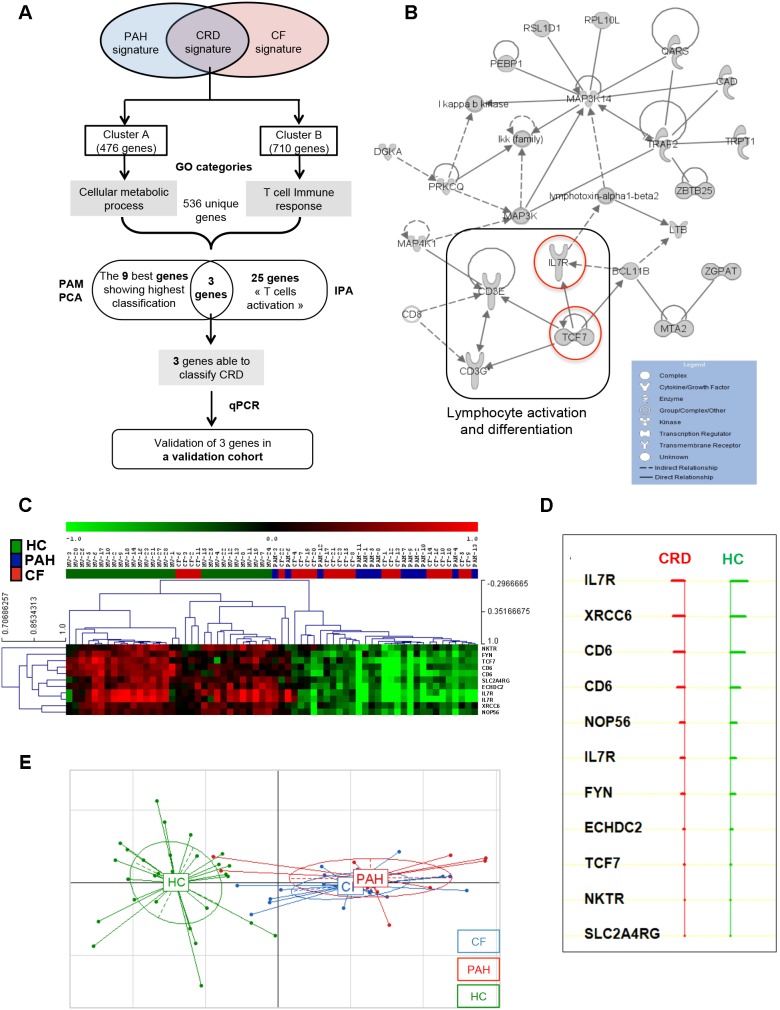
Microarray data highlighted two under-expressed genes signatures in both diseases (Figure 3A). Cluster A was mainly related to cellular metabolic processes (GO:0044237) (Table 2A) including genes involved in the cell cycle (cyclin-dependent kinase 9 (CDK9), ataxia telangiectasia mutated (ATM) and B-cell CLL/lymphoma 2 (BCL2)). Concerning cluster B, we found GO categories related to T cell signalling (such as “T cell receptor signalling pathway”, GO:0050852 and “antigen receptor-mediated signalling pathway”, GO:0050851) (Table 2B). Among genes from cluster B (associated with CRD) and from enriched GO categories (mainly related to gene expression and T cell signalling), we identified a main network of 25 genes associated with lymphocyte survival including 4 major genes under-expressed in both CRDs: CD3 gamma (CD3G), CD3 Epsilon (CD3E), Transcription factor 7 (TCF-7) and Interleukin-7 Receptor (IL-7R) (Figure 3B).
Figure 3. Characterization of under-expressed genes in the CRD signature.
A) Tree analysis of the CRD signature. Identification of the most representative genes by Gominer, PAM, IPA analysis and validation of the candidate genes in the validation cohort by qPCR; B) Network generated by IPA on the most significant GO categories in the CRD signature. Solid lines indicate direct interactions and dashed lines represent indirect interactions. Under-expressed genes in CRD are in gray. C) PAM analysis based on the most representative GO categories of Cluster A and B. Green represents relatively low expression, and red indicates relatively high expression. D) List of 9 genes able to classify correctly CRD and HC. E) The PCA graph of the 9 genes identified by PAM analysis indicated a clear separation between HC and patients with CRD (CF and PAH).
Table 2. Gene Ontology (GO categories) for genes in CRD signature versus HC.
| GO ID | GO category | Number of significant genes | Enrichment | FDR |
| A. | Cluster A - CRD signature | |||
| GO:0044260 | cellular macromolecule metabolic process | 165 | 1.39 | 0.00 |
| GO:0043170 | macromolecule metabolic process | 169 | 1.32 | 0.00 |
| GO:0090304 | nucleic acid metabolic process | 117 | 1.49 | 0.00 |
| GO:0006139 | nucleobase nucleoside nucleotide and nucleic acid metabolic process | 128 | 1.42 | 0.00 |
| GO:0044237 | cellular metabolic process | 189 | 1.23 | 0.00 |
| GO:0044238 | primary metabolic process | 188 | 1.23 | 0.00 |
| GO:0034641 | cellular nitrogen compound metabolic process | 132 | 1.36 | 0.00 |
| GO:0006807 | nitrogen compound metabolic process | 133 | 1.34 | 0.00 |
| GO:0008152 | metabolic process | 199 | 1.17 | 0.00 |
| GO:0010467 | gene expression | 108 | 1.38 | 0.00 |
| GO:0016070 | RNA metabolic process | 81 | 1.48 | 0.01 |
| GO:0010556 | regulation of macromolecule biosynthetic process | 83 | 1.46 | 0.01 |
| GO:0031323 | regulation of cellular metabolic process | 101 | 1.37 | 0.01 |
| GO:0031326 | regulation of cellular biosynthetic process | 84 | 1.42 | 0.01 |
| GO:2000112 | regulation of cellular macromolecule biosynthetic process | 80 | 1.44 | 0.02 |
| GO:0019222 | regulation of metabolic process | 107 | 1.33 | 0.01 |
| B. | Cluster B - CRD signature | |||
| GO:0006414 | translational elongation | 26 | 4.93 | 0.00 |
| GO:0006415 | translational termination | 23 | 4.74 | 0.00 |
| GO:0031018 | endocrine pancreas development | 23 | 4.69 | 0.00 |
| GO:0031016 | pancreas development | 23 | 4.45 | 0.00 |
| GO:0035270 | endocrine system development | 23 | 3.80 | 0.00 |
| GO:0006364 | rRNA processing | 18 | 3.79 | 0.00 |
| GO:0016072 | rRNA metabolic process | 18 | 3.67 | 0.00 |
| GO:0043624 | cellular protein complex disassembly | 24 | 3.56 | 0.00 |
| GO:0008033 | tRNA processing | 12 | 3.54 | 0.01 |
| GO:0006399 | tRNA metabolic process | 19 | 3.53 | 0.00 |
| GO:0019080 | viral genome expression | 24 | 3.51 | 0.00 |
| GO:0019083 | viral transcription | 24 | 3.51 | 0.00 |
| GO:0043241 | protein complex disassembly | 24 | 3.48 | 0.00 |
| GO:0034470 | ncRNA processing | 30 | 3.44 | 0.00 |
| GO:0050852 | T cell receptor signaling pathway | 14 | 3.39 | 0.00 |
| GO:0034623 | cellular macromolecular complex disassembly | 24 | 3.36 | 0.00 |
| GO:0042254 | ribosome biogenesis | 21 | 3.32 | 0.00 |
| GO:0032984 | macromolecular complex disassembly | 24 | 3.28 | 0.00 |
| GO:0034660 | ncRNA metabolic process | 39 | 3.23 | 0.00 |
| GO:0022613 | ribonucleoprotein complex biogenesis | 28 | 3.00 | 0.00 |
| GO:0050851 | antigen receptor-mediated signaling pathway | 15 | 2.93 | 0.01 |
| GO:0006412 | translation | 56 | 2.88 | 0.00 |
| GO:0071843 | cellular component biogenesis at cellular level | 28 | 2.88 | 0.00 |
| GO:0019058 | viral infectious cycle | 27 | 2.84 | 0.00 |
| GO:0071845 | cellular component disassembly at cellular level | 27 | 2.74 | 0.00 |
| GO:0002429 | immune response-activating cell surface receptor signaling pathway | 15 | 2.74 | 0.01 |
| GO:0022411 | cellular component disassembly | 27 | 2.69 | 0.00 |
| GO:0022415 | viral reproductive process | 27 | 2.61 | 0.00 |
| GO:0048610 | cellular process involved in reproduction | 31 | 2.58 | 0.00 |
| GO:0006396 | RNA processing | 62 | 2.32 | 0.00 |
Only Gene Ontology (GO) categories enriched in cluster A (A) and in cluster B (B) with False Discovery rate (FDR) less than 1% and with more than 10 genes are displayed.
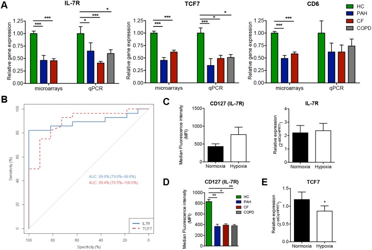
We performed a Prediction Analysis of Microarrays (PAM) based on genes from enriched GO categories for clusters A and B (536 unique genes) in order to define the genes specifically modulated in CRD (Figure 3C). A combination of 11 probes corresponding to 9 unique genes successfully classified CRD patients, with only 4 out of 28 HC misclassified (PAM overall error = 14%) (Figure 3D). PCA analysis based on the expression of these 9 genes clearly separated CRD from HC, suggesting a strong involvement of these genes in both CRDs (Figure 3E). Based on PAM and IPA analysis, we focused on CD6, IL-7R and TCF-7, 3 genes involved in lymphocyte activation. Transcript levels of these 3 genes were measured by qPCR in the validation cohort of 9 CF, 7 PAH, and 11 HC. To confirm the link between these genes and CRD, regardless of the primary disease, blood samples from 12 COPD patients were also analysed. We confirmed the under-expression of TCF-7 and IL-7R in the three CRD groups expect for CD6 (Figure 4A). The ROC analysis indicated that IL-7R and TCF-7 discriminated CRD with high sensitivity and specificity (AUC = 89.6%; p<0.001 and AUC = 89.4%; p<0.001, respectively) (Figure 4B).
Figure 4. Validation of the most representative genes in the validation cohort.
A) Quantitative PCR validation of 3 genes from the PAM analysis (IL-7R, TCF-7 and CD6) using the validation cohort; B) based on these qPCR values, IL-7R and TCF-7 enabled good discrimination between patients with CRD and HC, according to a receiver operating characteristic (ROC) analysis (AUC = 89.6% with p<0.001 and AUC = 89.4% with p<0.001, respectively); C) Median intensity fluorescence (MFI) and mRNA expression of IL-7R in PBMCs from healthy controls (HC) cultivated 12 hours under hypoxic and normoxic condition; D) MFI of IL-7R on PBMCs from CRD patients compared to HC; E) TCF-7 expression in PBMCs from HC under hypoxia or normoxia.
Effect of hypoxia on TCF-7 and IL-7R expression
Finally, we investigated whether hypoxia itself, a hallmark of peripheral tissues in all end-stage CRD patients, regulates TCF-7 and IL-7R genes. For this, we studied variation in the fluorescence intensity of IL-7R and expression of TCF-7 and IL-7R in PBMCs from HC incubated 12h under hypoxic or normoxic conditions. No difference in median fluorescence intensity (MFI) or expression of IL-7R was observed, supposing a long-term action of hypoxia on its regulation (Figure 4C). However IL-7R was significantly downregulated in PBMC of CRD patients compared to HC (Figure 4D). Similarly, a significant decrease in TCF-7 expression was found under hypoxia (Figure 4E). These results might suggest a possible modulation of TCF-7 gene and IL-7R protein in response to the hypoxic state of patients with CRD.
Identification and validation of genes associated with PAH
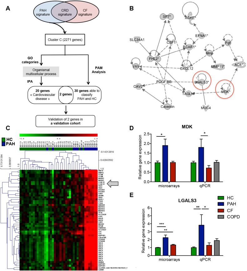
Microarray analysis highlighted one cluster (cluster C) associated with PAH (Figure 5A). GO ontology analysis allowed us to identify genes related to “organismal multicellular process” (GO:0032501), “G-protein coupled receptor protein signaling pathway” (GO:0007186) and “sensory perception of smell” (GO:0007608) (Table 3). Using IPA analysis, we characterized several over-expressed genes in PAH, in particular related to cardiovascular diseases, including the genes coding for caveolin-2 (CAV2), the vasoconstrictor angiotensin-converting enzyme gene (ACE), as well as molecules involved in angiogenesis and adhesion, such as midkine (MDK), and lectin galactoside-binding soluble 3 (LGALS3) (Figure 5B). Similarly, using a PAM analysis based on the 2,271 genes for cluster C, we identified the most informative genes in the PAH-specific signature. A combination of 30 unique genes (corresponding to 53 probes) classified PAH accurately, with only 1 out of 28 HC misclassified (Figure 5C). Based on PAM and IPA analysis, we validated by qPCR two genes: MDK and LGALS3 (Figure 5D, E). MDK was clearly over-expressed in PAH compared to the 3 other groups, but difference was only significant with CF (p<0.05) (Figure 5D). LGALS3 was significantly over-expressed in PAH compared to HC and CF (p<0.01 and p<0.05, respectively) implying a strong contribution of this gene to PAH (Figure 5E).
Figure 5. Characterization of over-expressed genes in the PAH signature.
A) Tree analysis of the PAH signature. Identification of the most representative genes by Gominer, PAM, IPA analysis and validation by qPCR of the candidate genes in the validation cohort; B) Network generated by IPA on the most significant GO categories in PAH signature. Over-expressed genes in PAH are in gray. C) PAM analysis based on the cluster, Green represents relatively low expression, and red indicates relatively high expression; D and E) Validation by qPCR of MDK and LGALS3 in the validation cohort, respectively.
Table 3. Gominer Analysis based for over-expressed genes in PAH signature versus HC.
| GO ID | GO category | Number of significant genes | Enrichment | FDR |
| GO:0032501 | multicellular organismal process | 323 | 1.29 | 0.00 |
| GO:0007608 | sensory perception of smell | 18 | 4.28 | 0.00 |
| GO:0003008 | system process | 104 | 1.65 | 0.00 |
| GO:0007275 | multicellular organismal development | 243 | 1.33 | 0.00 |
| GO:0007606 | sensory perception of chemical stimulus | 20 | 3.40 | 0.00 |
| GO:0032502 | developmental process | 259 | 1.28 | 0.00 |
| GO:0007186 | G-protein coupled receptor protein signaling pathway | 57 | 1.85 | 0.00 |
| GO:0048731 | system development | 201 | 1.33 | 0.00 |
| GO:0048856 | anatomical structure development | 219 | 1.30 | 0.00 |
| GO:0050877 | neurological system process | 74 | 1.65 | 0.00 |
| GO:0009187 | cyclic nucleotide metabolic process | 21 | 2.61 | 0.00 |
| GO:0009190 | cyclic nucleotide biosynthetic process | 19 | 2.75 | 0.01 |
| GO:0048513 | organ development | 141 | 1.36 | 0.01 |
| GO:0030154 | cell differentiation | 150 | 1.34 | 0.01 |
| GO:0030218 | erythrocyte differentiation | 16 | 2.90 | 0.01 |
| GO:0031279 | regulation of cyclase activity | 14 | 3.12 | 0.01 |
| GO:0030802 | regulation of cyclic nucleotide biosynthetic process | 16 | 2.85 | 0.01 |
| GO:0030808 | regulation of nucleotide biosynthetic process | 16 | 2.85 | 0.01 |
| GO:0007155 | cell adhesion | 65 | 1.58 | 0.01 |
Only GO categories enriched in cluster C with FDR less than 1%, with enrichment superior to 2 and with more than 10 genes are displayed.
Identification and validation of genes associated with CF
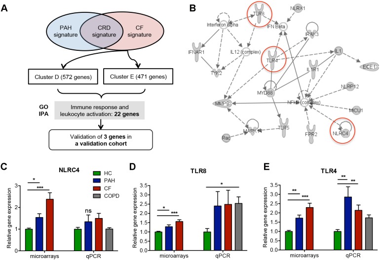
The CF signature was composed of two clusters (clusters D and E) of over-expressed genes involved in “cellular localization” (GO:0051641) (Table 4A) and in “Immune Response” (GO:0006955) and more specifically in “leukocyte activation” (GO:0002366) (Table 4B, Figure 6A). In addition, we found a set of 22 over-expressed genes associated with innate immunity, especially genes coding toll-like receptor 4 (TLR4), TLR8, NLR family CARD domain-containing protein 4 (NLRC4) and interleukin 1 (IL1) (Figure 6B). Using IPA, we measured the level of gene transcripts involved in CF, notably NLRC4, TLR4 and TLR8, in the independent validation cohort. Whereas the over-expression of NLRC4 and TLR8 were not confirmed (Figure 6C, D), we found a significant increase in TLR4 gene expression in CF patients (p<0.01 vs HC) (Figure 6E). Interestingly our investigation showed a significant expression of TLR8 in COPD (p<0.05 vs HC) and TLR4 in PAH, suggesting that these genes are not specific to CF. Altogether, these results still confirm the stimulation of innate immune response during CF.
Table 4. Gominer Analysis based for over-expressed genes in CF signature versus HC.
| GO ID | GO category | Number of significant genes | Enrichment | FDR |
| A. | Cluster D - CF signature | |||
| GO:0008104 | protein localization | 73 | 1.74 | 0.00 |
| GO:0033036 | macromolecule localization | 82 | 1.63 | 0.00 |
| GO:0045184 | establishment of protein localization | 63 | 1.73 | 0.01 |
| GO:0016050 | vesicle organization | 12 | 4.46 | 0.01 |
| GO:0015031 | protein transport | 62 | 1.73 | 0.00 |
| GO:0006464 | protein modification process | 97 | 1.49 | 0.01 |
| GO:0046907 | intracellular transport | 56 | 1.75 | 0.01 |
| GO:0051641 | cellular localization | 77 | 1.57 | 0.01 |
| GO:0034613 | cellular protein localization | 41 | 1.94 | 0.01 |
| GO:0070727 | cellular macromolecule localization | 41 | 1.93 | 0.01 |
| GO:0043412 | macromolecule modification | 99 | 1.45 | 0.01 |
| GO:0016192 | vesicle-mediated transport | 49 | 1.78 | 0.01 |
| GO:0044260 | cellular macromolecule metabolic process | 242 | 1.19 | 0.01 |
| GO:0006886 | intracellular protein transport | 35 | 1.92 | 0.01 |
| B. | Cluster E - CF signature | |||
| GO:0009611 | response to wounding | 54 | 2.48 | 0.00 |
| GO:0006952 | defense response | 53 | 2.45 | 0.00 |
| GO:0023052 | signaling | 148 | 1.54 | 0.00 |
| GO:0023033 | signaling pathway | 121 | 1.63 | 0.00 |
| GO:0023046 | signaling process | 112 | 1.60 | 0.00 |
| GO:0023060 | signal transmission | 111 | 1.58 | 0.00 |
| GO:0007165 | signal transduction | 101 | 1.62 | 0.00 |
| GO:0006955 | immune response | 51 | 2.13 | 0.00 |
| GO:0002376 | immune system process | 70 | 1.84 | 0.00 |
| GO:0048583 | regulation of response to stimulus | 47 | 2.17 | 0.00 |
| GO:0035466 | regulation of signaling pathway | 60 | 1.94 | 0.00 |
| GO:0035556 | intracellular signal transduction | 73 | 1.78 | 0.00 |
| GO:0007243 | intracellular protein kinase cascade | 41 | 2.28 | 0.00 |
| GO:0023014 | signal transmission via phosphorylation event | 41 | 2.28 | 0.00 |
| GO:0006954 | inflammatory response | 27 | 2.85 | 0.00 |
| GO:0002263 | cell activation involved in immune response | 14 | 4.78 | 0.00 |
| GO:0002366 | leukocyte activation involved in immune response | 14 | 4.78 | 0.00 |
| GO:0006950 | response to stress | 100 | 1.57 | 0.00 |
| GO:0007169 | transmembrane receptor protein tyrosine kinase signaling pathway | 36 | 2.37 | 0.00 |
| GO:0007166 | cell surface receptor linked signaling pathway | 74 | 1.73 | 0.00 |
| GO:0023034 | intracellular signaling pathway | 80 | 1.65 | 0.00 |
| GO:0050817 | coagulation | 28 | 2.61 | 0.00 |
Only GO categories enriched in cluster D (A) and in cluster E (B) with FDR less than 1%, with enrichment superior to 2 and with more than 10 genes are displayed.
Figure 6. Characterization of over-expressed genes in the CF signature.
A) Tree analysis of CF signature. Identification of the most representative genes by Gominer and IPA analysis and validation by qPCR of these genes in the validation cohort; B) Network generated by IPA on the most significant GO categories in CF signature. Over-expressed genes in CF are in gray; C, D, E) qPCR validation on the new cohort for NLRC4, TLR8 and TLR4 genes.
Discussion
The objectives of this research were to discover genes potentially involved in the peripheral damages seen in end-stage CRD by performing a systematic analysis of the blood mRNA from CRD patients. To make sure that genes identified as related to CRD were independent of the aetiology, we included in the screening analysis 2 diseases completely distinct in their pathophysiology, CF and PAH. We added a third unrelated disease, COPD, in the validation analysis, again increasing the probability that any link found to CRD was independent of the underlying diseases. This strategy not only provided CRD-related signatures, but also allowed the identification of genes specific of CF and PAH, some of which had not been suspected previously. The relevance of these disease-specific, highly contrasted signatures was reinforced by the concomitant study of 3 diseases that allowed each of them to serve as a control for the others and eliminate non-specific genes. COLT, a lung transplant cohort of 360 patients at time of this study, offered a unique opportunity to select homogeneous groups in terms of age, sex, underlying care and treatment, thus increasing the chances of detecting reliable signatures.
First, based on a microarray analysis using a combined hierarchical clustering approach, we identified a signature for CRD. The GO terms analysis in cluster A and B highlighted large families of genes associated with “metabolic process” and “T cell receptor signalling pathway”, respectively. A set composed of under-expressed genes was determined, including genes involved in T-cell receptor (TCR) signalling, namely CD3E, CD3G, IL-7R and TCF-7 (also known as TCF-1). TCF-7 and IL-7R were identified in the gene network related to lymphocyte activation and were among the 9 genes selected in the PAM analysis. Their significant under-expression was confirmed by qPCR in independent CF and PAH groups and in individuals with COPD. Interestingly, these two genes are dependent on activation of the Wnt/β-catenin pathway and are pivotal in the control of T lymphocyte survival [19], [20]. IL-7R composed of 2 chains, the common γ chain and the α chain (or CD127), mediates signalling of IL-7. IL-7R neutralization delays post-depletional T cell recovery through both the suppression of thymopoiesis and the inhibition of T cell homeostatic proliferation [19]. As for IL-7R, a number of works have established TCF-7 as a critical regulator necessary for the maintenance of normal T-cell development, but also for the induction of many components of T-cell identity [20], [21]. Among the genes induced by TCF-7, there are T-cell essential transcription factors such as Gata3, as well as components involved in the regulation of TCR such as IL-7R [22]–[24]. Recently TCF-7 has been shown to induce Th2 and Th17 inflammation, supporting the hypothesis that dysfunction of this pathway at any stage of T cell differentiation could lead to immune deficiency [21]. In light of the above, the down-regulation of IL-7R and TCF-7 genes implies decreased adaptive immunity in end-stage CRD patients and could indirectly explain some infections and complications. A recent study described a systemic gene expression profile in patients with COPD [25]. Among the candidate genes found, TCF-7 was a biomarker for COPD. However, our study clearly shows that TCF-7 is not specific of COPD, but is rather an important marker of CRD. Indeed TCF-7 was down regulated in COPD, but also in CF and PAH, evidencing a modulation of TCF-7 in response to respiratory failure, whatever its cause. Since functional immunity is maintained by the metabolic requirement of proteins [26], the alteration of the immune response in end-stage CRD could be related to under-nutrition.
Whether the signature is related to CRD whatever the stage, or is specific to advanced respiratory failure, should be elucidated in cohorts with less developed respiratory diseases. In addition, to make sure that the signature is specific to CRD, other chronic invalidating diseases involving under-nutrition should be investigated. However the direct hypoxia-driven down-regulation of TCF-7 in PBMC suggests that it is a respiratory-related signature. The consequences of hypoxia were also confirmed by the down-modulation of IL-7R on the surface of PBMC from CRD patients. An improvement in the gene or protein expression after lung transplant would confirm the direct link with respiratory disorders.
In addition, we identified a specific signature for PAH. The GO and IPA analysis identified a number of genes already described in the pathophysiology of PAH: proteins coded by CAV2 regulating lung endothelial cell proliferation and differentiation, and by ACE, a key enzyme in cardiovascular pathobiology, which serum levels are correlated with lung endothelial injury [27], [28]. Furthermore, the midkine (MDK) gene, a heparin-binding growth factor linked to ACE, was identified in our gene network. MDK is known to promote vascular leukocyte infiltration and migration and proliferation of smooth muscle cells. MDK levels are increased in systemic hypertension and MDK interacts with ACE in the renin-angiotensin system [29], [30].
The gene most contributing to PAH according to the PAM analysis was LGALS3, a member of the galectin family of carbohydrate binding proteins. We validated its over-expression in an independent PAH group by qPCR. Galectin-3 is described as a multifunctional protein involved in a variety of biological processes including fibrosis, angiogenesis and the activation of various immune cells, such as macrophages, neutrophils, mast cells and lymphocytes [31], [32]. Interestingly, several works have shown that the upregulation of LGALS3 is linked to heart failure and is an independent blood biomarker for ventricular remodeling and mortality [33]–[35]. Our results suggest that LGALS3 may be involved in right heart failure, the most common cause of death in PAH [36]. Further investigations are required to decipher the functional role of galectin-3 in PAH.
The over-expression of genes related to innate immunity identified in CF through GO analysis was consistent with inflammation in this disease [37]. In accord with this, the IPA analysis showed many genes involved in inflammatory functions. Among these, Pattern Recognition Receptor (PRR) family genes, including TLR, notably TLR4 and TLR8, were overexpressed. Their altered expression is directly associated with immune-deregulation [38], [39]. We validated the TLR4 over-expression by qPCR in blood from other CF patients. Most interestingly, TLR4 was also significantly increased in uninfected PAH patients, suggesting that TLR4 upregulation in CF patients is not related to infections. Additional PRRs were present in this network, including a member of the Nod-Like Receptor family, NLRC4. The NLRC4 inflammasome is essential for host immunity against extracellular pathogens, such as Pseudomonas aeruginosa, a frequent pulmonary pathogen in CF [40]. Thus, NLRC4 over-expression in the blood of CF could relate to their infectious status.
The number of patients per group is small, which can be seen as a limitation of the study. Indeed as showed on Figure 1 the selection strategy that aimed to get homogeneity of the patients populations within groups regarding type of the disease, treatments, experimental process (RNA and cDNA qualities) led to eliminating many samples from the analysis. The number of analysed patients was further decreased by elimination of unsuitable RNA. This strategy reduced the power of the study to detect genes relevant for each disease, but also lowered the risk of misclassifying the patients because of comorbidities. In addition, confounding factors such as age, lower in CF, or specific treatments, still cannot be eliminated. Nevertheless, we tried to overcome this by matching the age of HC population with these of CF and PAH groups. A systematic strategy of propensity score matching would have stratified group comparisons on such covariates. However the study was mainly designed to detect a signature common to CF and PAH that could be validated in new sets of patients and COPD, rather than identifying genes specific of each disease. A different strategy of selection aiming to eliminate confounding factors would have been applied if the discovery of genes specific for each disease had been the primary objective. Despite this limitation, clustering analysis discriminated groups accurately, provided functional clusters, and most significant genes were validated in independent samples. Moreover, the risk of bias is lowered by that overexpressed genes are related to pathophysiological pathways already known to be disturbed in the respective diseases. It is the case of innate immunity genes in CF.
The blood gene expression profiling of patients with CRD enabled us providing a systematic description of peripheral molecular events related to CRD, CF and PAH. Notably, a common pattern associated with respiratory diseases, mainly under-expressed genes playing a role in immune functions is described. The relevance of these genes in the immunodeficiency of CRD patients is potentially important and would benefit from being investigated in functional studies. Our study further demonstrates the interest of systematic gene screening in order to detect unexplored mechanisms. The peripheral signatures found strengthen the arguments for a global approach of respiratory diseases in a systemic medical strategy. Elucidation of the molecular mechanisms involved in these changes in gene expression will require further investigations.
Supporting Information
Blood cell count in Giga/L and in percentage in the microarray cohort (A and B) and the validation cohort (C and D). Results are given as mean ± standard error (SEM). PAH = pulmonary arterial hypertension; CF = cystic fibrosis; COPD: Chronic Obstructive Respiratory Disease; Leu: Leukocytes; PMN: Polymorphonuclear Neutrophil; Ly: Lymphocytes; Mo: Monocytes; Eo: Eosinophils; Bas: Basophils.
(TIF)
Acknowledgments
The authors thank the Integrative genomics platform of Nantes, in particular Catherine Chevalier and Edouard Hirchaud for assistance with the microarray data. The authors also thank the Cytocell platform of Nantes for assistance in the flow cytometry analysis and Béatrice Delasalle for statistics help. The authors are also grateful to the members of the COLT consortium. Membership of the Cohort of Lung Transplantation–COLT Consortium (associating surgeons; anaesthetists,-intensivists, physicians, research staff): Leader: Magnan Antoine; antoine.magnan@univ-nantes.fr; Bordeaux: J. Jougon, J.-F. Velly; H. Rozé; E. Blanchard, C. Dromer; Bruxelles: M. Antoine, M. Cappello, R. Souilamas, M. Ruiz, Y. Sokolow, F. Vanden Eynden, G. Van Nooten; L. Barvais, J. Berré, S. Brimioulle, D. De Backer, J. Créteur, E. Engelman, I. Huybrechts, B. Ickx, T. J.C. Preiser, T. Tuna, L. Van Obberghe, N. Vancutsem, J.-L. Vincent; P. De Vuyst, I. Etienne, F. Féry, F. Jacobs, C. Knoop, J.L. Vachiéry, P. Van den Borne, I. Wellemans; G. Amand, L. Collignon, M. Giroux; Grenoble: E. Arnaud-Crozat, V. Bach, P.-Y. Brichon, P. Chaffanjon, O. Chavanon, A. de Lambert, J.-P. Fleury, S. Guigard, K. Hireche, A. Pirvu, P. Porcu, R. Hacini; P. Albaladejo, C. Allègre, D. Anglade, D. Bedague, P. Bouzat, E. Briot, O. Carle, M. Casez-Brasseur, D. Colas, G. Dessertaine, M. Durand, J. Duret, M.C. Fèvre, G. Francony, S. Gay, M.R Marino, B. Oummahan, D. Protar, D. Rehm, S. Robin, M. Rossi-Blancher, L. Saunier; P. Bedouch, A. Boignard, H. Bouvaist, A. Briault, B. Camara, S. Chanoine, M. Dubuc, S. Lantuéjoul, S. Quêtant, J. Maurizi, P. Pavèse, C. Pison, C. Saint-Raymond, N. Wion; C. Chérion; Lyon: R. Grima, O. Jegaden, J.-M. Maury, F. Tronc; C. Flamens, S. Paulus; J.-F. Mornex, F. Philit, A. Senechal, J.-C. Glérant, S. Turquier; D. Gamondes; L. Chalabresse, F. Thivolet-Bejui; C Barnel, C. Dubois, A. Tiberghien; Paris, Hôpital Européen Georges Pompidou: F. Le Pimpec-Barthes, A. Bel, P. Mordant, P. Achouh; V. Boussaud; R. Guillemain, D. Méléard, M.O. Bricourt, B. Cholley; V. Pezella; Marseille: M. Adda, M. Badier, F. Bregeon, B. Coltey, X.B. D’Journo, S. Dizier, C. Doddoli, N. Dufeu, H. Dutau, JM. Forel, JY. Gaubert, C. Gomez, M. Leone, A. Nieves, B. Orsini, L. Papazian L, C. Picard, M. Reynaud-Gaubert, A. Roch, JM. Rolain, E. Sampol, V. Secq, P. Thomas, D. Trousse; Yahyaoui M; Nantes: O. Baron, P. Lacoste, C. Perigaud, J.C. Roussel; I. Danner, A Haloun A. Magnan, A Tissot; T. Lepoivre, M. Treilhaud; K. Botturi-Cavaillès, S. Brouard, R. Danger, J. Loy, M. Morisset, M. Pain, S. Pares, D. Reboulleau, P.-J. Royer; Paris, Hôpital Marie Lannelongue: P. Dartevelle, E. Fadel, S. Mussot, D. Fabre, O. Mercier; P. Viard, S. François; J. Cerrina, P. Hervé, J. Le Pavec, F. Le Roy Ladurie; l. lamran; Paris, Hôpital Bichat: Y. Castier, P. Cerceau, F. Francis, G. Lesèche; N. Allou, P. Augustin, S. Boudinet, M. Desmard, G. Dufour, P. Montravers; O. Brugière, G. Dauriat, G. Jébrak, H. Mal, A. Marceau, A.-C. Métivier, G. Thabut; B. Ait Ilalne; Strasbourg: P. Falcoz, G. Massard, N. Santelmo; G. Ajob, O. Collange O. Helms, J. Hentz, A. Roche; B. Bakouboula, T. Degot, A. Dory, S. Hirschi, S. Ohlmann-Caillard, L. Kessler, R. Kessler, A. Schuller; K. Bennedif, S. Vargas; Suresnes: P. Bonnette, A. Chapelier, P. Puyo, E. Sage; J. Bresson, V. Caille, C. Cerf, J. Devaquet, V. Dumans-Nizard, ML. Felten, M. Fischler, AG. Si Larbi, M. Leguen, L. Ley, N. Liu, G. Trebbia; S. De Miranda, B. Douvry, F. Gonin, D. Grenet, A.M. Hamid, H. Neveu, F. Parquin, C. Picard, A. Roux, M. Stern; F. Bouillioud, P. Cahen, M. Colombat, C. Dautricourt, M. Delahousse, B. D’Urso, J. Gravisse, A. Guth, S. Hillaire, P. Honderlick, M. Lequintrec, E. Longchampt, F. Mellot, A. Scherrer, L. Temagoult, L. Tricot; M. Vasse, C. Veyrie, L. Zemoura; Toulouse: J. Berjaud, L. Brouchet, M. Dahan; F. Le Balle, O. Mathe; H. Benahoua, A. Didier, A.L. Goin, M. Murris; L. Crognier, O. Fourcade.
Data Availability
The authors confirm that all data underlying the findings are fully available without restriction. Data are available from the GEO database at: http://www.ncbi.nlm.nih.gov/geo/query/acc.cgi?token=epkbaewellmjhuv&acc=GSE38267.
Funding Statement
This study was funded mainly by a grant from Vaincre La Mucoviscidose, the Programme National Hospitalier de Recherche Clinique, a CENTAURE foundation grant, an Agence de la Biomédecine Française (ABM) grant, and an ESOT grant. This work was realized in the context of the IHU-Cesti project thanks to French government financial support managed by the National Research Agency via the “Investment Into The Future” program ANR-10-IBHU-005. The IHU-Cesti project is also supported by Nantes Metropole and the Pays de la Loire Region. The funders had no role in study design, data collection and analysis, decision to publish, or preparation of the manuscript.
References
- 1. Chailleux E, Fauroux B, Binet F, Dautzenberg B, Polu JM (1996) Predictors of survival in patients receiving domiciliary oxygen therapy or mechanical ventilation. A 10-year analysis of ANTADIR Observatory. CHEST 109: 741–749. [DOI] [PubMed] [Google Scholar]
- 2. Barnes PJ (2000) Chronic obstructive pulmonary disease. N Engl J Med 343: 269–280 10.1056/NEJM200007273430407 [DOI] [PubMed] [Google Scholar]
- 3. Ratjen F, Döring G (2003) Cystic fibrosis. Lancet 361: 681–689 10.1016/S0140-6736(03)12567-6 [DOI] [PubMed] [Google Scholar]
- 4. Humbert M, Sitbon O, Simonneau G (2004) Treatment of pulmonary arterial hypertension. N Engl J Med 351: 1425–1436 10.1056/NEJMra040291 [DOI] [PubMed] [Google Scholar]
- 5. Price LC, Wort SJ, Perros F, Dorfmüller P, Huertas A, et al. (2012) Inflammation in pulmonary arterial hypertension. CHEST 141: 210–221 10.1378/chest.11-0793 [DOI] [PubMed] [Google Scholar]
- 6. de Theije C, Costes F, Langen RC, Pison C, Gosker HR (2011) Hypoxia and muscle maintenance regulation: implications for chronic respiratory disease. Curr Opin Clin Nutr Metab Care 14: 548–553 10.1097/MCO.0b013e32834b6e79 [DOI] [PubMed] [Google Scholar]
- 7. Cano NJM, Pichard C, Roth H, Court-Fortuné I, Cynober L, et al. (2004) C-reactive protein and body mass index predict outcome in end-stage respiratory failure. CHEST 126: 540–546 10.1378/chest.126.2.540 [DOI] [PubMed] [Google Scholar]
- 8. Bull TM, Coldren CD, Moore M, Sotto-Santiago SM, Pham DV, et al. (2004) Gene Microarray Analysis of Peripheral Blood Cells in Pulmonary Arterial Hypertension. Am J Respir Crit Care Med 170: 911–919 10.1164/rccm.200312-1686OC [DOI] [PubMed] [Google Scholar]
- 9. Adib-Conquy M, Pedron T, Petit-Bertron A-F, Tabary O, Corvol H, et al. (2008) Neutrophils in cystic fibrosis display a distinct gene expression pattern. Mol Med 14: 36–44 10.2119/2007-00081.Adib-Conquy [DOI] [PMC free article] [PubMed] [Google Scholar]
- 10. Chen H, Wang Y, Bai C, Wang X (2012) Alterations of plasma inflammatory biomarkers in the healthy and chronic obstructive pulmonary disease patients with or without acute exacerbation. J Proteomics 75: 2835–2843 10.1016/j.jprot.2012.01.027 [DOI] [PubMed] [Google Scholar]
- 11. Le Meur N, Lamirault G, Bihouée A, Steenman M, Bédrine-Ferran H, et al. (2004) A dynamic, web-accessible resource to process raw microarray scan data into consolidated gene expression values: importance of replication. Nucleic Acids Res 32: 5349–5358 10.1093/nar/gkh870 [DOI] [PMC free article] [PubMed] [Google Scholar]
- 12. Chopard A, Lecunff M, Danger R, Lamirault G, Bihouee A, et al. (2009) Large-scale mRNA analysis of female skeletal muscles during 60 days of bed rest with and without exercise or dietary protein supplementation as countermeasures. Physiol Genomics 38: 291–302 10.1152/physiolgenomics.00036.2009 [DOI] [PubMed] [Google Scholar]
- 13. Applied Biosystems (1997) ABI PRISM 7900 user bulletin. 2: 11–24 Available: http://www3.appliedbiosystems.com/cms/groups/mcb_support/documents/generaldocuments/cms_040980.pdf Accessed 2014 Sep 29.. [Google Scholar]
- 14. Dray S, Dufour AB (2007) The ade4 package: implementing the duality diagram for ecologists. Journal of Statistical Software 22(4): 1–20. [Google Scholar]
- 15.Kuimelis RG, Livak KJ, Mullah B, Andrus A (1997) Structural analogues of TaqMan probes for real-time quantitative PCR. Nucleic Acids Symp Ser: 255–256. [PubMed]
- 16. Alizadeh AA, Eisen MB, Davis RE, Ma C, Lossos IS, et al. (2000) Distinct types of diffuse large B-cell lymphoma identified by gene expression profiling. Nature 403: 503–511 10.1038/35000501 [DOI] [PubMed] [Google Scholar]
- 17. Qiu W, Lee M-LT (2006) SPCalc: A web-based calculator for sample size and power calculations in micro-array studies. Bioinformation 1: 251–252. [DOI] [PMC free article] [PubMed] [Google Scholar]
- 18. Eisen MB, Spellman PT, Brown PO, Botstein D (1998) Cluster analysis and display of genome-wide expression patterns. Proc Natl Acad Sci USA 95: 14863–14868. [DOI] [PMC free article] [PubMed] [Google Scholar]
- 19. Mai H-L, Boeffard F, Longis J, Danger R, Martinet B, et al. (2014) IL-7 receptor blockade following T cell depletion promotes long-term allograft survival. J Clin Invest 124: 1723–1733 10.1172/JCI66287 [DOI] [PMC free article] [PubMed] [Google Scholar]
- 20. Ioannidis V, Beermann F, Clevers H, Held W (2001) The beta-catenin–TCF-1 pathway ensures CD4(+)CD8(+) thymocyte survival. Nat Immunol 2: 691–697 10.1038/90623 [DOI] [PubMed] [Google Scholar]
- 21. Ma J, Wang R, Fang X, Sun Z (2012) β-catenin/TCF-1 pathway in T cell development and differentiation. J Neuroimmune Pharmacol 7: 750–762 10.1007/s11481-012-9367-y [DOI] [PMC free article] [PubMed] [Google Scholar]
- 22. Weber BN, Chi AW-S, Chavez A, Yashiro-Ohtani Y, Yang Q, et al. (2011) A critical role for TCF-1 in T-lineage specification and differentiation. Nature 476: 63–68 10.1038/nature10279 [DOI] [PMC free article] [PubMed] [Google Scholar]
- 23. Yu Q, Xu M, Sen JM (2007) Beta-catenin expression enhances IL-7 receptor signaling in thymocytes during positive selection. J Immunol 179: 126–131. [DOI] [PMC free article] [PubMed] [Google Scholar]
- 24. Peschon JJ, Morrissey PJ, Grabstein KH, Ramsdell FJ, Maraskovsky E, et al. (1994) Early lymphocyte expansion is severely impaired in interleukin 7 receptor-deficient mice. J Exp Med 180: 1955–1960. [DOI] [PMC free article] [PubMed] [Google Scholar]
- 25. Bahr TM, Hughes GJ, Armstrong M, Reisdorph R, Coldren CD, et al. (2013) Peripheral Blood Mononuclear Cell Gene Expression in Chronic Obstructive Pulmonary Disease. Am J Respir Cell Mol Biol 49: 316–323 10.1165/rcmb.2012-0230OC [DOI] [PMC free article] [PubMed] [Google Scholar]
- 26. Iyer SS, Chatraw JH, Tan WG, Wherry EJ, Becker TC, et al. (2012) Protein energy malnutrition impairs homeostatic proliferation of memory CD8 T cells. The Journal of Immunology 188: 77–84 10.4049/jimmunol.1004027 [DOI] [PMC free article] [PubMed] [Google Scholar]
- 27. Orfanos SE, Armaganidis A, Glynos C, Psevdi E, Kaltsas P, et al. (2000) Pulmonary capillary endothelium-bound angiotensin-converting enzyme activity in acute lung injury. Circulation 102: 2011–2018. [DOI] [PubMed] [Google Scholar]
- 28. Xie L, Vo-Ransdell C, Abel B, Willoughby C, Jang S, et al. (2011) Caveolin-2 is a negative regulator of anti-proliferative function and signaling of transforming growth factor-β in endothelial cells. Am J Physiol, Cell Physiol 301: C1161–C1174 10.1152/ajpcell.00486.2010 [DOI] [PMC free article] [PubMed] [Google Scholar]
- 29. Hobo A, Yuzawa Y, Kosugi T, Kato N, Asai N, et al. (2009) The growth factor midkine regulates the renin-angiotensin system in mice. J Clin Invest 119: 1616–1625 10.1172/JCI37249 [DOI] [PMC free article] [PubMed] [Google Scholar]
- 30. Weckbach LT, Muramatsu T, Walzog B (2011) Midkine in inflammation. ScientificWorldJournal 11: 2491–2505 10.1100/2011/517152 [DOI] [PMC free article] [PubMed] [Google Scholar]
- 31. Taniguchi T, Asano Y, Akamata K, Noda S, Masui Y, et al. (2012) Serum levels of galectin-3: possible association with fibrosis, aberrant angiogenesis, and immune activation in patients with systemic sclerosis. J Rheumatol 39: 539–544 10.3899/jrheum.110755 [DOI] [PubMed] [Google Scholar]
- 32. Tribulatti MV, Figini MG, Carabelli J, Cattaneo V, Campetella O (2012) Redundant and Antagonistic Functions of Galectin-1, -3, and -8 in the Elicitation of T Cell Responses. The Journal of Immunology 188: 2991–2999 10.4049/jimmunol.1102182 [DOI] [PubMed] [Google Scholar]
- 33. de Boer RA, Yu L, van Veldhuisen DJ (2010) Galectin-3 in cardiac remodeling and heart failure. Curr Heart Fail Rep 7: 1–8 10.1007/s11897-010-0004-x [DOI] [PMC free article] [PubMed] [Google Scholar]
- 34. Chowdhury P, Kehl D, Choudhary R, Maisel A (2013) The Use of Biomarkers in the Patient with Heart Failure. Curr Cardiol Rep 15: 372 10.1007/s11886-013-0372-4 [DOI] [PubMed] [Google Scholar]
- 35. Gaggin HK, Januzzi JL Jr (2013) Biochimica et Biophysica Acta. BBA - Molecular Basis of Disease 1832: 2442–2450 10.1016/j.bbadis.2012.12.014 [DOI] [PubMed] [Google Scholar]
- 36. Schermuly RT, Ghofrani HA, Wilkins MR, Grimminger F (2011) Mechanisms of disease: pulmonary arterial hypertension. Nat Rev Cardiol 8: 443–455 10.1038/nrcardio.2011.87 [DOI] [PMC free article] [PubMed] [Google Scholar]
- 37. Cohen TS, Prince A (2012) Cystic fibrosis: a mucosal immunodeficiency syndrome. Nat Med 18: 509–519 10.1038/nm.2715 [DOI] [PMC free article] [PubMed] [Google Scholar]
- 38. Krieg AM, Vollmer J (2007) Toll-like receptors 7, 8, and 9: linking innate immunity to autoimmunity. Immunol Rev 220: 251–269 10.1111/j.1600-065X.2007.00572.x [DOI] [PubMed] [Google Scholar]
- 39. Sturges NC, Wikström ME, Winfield KR, Gard SE, Brennan S, et al. (2010) Monocytes from children with clinically stable cystic fibrosis show enhanced expression of Toll-like receptor 4. Pediatr Pulmonol 45: 883–889 10.1002/ppul.21230 [DOI] [PubMed] [Google Scholar]
- 40. Cai S, Batra S, Wakamatsu N, Pacher P, Jeyaseelan S (2012) NLRC4 inflammasome-mediated production of IL-1β modulates mucosal immunity in the lung against gram-negative bacterial infection. The Journal of Immunology 188: 5623–5635 10.4049/jimmunol.1200195 [DOI] [PMC free article] [PubMed] [Google Scholar]
Associated Data
This section collects any data citations, data availability statements, or supplementary materials included in this article.
Supplementary Materials
Blood cell count in Giga/L and in percentage in the microarray cohort (A and B) and the validation cohort (C and D). Results are given as mean ± standard error (SEM). PAH = pulmonary arterial hypertension; CF = cystic fibrosis; COPD: Chronic Obstructive Respiratory Disease; Leu: Leukocytes; PMN: Polymorphonuclear Neutrophil; Ly: Lymphocytes; Mo: Monocytes; Eo: Eosinophils; Bas: Basophils.
(TIF)
Data Availability Statement
The authors confirm that all data underlying the findings are fully available without restriction. Data are available from the GEO database at: http://www.ncbi.nlm.nih.gov/geo/query/acc.cgi?token=epkbaewellmjhuv&acc=GSE38267.


