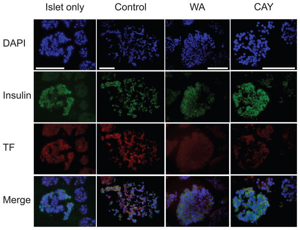
FIGURE 5.
Tissue factor (TF) expression in islets after exposure to autologous blood. The expression of TF was evaluated with fluorescence stain (red) where nuclei and insulin were stained as blue and green. The autologous islet-blood samples were taken at 3 hours after the culture initiation. White bar indicates 50 μm. Representative sections are shown. WA indicates withaferin A; CAY, CAY10512.

