

Abstract
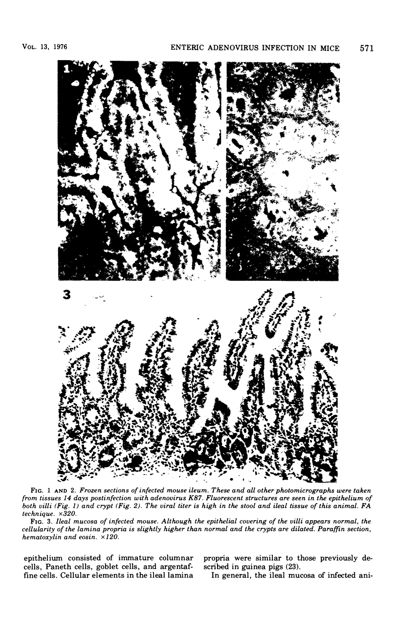
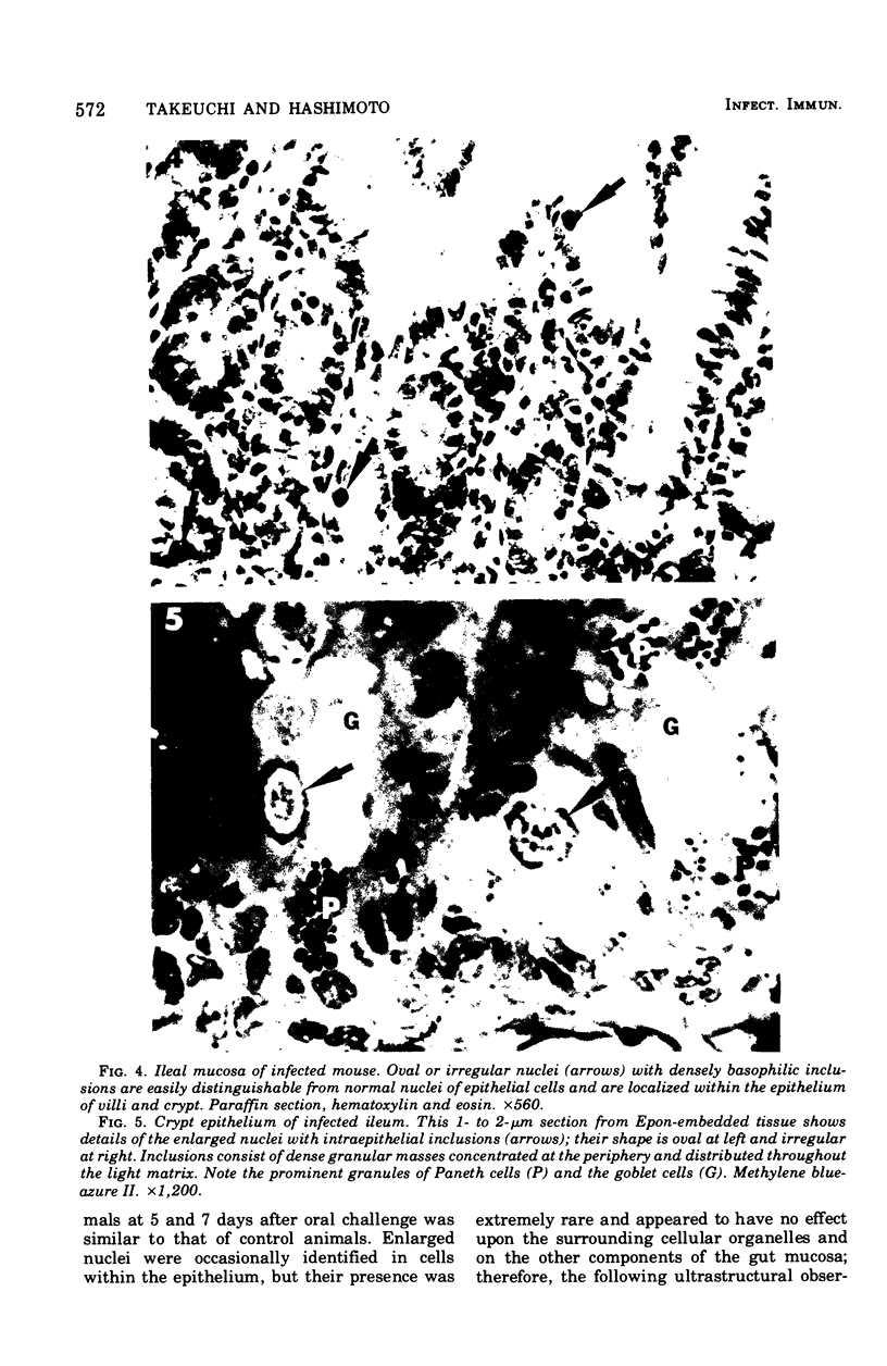
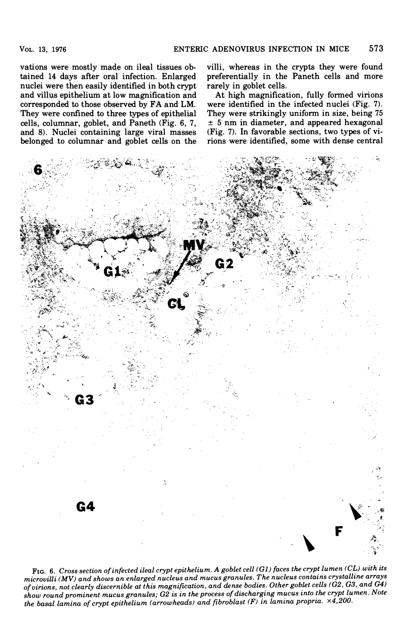
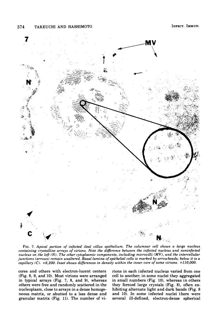
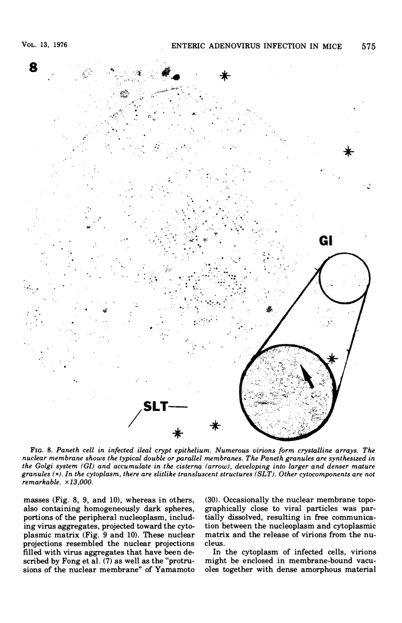
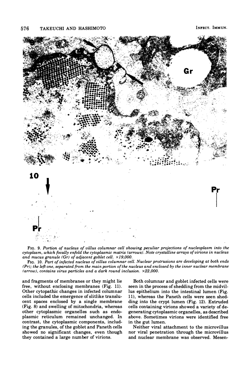
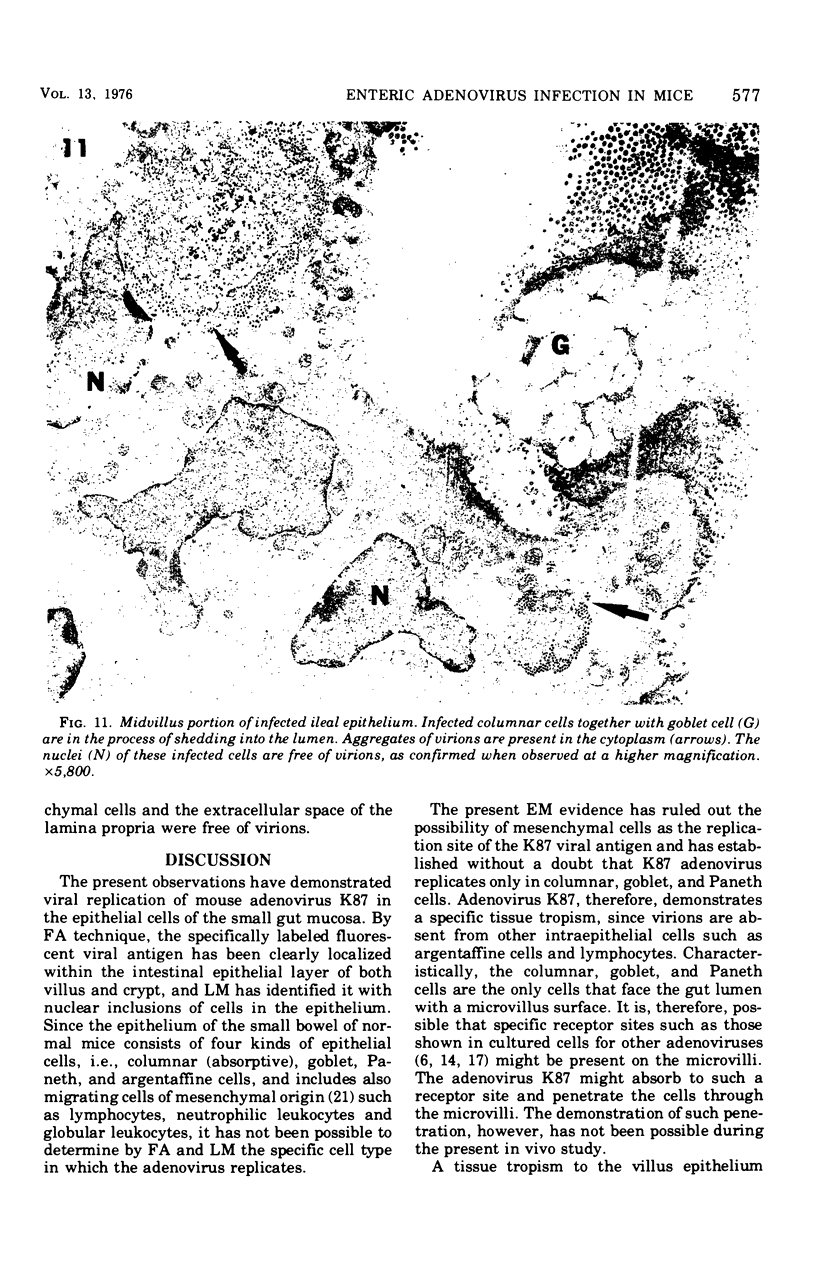
Using fluorescent antibody techniques (FA) and light microscopy (LM) and electron microscopy (EM), this paper describes the morphological features of the ileum in the DK1 mouse orally challenged with adenovirus K87. At the peak of infection, virus is easily identified by FA in the epithelium of the villi and crypts of the ileum. LM shows that fluorescent cells have large, bizarre, uniformly basophilic nuclei containing deoxyribonucleic acid, as indicated by histochemical tests. EM further identifies these nuclei as belonging to columnar, goblet, or Paneth cells, all epithelial cells facing the lumen with a microvillus border. The basophilic material in the nuclei consists of virus particles 75 nm in diameter arranged in crystalline arrays. When found in the cell cytoplasm, the virions do not form arrays but are scattered or form irregular aggregates, which may or may not be enclosed by single membranes. Infected columnar cells show mild cytopathic effects with no cell degeneration and necrosis, whereas the goblet and Paneth cells appear normal and maintain synthetic and secretory functions. All infected cells, however, share an abnormally accelerated extrusion rate, with columnar and goblet cells often being shed from the side rather than from the tip of the villi. The Paneth cells, which do not migrate out of the crypts, show a higher than normal rate of extrusion in the crypt lumen.
Full text
PDF

Images in this article
Selected References
These references are in PubMed. This may not be the complete list of references from this article.
- CLEMMER D. L. EXPERIMENTAL ENTERIC INFECTION OF CHICKENS WITH AN AVIAN ADENO-VIRUS (STRAIN 93). Proc Soc Exp Biol Med. 1965 Apr;118:943–948. doi: 10.3181/00379727-118-30013. [DOI] [PubMed] [Google Scholar]
- Cairnie A. B. Renewal of goblet and Paneth cells in the small intestine. Cell Tissue Kinet. 1970 Jan;3(1):35–45. doi: 10.1111/j.1365-2184.1970.tb00250.x. [DOI] [PubMed] [Google Scholar]
- Chardonnet Y., Dales S. Early events in the interaction of adenoviruses with HeLa cells. II. Comparative observations on the penetration of types 1, 5, 7, and 12. Virology. 1970 Mar;40(3):478–485. doi: 10.1016/0042-6822(70)90190-x. [DOI] [PubMed] [Google Scholar]
- Cheng H., Merzel J., Leblond C. P. Renewal of Paneth cells in the small intestine of the mouse. Am J Anat. 1969 Dec;126(4):507–525. doi: 10.1002/aja.1001260409. [DOI] [PubMed] [Google Scholar]
- Clemmer D. I., Ichinose H. The cellular site of virus replication in the intestine of chicks infected with an avian adenovirus. Arch Gesamte Virusforsch. 1968;25(3):277–287. doi: 10.1007/BF01556556. [DOI] [PubMed] [Google Scholar]
- Dales S. Early events in cell-animal virus interactions. Bacteriol Rev. 1973 Jun;37(2):103–135. doi: 10.1128/br.37.2.103-135.1973. [DOI] [PMC free article] [PubMed] [Google Scholar]
- Fong C. K., Bensch K. G., Hsiung G. D. Productive and abortive infections of simian and nonsimian cells with a simian adenovirus SV15. I. Microscopic observations. Virology. 1968 Jun;35(2):297–310. doi: 10.1016/0042-6822(68)90270-5. [DOI] [PubMed] [Google Scholar]
- Hashimoto K., Sugiyama T., Sasaki S. An adenovirus isolated from the feces of mice I. Isolation and identification. Jpn J Microbiol. 1966 Jul;10(2):115–125. doi: 10.1111/j.1348-0421.1966.tb00298.x. [DOI] [PubMed] [Google Scholar]
- Hashimoto K., Sugiyama T., Yoshikawa M., Sasaki S. Intestinal resistance in the experimental enteric infection of mice with a mouse adenovirus. I. Growth of the virus and appearance of a neutralizing substance in the intestinal tract. Jpn J Microbiol. 1970 Sep;14(5):381–395. doi: 10.1111/j.1348-0421.1970.tb00538.x. [DOI] [PubMed] [Google Scholar]
- Henry C. J., Slifkin M., Merkow L. P., Pardo M. The ultrastructure and nature of adenovirus type 2-induced paracrystalline formations. Virology. 1971 Apr;44(1):215–218. doi: 10.1016/0042-6822(71)90167-x. [DOI] [PubMed] [Google Scholar]
- Hoenig E. M., Margolis G., Kilham L. Experimental adenovirus infection of the mouse adrenal gland. II. Electron microscopic observations. Am J Pathol. 1974 May;75(2):375–394. [PMC free article] [PubMed] [Google Scholar]
- Kalnins V. I., Stich H. F., Yohn D. S. Electron microscopic localization of virus-associated antigens in human amnion cells (AV-3) infected with human adenovirus, type 12. Virology. 1966 Apr;28(4):751–754. doi: 10.1016/0042-6822(66)90259-5. [DOI] [PubMed] [Google Scholar]
- Lonberg-Holm K., Philipson L. Early events of virus-cell interaction in an adenovirus system. J Virol. 1969 Oct;4(4):323–338. doi: 10.1128/jvi.4.4.323-338.1969. [DOI] [PMC free article] [PubMed] [Google Scholar]
- Margolis G., Kilham L., Hoenig E. M. Experimental adenovirus infection of the mouse adrenal gland. I. Light microscopic observations. Am J Pathol. 1974 May;75(2):363–374. [PMC free article] [PubMed] [Google Scholar]
- Merzel J., Leblond C. P. Origin and renewal of goblet cells in the epithelium of the mouse small intestine. Am J Anat. 1969 Mar;124(3):281–305. doi: 10.1002/aja.1001240303. [DOI] [PubMed] [Google Scholar]
- Philipson L., Lonberg-Holm K., Pettersson U. Virus-receptor interaction in an adenovirus system. J Virol. 1968 Oct;2(10):1064–1075. doi: 10.1128/jvi.2.10.1064-1075.1968. [DOI] [PMC free article] [PubMed] [Google Scholar]
- Phillips D. M., Raskas H. J. Ultrastructural changes in KB cultures infected with adenovirus type 2. Virology. 1972 Apr;48(1):156–169. doi: 10.1016/0042-6822(72)90123-7. [DOI] [PubMed] [Google Scholar]
- Sugiyama T., Hashimoto K., Sasaki S. An adenovirus isolated from the feces of mice. II. Experimental infection. Jpn J Microbiol. 1967 Mar;11(1):33–42. doi: 10.1111/j.1348-0421.1967.tb00318.x. [DOI] [PubMed] [Google Scholar]
- TRIER J. S. STUDIES ON SMALL INTESTINAL CRYPT EPITHELIUM. I. THE FINE STRUCTURE OF THE CRYPT EPITHELIUM OF THE PROXIMAL SMALL INTESTINE OF FASTING HUMANS. J Cell Biol. 1963 Sep;18:599–620. doi: 10.1083/jcb.18.3.599. [DOI] [PMC free article] [PubMed] [Google Scholar]
- Takeuchi A., Formal S. B., Sprinz H. Exerimental acute colitis in the Rhesus monkey following peroral infection with Shigella flexneri. An electron microscope study. Am J Pathol. 1968 Mar;52(3):503–529. [PMC free article] [PubMed] [Google Scholar]
- Takeuchi A., Jervis H. R., Sprinz H. The globule leucocyte in the intestinal mucosa of the cat: a histochemical, light and electron microscopic study. Anat Rec. 1969 May;164(1):79–99. doi: 10.1002/ar.1091640106. [DOI] [PubMed] [Google Scholar]
- Takeuchi A., Phillips B. P. Electron microscope studies of experimental Entamoeba histolytica infection in the guinea pig. I. Penetration of the intestinal epithelium by trophozoites. Am J Trop Med Hyg. 1975 Jan;24(1):34–48. doi: 10.4269/ajtmh.1975.24.34. [DOI] [PubMed] [Google Scholar]
- Takeuchi A., Sprinz H. Electron-Microscope Studies of Experimental Salmonella Infection in the Preconditioned Guinea Pig: II. Response of the Intestinal Mucosa to the Invasion by Salmonella typhimurium. Am J Pathol. 1967 Jul;51(1):137–161. [PMC free article] [PubMed] [Google Scholar]
- Takeuchi A., Sprinz H., LaBrec E. H., Formal S. B. Experimental bacillary dysentery. An electron microscopic study of the response of the intestinal mucosa to bacterial invasion. Am J Pathol. 1965 Dec;47(6):1011–1044. [PMC free article] [PubMed] [Google Scholar]
- Takeuchi A., Zeller J. A. Ultrastructural identification of spirochetes and flagellated microbes at the brush border of the large intestinal epithelium of the rhesus monkey. Infect Immun. 1972 Dec;6(6):1008–1018. doi: 10.1128/iai.6.6.1008-1018.1972. [DOI] [PMC free article] [PubMed] [Google Scholar]
- Thrasher J. D., Greulich R. C. The duodenal progenitor population. 3. The progenitor cell cycle of principal, goblet and paneth cells. J Exp Zool. 1966 Feb;161(1):9–19. doi: 10.1002/jez.1401610103. [DOI] [PubMed] [Google Scholar]
- Windisch M. C. Etude morphologique des cellules de Paneth dans diverses conditions physiologiques. Rev Can Biol. 1966 Sep;25(3):167–177. [PubMed] [Google Scholar]