Abstract
OBJECTIVE
Diabetic nephropathy (DN) remains the most common cause of end-stage renal disease and is a major cause of mortality in type 2 diabetes. Insulin sensitivity is an important determinant of renal health in adults with type 2 diabetes, but limited data exist in adolescents. We hypothesized that measured insulin sensitivity (glucose infusion rate [GIR]) would be associated with early markers of DN reflected by estimated glomerular filtration rate (eGFR) and albumin-creatinine ratio (ACR) in adolescents with type 2 diabetes.
RESEARCH DESIGN AND METHODS
Type 2 diabetic (n = 46), obese (n = 29), and lean (n = 19) adolescents (15.1 ± 2.2 years) had GIR measured by hyperinsulinemic-euglycemic clamps. ACR was measured and GFR was estimated by the Bouvet equation (combined creatinine and cystatin C).
RESULTS
Adolescents with type 2 diabetes had significantly lower GIR, and higher eGFR and ACR than obese or lean adolescents. Moreover, 34% of type 2 diabetic adolescents had albuminuria (ACR ≥30 mg/g), and 24% had hyperfiltration (≥135 mL/min/1.73 m2). Stratifying ACR and eGFR into tertiles, adolescents with type 2 diabetes in the highest tertiles of ACR and eGFR had respectively lower GIR than those in the mid and low tertiles, after adjusting for age, sex, Tanner stage, BMI, and HbA1c (P = 0.02 and P = 0.04). GIR, but not HbA1c, LDL, or systolic blood pressure, was also associated with eGFR after adjusting for sex and Tanner stage (β ± SE: −2.23 ± 0.87; P = 0.02).
CONCLUSIONS
A significant proportion of adolescents with type 2 diabetes showed evidence of early DN, and insulin sensitivity, rather than HbA1c, blood pressure, or lipid control, was the strongest determinant of renal health.
Introduction
Diabetic nephropathy (DN) remains the most important cause of end-stage renal disease (ESRD) in North America and Europe and also one of the major causes of mortality in type 2 diabetes. The 2011 U.S. Renal Data System showed that DN accounted for 44.5% of all cases of ESRD in the U.S. in 2009 (1). DN is also an important risk factor for the development of future cardiovascular disease. Unfortunately, the initiation of early DN is clinically silent for many years, and during this time, renal parenchymal damage progresses (2). Furthermore, early markers of DN prior to the loss of renal function, such as hyperfiltration (glomerular filtration rate [GFR] ≥135 mL/min/1.73 m2) and albuminuria within the normal range, can manifest in adolescents with type 2 diabetes and are also associated with early cardiovascular abnormalities (3,4). In fact, adolescents with type 2 diabetes have a twofold increased risk of microalbuminuria compared with youth with type 1 diabetes (5–7). In addition to early renal disease being a determinant of future cardiovascular risk, insulin sensitivity is associated with renal health in adults with and without type 2 diabetes (8,9). Moreover, prediabetic conditions are associated with early preclinical manifestations of kidney dysfunction, including hyperfiltration, in adults (10). However, limited data exist in adolescents with type 2 diabetes.
Accordingly, our aim was to describe the prevalence of hyperfiltration and albuminuria in a contemporary adolescent cohort of patients with type 2 diabetes. Moreover, we sought to investigate the associations among measured insulin sensitivity, estimated GFR (eGFR), and albumin-creatinine ratio (ACR). In light of associations between insulin insensitivity and early DN in adults, we hypothesized that greater insulin sensitivity would be associated with renal health, reflected by eGFR and ACR within normal ranges, in adolescents with type 2 diabetes.
Research Design and Methods
Participants
A total of 94 pubertal adolescents between the ages of 12 and 19 years were recruited for a study of diabetes and insulin resistance in youth and had insulin sensitivity assessed by hyperinsulinemic-euglycemic clamp, as well as data available to calculate ACR and eGFR by creatinine and cystatin C. Of the 94 adolescents, 46 were diagnosed with type 2 diabetes, 29 with obesity (BMI >95th percentile) but no diabetes, and 19 were normal weight controls (BMI >5th percentile and <85th percentile). Family history of diabetes was similar in obese and type 2 diabetic participants, but negative in control participants. Obese participants were chosen to have similar BMI percentile and fat mass as participants with type 2 diabetes. All three subject groups were chosen to be of similar age, level of physical activity, and sex distribution. The study was approved by the University of Colorado Denver institutional review board, and appropriate consent and assent were obtained.
Height and weight were measured for determination of BMI. BMI z-score was calculated using BMI, sex, and age. Absence of diabetes was confirmed in the nondiabetic groups by a 2-h, 75-g oral glucose tolerance test. Type 2 diabetes was defined by American Diabetes Association’s criteria and the absence of glutamic acid decarboxylase, islet cell or insulin autoantibodies, insulin requirement, or secondary causes of diabetes, as previously described (11). Inclusion criteria included pubertal status (Tanner stage >1) and sedentary status (<3 h of exercise/week) to minimize training effects. Exclusions included body weight >300 pounds, blood pressure >140/90 mmHg at rest or >190/100 mmHg during exercise, hemoglobin <9 mg/dL, serum creatinine >1.5 mg/dL, smoking, antihypertensive drugs, pregnancy, breastfeeding, ≥3 h of physical activity per week, or plans to alter exercise or diet during the study. For participants with type 2 diabetes, additional exclusion criteria included HbA1c ≥12%, medications known to affect insulin sensitivity other than metformin (oral or inhaled steroids, thiazolidinediones, and atypical antipsychotics), and other antidiabetes drugs except insulin. For nondiabetic participants, additional exclusions included medications known to affect insulin sensitivity (metformin, oral or inhaled steroids, thiazolidinediones, and atypical antipsychotics), other antidiabetes drugs, and insulin.
Pubertal development was assessed by a single pediatric endocrinologist using the criteria established by Tanner and Marshall for pubic hair and breast development. Testicular volume was measured using an orchidometer.
Laboratory Measures
For the 3 days prior to admission, participants were asked to refrain from all strenuous physical activity due to the impact on insulin sensitivity and albuminuria. They were also provided with a Pediatric Clinical and Translational Research Center–prepared weight maintenance, 3-day, fixed macronutrient diet to limit impacts of macronutrient variation on insulin sensitivity and renal function, as previously described in detail (11). Participants were admitted overnight to the Pediatric Clinical and Translational Research Center to ensure fasting. Type 2 diabetic participants (n = 22) on insulin were instructed to replace their long-acting insulin (Lantus or Levemir) as needed with intermediate-acting insulin (NPH) and short-acting insulin (Humalog or Novolog) to ensure that their last long-acting insulin injection was at least 24 h prior to admission (36 h prior to the clamp). The evening of admission, all subcutaneous insulin was replaced by an insulin drip. Participants were then maintained overnight on intravenous regular insulin with adjustments by a standard protocol to maintain near euglycemia (goal blood glucose 100–110 mg/dL) until starting the clamp the next morning. Metformin was not taken within 72 h of the clamp to wash out acute effects on insulin sensitivity. No other antidiabetes drugs except insulin were taken per exclusion criteria (see above). Fasting laboratory evaluation included: total cholesterol, LDL cholesterol (LDL-C), HDL cholesterol (HDL-C), triglycerides, glucose, and HbA1c (Diabetes Control and Complications Trial-calibrated); assays were performed by standard methods. Insulin sensitivity (glucose infusion rate [GIR]) was calculated from a 3-h hyperinsulinemic-euglycemic clamp (80 mU * m−2 * min−1 insulin) as previously described (11,12). Serum creatinine and cystatin C were measured from postclamp samples, which were collected after 3 to 4 h of euglycemia, eliminating the effects of acute glycemia on eGFR (13). Due to the absence of chronic kidney disease and expected normal to elevated GFRs for age, we used the Bouvet equation to estimate GFR (eGFR = 63.2 * [serum creatinine/96]−0.35 * [serum cystatin C/1.2]−0.56 * [weight/45]0.30 * [age/14]0.40) (14,15). This equation has high accuracy when compared with gold-standard measurements in adolescents with eGFR >90 mL/min/1.73 m2 (14). Hyperfiltration was defined as eGFR ≥135 mL/min/1.73 m2 (4,16). Spot urine was collected upon admission for urinary albumin and creatinine, and ACR was calculated. Albuminuria was defined as microalbuminuria or greater with ACR ≥30 mg/g.
Imaging
Body composition (e.g., adiposity) by DEXA was performed by standard methods on a Hologic device (Hologic, Waltham, MA) (17).
Statistical Analysis
Analyses were performed in SAS (version 9.3 for Windows; SAS Institute, Cary, NC). Variables were checked for the distributional assumption of normality using normal plots. The distributions of ACR, triglycerides, and insulin dose were skewed. Therefore, natural log transformations were applied. eGFR by Bouvet and ACR were stratified into tertiles. ANOVA with a Tukey-Kramer P value adjustment was used for comparison of continuous variables across the three groups (mid, low, and high tertiles), and least square means were calculated for the tertile groups. The χ2 test was used to compare categorical variables in the tertile groups.
Univariate and multivariable linear regression models were used to examine the associations between measured insulin sensitivity, HbA1c, LDL-C, non–HDL-C, systemic blood pressure (SBP), diastolic blood pressure, natural log of ACR (LnACR), lean mass and fat mass with eGFR by Bouvet, unadjusted and adjusted for Tanner stage, sex, HbA1c, BMI percentile, and diabetes duration. Logistic regression models were used to evaluate associations between measured insulin sensitivity with hyperfiltration (eGFR ≥135 mL/min/1.73 m2) and albuminuria (ACR ≥30 mg/g), unadjusted and adjusted for Tanner stage, sex, HbA1c, BMI percentile, and diabetes duration. Significance was based on an α-level of 0.05.
Results
Participant Characteristics
Table 1 shows participant characteristics stratified by group (lean, obese, and type 2 diabetic), and Table 2 shows measured insulin sensitivity, serum creatinine, serum cystatin C, eGFR, and ACR adjusted for Tanner stage and sex. By design, there were no significant differences across groups either in age or sex distribution or between the type 2 diabetic and obese groups for BMI percentile or percent fat mass (Table 1). All groups had more females than males, reflective of the typical pediatric type 2 diabetic population. As expected, HbA1c was elevated in youth with type 2 diabetes and normal in obese and control youth. Average duration of diabetes in youth with type 2 diabetes was 2.0 ± 1.8 years. Of the 13 participants with albuminuria, all of them had a BMI >85th percentile. Thirty-four percent of participants with type 2 diabetes had albuminuria but only a single participant with obesity had albuminuria. Twenty-four percent of participants with type 2 diabetes exhibited renal hyperfiltration. None of the obese or lean participants exhibited hyperfiltration.
Table 1.
Participant characteristics for obese, type 2 diabetic, and lean adolescents
| Variable | Obese (N = 29) | Type 2 diabetic (N = 46) | Lean (N = 19) | P value |
|---|---|---|---|---|
| Male (%) | 34 | 29 | 41 | 0.55 |
| Age (years) | 14.8 ± 2.1 | 15.3 ± 2.3 | 14.7 ± 2.1 | 0.39 |
| Non-Hispanic white | 39% | 20% | 64% | 0.002 |
| Hispanic | 41% | 60% | 21% | |
| Black | 7% | 20% | 15% | |
| American-Indian | 3% | 0% | 0% | |
| Asian | 7% | 0% | 0% | |
| Other | 3% | 0% | 0% | |
| HbA1c (%) | 5.2 ± 0.3 | 7.9 ± 2.3 | 5.1 ± 0.3 | <0.0001 |
| HbA1c (mmol/mol) | 33.3 ± 2.1 | 62.8 ± 22.8 | 32.2 ± 2.1 | <0.0001 |
| Diabetes duration (years) | — | 2.0 ± 1.8 | — | NA |
| Height (cm) | 165.6 ± 7.8 | 165.1 ± 9.1 | 164.0 ± 7.5 | 0.78 |
| Weight (kg) | 87.7 ± 23.2 | 92.7 ± 21.2 | 54.7 ± 8.3 | <0.0001 |
| BMI percentile | 96 ± 3 | 97 ± 4 | 52 ± 22 | <0.0001 |
| Fat mass (%) | 35.0 ± 13.4 | 38.4 ± 12.6 | 11.9 ± 6.1 | <0.0001 |
| Waist-to-hip ratio | 0.90 ± 0.07 | 0.96 ± 0.09 | 0.82 ± 0.07 | <0.0001 |
| Waist circumference (cm) | 101.3 ± 21.4 | 106.4 ± 15.3 | 70.1 ± 6.1 | <0.0001 |
| Total cholesterol (mg/dL) | 172 ± 35 | 161 ± 35 | 145 ± 24 | 0.03 |
| LDL-C (mg/dL) | 99 ± 24 | 87 ± 24 | 82 ± 26 | 0.04 |
| HDL-C (mg/dL) | 42 ± 9 | 38 ± 11 | 46 ± 9 | 0.02 |
| Triglycerides§ (mg/dL) | 131 ± 2 | 146 ± 2 | 74 ± 1 | <0.0001 |
| Fasting glucose (mg/dL) | 88 ± 8 | 113 ± 29 | 89 ± 9 | <0.0001 |
| Fasting insulin§ (μU/mL) | 19 ± 2 | 31 ± 2 | 8 ± 2 | <0.0001 |
| Steady-state glucose (mg/dL) | 96 ± 6 | 98 ± 5 | 99 ± 9 | 0.27 |
| Steady-state insulin§ (μU/mL) | 162 ± 2 | 148 ± 2 | 141 ± 2 | 0.68 |
| GIR (mg/kg/min) | 15.0 ± 6.2 | 7.6 ± 4.1 | 19.9 ± 3.7 | <0.0001 |
| BUN (mg/dL) | 12.9 ± 3.0 | 11.3 ± 2.1 | 12.4 ± 3.9 | 0.07 |
| ACR§ (mg/g) | 6.3 ± 2.1 | 22.6 ± 5.5 | 7.6 ± 2.8 | 0.001 |
| Serum creatinine (mg/dL) | 0.72 ± 0.20 | 0.60 ± 0.16 | 0.73 ± 0.15 | 0.006 |
| Serum cystatin C (mg/L) | 0.94 ± 0.12 | 0.88 ± 0.16 | 1.00 ± 0.17 | 0.02 |
| eGFR by Bouvet (mL/min/1.73 m2) | 103.0 ± 11.3 | 124.4 ± 24.2 | 87.6 ± 14.2 | <0.0001 |
| SBP (mmHg) | 117 ± 9 | 123 ± 13 | 111 ± 8 | 0.0003 |
| Diastolic blood pressure (mmHg) | 71 ± 7 | 72 ± 11 | 66 ± 7 | 0.003 |
| Tanner 1 | 0 (0%) | 0 (0%) | 0 (0%) | 0.0002 |
| Tanner 2 | 0 (0%) | 1 (2.2%) | 1 (5.3%) | |
| Tanner 3 | 3 (10.3%) | 3 (6.5%) | 1 (5.3%) | |
| Tanner 4 | 6 (20.7%) | 3 (6.5%) | 10 (52.6%) | |
| Tanner 5 | 20 (69.0%) | 39 (84.8%) | 7 (36.8%) |
BUN, blood urea nitrogen; NA, not applicable.
§Geometric means ± SE.
Table 2.
Tanner and sex-adjusted means for insulin sensitivity and variables of renal health by group for adolescents with type 2 diabetes
| Variable | Obese (N = 29) | Type 2 diabetic (N = 46) | Lean (N = 19) |
|---|---|---|---|
| Steady-state insulin§ (μU/mL) | 162 ± 1 | 147 ± 1 | 142 ± 1 |
| Steady-state glucose (mg/dL) | 96 ± 1 | 98 ± 1 | 100 ± 2 |
| GIR (mg/kg/min) | 14.9 ± 1.0* | 7.5 ± 0.8* | 19.6 ± 1.0* |
| Serum creatinine (mg/dL) | 0.75 ± 0.03 | 0.62 ± 0.03** | 0.74 ± 0.03 |
| Serum cystatin C (mg/L) | 0.95 ± 0.03 | 0.92 ± 0.02 | 0.99 ± 0.04 |
| eGFR by Bouveta (mL/min/1.73 m2) | 101.0 ± 4.4 | 120.6 ± 3.4*** | 88.5 ± 5.2 |
| ACR§ (mg/g) | 6.6 ± 1.3 | 22.2 ± 1.2*** | 7.5 ± 1.3 |
Data presented as least square means ± SE.
aNot adjusted for age as age is part of Bouvet eGFR equation.
§Geometric means.
*P < 0.01 in all pairwise comparisons.
**P < 0.05 compared with obese and lean.
***P < 0.01 compared with obese and lean.
eGFR and Insulin Sensitivity
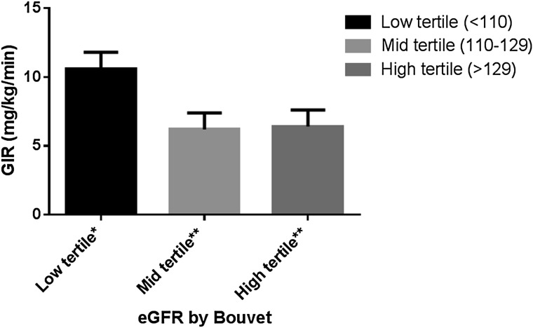
Insulin sensitivity was lowest in youth with type 2 diabetes and intermediate in obese nondiabetic youth, with no differences in steady-state insulin or glucose values (Table 2). In participants with type 2 diabetes, measured insulin sensitivity explained 13.0% (r = −0.36; P = 0.03) of eGFR, and measured insulin sensitivity was inversely associated with eGFR after adjusting for Tanner stage and sex (β ± SE: −2.23 ± 0.87; P = 0.02). HbA1c (P = 0.09), LDL-C (P = 0.78), SBP (P = 0.75), and adiposity (P = 0.18) were not significantly associated with eGFR. Stratifying eGFR into tertiles for participants with type 2 diabetes (low [<110 mL/min/1.73 m2], middle [110–128 mL/min/1.73 m2], and high tertiles [≥129 mL/min/1.73 m2]), participants in the highest tertile had significantly lower measured insulin sensitivity than participants in the middle and low tertiles after adjusting for sex and Tanner stage (P < 0.05; Fig. 1). The difference between the low and middle tertile remained significant after further adjustments for HbA1c, BMI, and diabetes duration (P = 0.04). No significant differences in HbA1c, LDL-C, SBP, or adiposity were observed among the tertiles of eGFR.
Figure 1.
Adjusted means of GIR stratified by tertiles of eGFR in adolescents with type 2 diabetes. Data presented as least square means ± SE adjusted for Tanner and sex (age is part of Bouvet eGFR equation) for adolescents with type 2 diabetes. The difference between low and mid tertiles remained significant after further adjustments for BMI, HbA1c, and duration (P = 0.04). *P < 0.05 compared with mid and high tertiles; **P < 0.05 compared with low tertile.
ACR and Insulin Sensitivity
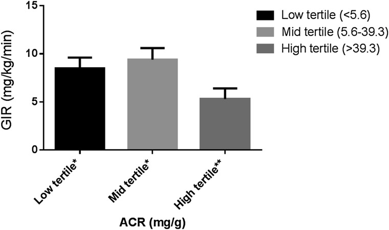
In participants with type 2 diabetes, measured insulin sensitivity was not significantly associated with LnACR (β ± SE: −0.10 ± 0.07; P = 0.14) in a linear regression model. Similarly, HbA1c, LDL-C, SBP, and adiposity were not significantly associated with LnACR in type 2 diabetic participants. Stratifying ACR into tertiles for participants with type 2 diabetes (low [<5.6 mg/g], mid [5.6–39.3 mg/g], and high [≥39.3 mg/g]), participants in the high ACR tertile had significantly lower insulin sensitivity compared with participants in the low and middle tertile (P < 0.05; Fig. 2). The difference between the high and middle tertile remained significant after further adjustments for Tanner stage, BMI, HbA1c, and diabetes duration (P = 0.02). In contrast, there were no significant differences in HbA1c, LDL-C, SBP, or adiposity between the ACR tertiles.
Figure 2.
Adjusted means of GIR stratified by tertiles of ACR in adolescents with type 2 diabetes. Data presented as least square means ± SE for tertiles of ACR adjusted for age and sex for adolescents with type 2 diabetes. The difference between high and mid tertiles remained significant after further adjustments for Tanner, BMI, HbA1c, and duration (P = 0.02). *P < 0.05 compared with high tertile; **P < 0.05 compared with low or mid tertiles.
Hyperfiltration, Albuminuria, and Insulin Sensitivity
After adjusting for sex, Tanner stage, and diabetes duration, 1-SD increase in measured insulin sensitivity was associated with a lower odds of having albuminuria (odds ratio [OR] 0.41 [95% CI 0.17–0.99]; P = 0.047, per 1 SD [4.23 mg/kg/min]) in participants with type 2 diabetes. Further adjustments for HbA1c and BMI attenuated the association (P = 0.22). In contrast, 1-SD increase in measured insulin sensitivity was not significantly associated with lower odds of having hyperfiltration (OR 0.54 [95% CI 0.20–1.47]; P = 0.22, per 1 SD) after adjusting for sex, Tanner stage, and diabetes duration, possibly due to the limited number of observations (hyperfiltration: n = 9).
Conclusions
Hyperfiltration and albuminuria were present in a significant proportion of adolescents with type 2 diabetes in our cohort, despite their short duration of diabetes. We report lower ACR and eGFR in the normal range in adolescents with higher insulin sensitivity, and insulin sensitivity also appears to be associated with lower odds of having albuminuria. These findings suggest a renoprotective role of insulin sensitivity in adolescents with type 2 diabetes.
Type 2 diabetes remains the leading cause of ESRD in the Western world (18), and most patients with type 2 diabetes develop some degree of renal dysfunction during their lifetime (8). The natural history of DN is characterized by a long silent period without overt clinical signs or symptoms of nephropathy. For that reason, a major challenge in preventing DN in type 2 diabetes is the difficulty in accurately identifying high-risk patients with preclinical disease. From our data, conventional risk factors including HbA1c, LDL-C, and SBP appear less important than insulin sensitivity in determining renal health in adolescents with type 2 diabetes. Moreover, there was no significant difference in adiposity by using standardized DEXA between the tertiles of ACR and eGFR.
Hyperfiltration and microalbuminuria are early, preclinical phenotypes of DN and are also associated with early cardiovascular abnormalities (3,4). In the TODAY study with an average follow-up of only 3.9 years (7), the prevalence of microalbuminuria among youth with type 2 diabetes almost tripled (from 6.3 to 16.6%). In fact, microalbuminuria may precede the development of type 2 diabetes in insulin-resistant obese adolescents (19,20). Hyperfiltration is thought to be a major contributing factor for DN in type 2 diabetes, reflecting underlying increases in intraglomerular pressure, leading to structural changes over time such as mesangial expansion and glomerular basement membrane thickening (21). The traditional mechanisms underlying these pathological changes in adolescence are complex and involve growth factors, neurohormonal activation, and changes in renal tubuloglomerular feedback (22). More recently, insulin resistance has also been implicated in the progression of DN in type 2 diabetes (23,24). A growing body of data demonstrates that insulin resistance is associated with an elevation of glomerular hydrostatic pressure, causing increased renal vascular permeability and ultimately glomerular hyperfiltration (25–27). Another possible mechanistic pathway linking insulin resistance to DN is via effects on overall nonesterified fatty acid exposure and lipotoxicity, leading to the development of microangiopathy (28). Early signs of DN are present in adolescents with type 2 diabetes, but mechanisms still remain unclear.
The association between insulin resistance and hemodynamic changes in the kidney is increasingly recognized, especially in adults with type 2 diabetes. Parvanova et al. (29) reported a significant cross-sectional association between measured insulin sensitivity and albuminuria, while De Cosmo et al. (30) showed that adult males with the highest quartile of HOMA of insulin resistance were more likely to have albuminuria than those in the lowest quartile. Others have demonstrated a longitudinal relationship between insulin resistance by baseline HOMA of insulin resistance and incident microalbuminuria over 5 years (24). Finally, we have previously demonstrated that reduced estimated insulin sensitivity predicted incident microalbuminuria and rapid GFR decline (31) and that increased insulin sensitivity predicted regression of albuminuria (32) in adults with type 1 diabetes. To our knowledge, this report is one of the first to demonstrate an association between measured insulin sensitivity and early renal abnormalities in adolescents with type 2 diabetes. Although evidence from basic and clinical research suggests direct effects of insulin sensitivity on renal health, we cannot rule out the presence of underlying common risk factors being responsible for both worsening insulin sensitivity and renal pathology.
Since insulin sensitivity can be modified by both lifestyle changes (diet and exercise) and drugs such as metformin, improved insulin sensitivity holds promise as a therapeutic target to reduce DN in adolescents with type 2 diabetes. The BARI-2D study (33) showed no benefit of insulin sensitizing strategy on DN in older adults with coronary artery disease and type 2 diabetes, but with a mean age of 62 years and most participants having both hypertension and hyperlipidemia, this is a very different study population than ours (34). The renal injury in longstanding nephropathy in older adults with multiple cardiovascular risk factors may also be less responsive to changes in insulin sensitivity than the early alterations in renal function seen in adolescents with type 2 diabetes. Since signs of DN are already present in youth with type 2 diabetes, early interventions may have the greatest effect in preventing further progression of DN.
Our study does have limitations. First, our sample size was limited. However, to minimize the effect of the sample size, we used careful physiological measurements, including direct insulin sensitivity assessments by hyperinsulinemic-euglycemic clamp studies, accepted as the gold-standard method for determining insulin sensitivity. Moreover, we measured adiposity by using standardized DEXA and could therefore evaluate the association between adiposity and DN, and normalized glucose for an extensive period and standardized activity and diet to eliminate the confounding impacts of acute glycemia, exercise, and macronutrient content on renal measures (35). Although we attempted to remove the effect of acute glycemia on insulin sensitivity and the renal measures as much as possible, we cannot completely rule out the confounding effects of more chronic glucose toxicity. We did adjust for HbA1c in our analyses, but with HbA1c only available at a single time point, we cannot account for the glycemic control over the complete disease duration of our adolescents with type 2 diabetes. Conversely, we previously demonstrated that HbA1c was not associated with insulin sensitivity in adolescents with type 2 diabetes (11). The groups were also similar for age and activity level and, for participants with type 2 diabetes and obesity, for BMI and percentage fat. Another limitation to the current study included the cross-sectional design that prevents determining causality. Finally, we used an estimate of GFR to assess renal function. However, we used the Bouvet equation, which is state of the art and has been shown to have high accuracy in estimating GFR in children and adolescents with GFR >90 mL/min/1.73 m2 (14).
With type 2 diabetes being the most common cause of ESRD in the Western world, it is critical to develop a better understanding of the determinants of DN risk and progression to improve outcomes in youth diagnosed with type 2 diabetes. In particular, the onset of type 2 diabetes in adolescence and the high rates of microalbuminuria and hyperfiltration at presentation forecast early renal morbidity and mortality, making youth with type 2 diabetes a vital population to study. In this report, insulin sensitivity, rather than adiposity, blood pressure, lipid, and glycemic control, was associated with renal health in a contemporary cohort of adolescents with type 2 diabetes. Further research is required to further evaluate the role of insulin sensitivity in the pathogenesis of DN in adolescents with type 2 diabetes and response to treatment.
Article Information
Funding. Support for this study was provided by National Center for Research Resources grant K23-RR-020038-01, National Institutes of Health (NIH) BIRCWH K12-5K12-HD-057022-04, American Diabetes Association grant 7-11-CD-08, JDRF Award 11-2010-343, NIH/NIDDK 1R56-DK-088971, American Diabetes Association grant 1-11-JF-23, and NIH/National Center for Advancing Translational Sciences Colorado Clinical and Translational Sciences Institute grant UL1-TR-000154.
Duality of Interest. No potential conflicts of interest relevant to this article were reported.
Author Contributions. P.B. researched, wrote, and formulated the analytic plan, contributed to the discussion and analytic plan, and reviewed and edited the manuscript. D.M.M. and D.Z.C. contributed to discussion and analytic plan and reviewed and edited the manuscript. M.C.-G. and A.W. researched and reviewed and edited the manuscript. L.P. reviewed and edited the analysis plan and manuscript. K.J.N. researched, wrote, contributed to the discussion, and reviewed and edited the manuscript. P.B. and K.J.N. are the guarantors of this work and, as such, had full access to all the data in the study and take responsibility for the integrity of the data and the accuracy of the data analysis.
References
- 1.Collins AJ, Foley RN, Herzog C, et al. US Renal Data System 2010 Annual Data Report. Am J Kidney Dis 2011;57:A8e1–e526 [DOI] [PubMed] [Google Scholar]
- 2.Drummond K, Mauer M, International Diabetic Nephropathy Study Group . The early natural history of nephropathy in type 1 diabetes: II. Early renal structural changes in type 1 diabetes. Diabetes 2002;51:1580–1587 [DOI] [PubMed] [Google Scholar]
- 3.Cherney DZ, Reich HN, Jiang S, et al. Hyperfiltration and effect of nitric oxide inhibition on renal and endothelial function in humans with uncomplicated type 1 diabetes mellitus. Am J Physiol Regul Integr Comp Physiol 2012;303:R710–R718 [DOI] [PMC free article] [PubMed] [Google Scholar]
- 4.Cherney DZ, Miller JA, Scholey JW, et al. Renal hyperfiltration is a determinant of endothelial function responses to cyclooxygenase 2 inhibition in type 1 diabetes. Diabetes Care 2010;33:1344–1346 [DOI] [PMC free article] [PubMed] [Google Scholar]
- 5.Eppens MC, Craig ME, Cusumano J, et al. Prevalence of diabetes complications in adolescents with type 2 compared with type 1 diabetes. Diabetes Care 2006;29:1300–1306 [DOI] [PubMed] [Google Scholar]
- 6.Maahs DM, Snively BM, Bell RA, et al. Higher prevalence of elevated albumin excretion in youth with type 2 than type 1 diabetes: the SEARCH for Diabetes in Youth study. Diabetes Care 2007;30:2593–2598 [DOI] [PubMed] [Google Scholar]
- 7.TODAY Study Group . Rapid rise in hypertension and nephropathy in youth with type 2 diabetes: the TODAY clinical trial. Diabetes Care 2013;36:1735–1741 [DOI] [PMC free article] [PubMed] [Google Scholar]
- 8.Retnakaran R, Cull CA, Thorne KI, Adler AI, Holman RR, UKPDS Study Group . Risk factors for renal dysfunction in type 2 diabetes: U.K. Prospective Diabetes Study 74. Diabetes 2006;55:1832–1839 [DOI] [PubMed] [Google Scholar]
- 9.Chen J, Muntner P, Hamm LL, et al. Insulin resistance and risk of chronic kidney disease in nondiabetic US adults. J Am Soc Nephrol 2003;14:469–477 [DOI] [PubMed] [Google Scholar]
- 10.Sasson AN, Cherney DZ. Renal hyperfiltration related to diabetes mellitus and obesity in human disease. World J Diabetes 2012;3:1–6 [DOI] [PMC free article] [PubMed] [Google Scholar]
- 11.Nadeau KJ, Zeitler PS, Bauer TA, et al. Insulin resistance in adolescents with type 2 diabetes is associated with impaired exercise capacity. J Clin Endocrinol Metab 2009;94:3687–3695 [DOI] [PMC free article] [PubMed] [Google Scholar]
- 12.DeFronzo RA, Tobin JD, Andres R. Glucose clamp technique: a method for quantifying insulin secretion and resistance. Am J Physiol 1979;237:E214–E223 [DOI] [PubMed] [Google Scholar]
- 13.Cherney DZ, Reich HN, Miller JA, et al. Age is a determinant of acute hemodynamic responses to hyperglycemia and angiotensin II in humans with uncomplicated type 1 diabetes mellitus. Am J Physiol Regul Integr Comp Physiol 2010;299:R206–R214 [DOI] [PubMed] [Google Scholar]
- 14.Sharma AP, Yasin A, Garg AX, Filler G. Diagnostic accuracy of cystatin C-based eGFR equations at different GFR levels in children. Clin J Am Soc Nephrol 2011;6:1599–1608 [DOI] [PubMed] [Google Scholar]
- 15.Schwartz GJ, Muñoz A, Schneider MF, et al. New equations to estimate GFR in children with CKD. J Am Soc Nephrol 2009;20:629–637 [DOI] [PMC free article] [PubMed] [Google Scholar]
- 16.Cherney DZ, Miller JA, Scholey JW, et al. The effect of cyclooxygenase-2 inhibition on renal hemodynamic function in humans with type 1 diabetes. Diabetes 2008;57:688–695 [DOI] [PubMed] [Google Scholar]
- 17.Kamel EG, McNeill G, Han TS, et al. Measurement of abdominal fat by magnetic resonance imaging, dual-energy X-ray absorptiometry and anthropometry in non-obese men and women. Int J Obes Rel Metab Dis 1999;23:686–692 [DOI] [PubMed] [Google Scholar]
- 18.Ritz E, Orth SR. Nephropathy in patients with type 2 diabetes mellitus. N Engl J Med 1999;341:1127–1133 [DOI] [PubMed] [Google Scholar]
- 19.Adelman RD, Restaino IG, Alon US, Blowey DL. Proteinuria and focal segmental glomerulosclerosis in severely obese adolescents. J Pediatr 2001;138:481–485 [DOI] [PubMed] [Google Scholar]
- 20.Zeitler P, Hirst K, Pyle L, et al. TODAY Study Group . A clinical trial to maintain glycemic control in youth with type 2 diabetes. N Engl J Med 2012;366:2247–2256 [DOI] [PMC free article] [PubMed] [Google Scholar]
- 21.Remuzzi G, Benigni A, Remuzzi A. Mechanisms of progression and regression of renal lesions of chronic nephropathies and diabetes. J Clin Invest 2006;116:288–296 [DOI] [PMC free article] [PubMed] [Google Scholar]
- 22.Cherney DZ, Scholey JW, Miller JA. Insights into the regulation of renal hemodynamic function in diabetic mellitus. Curr Diabetes Rev 2008;4:280–290 [DOI] [PubMed] [Google Scholar]
- 23.Dart AB, Sellers EA, Martens PJ, Rigatto C, Brownell MD, Dean HJ. High burden of kidney disease in youth-onset type 2 diabetes. Diabetes Care 2012;35:1265–1271 [DOI] [PMC free article] [PubMed] [Google Scholar]
- 24.Hsu CC, Chang HY, Huang MC, et al. Association between insulin resistance and development of microalbuminuria in type 2 diabetes: a prospective cohort study. Diabetes Care 2011;34:982–987 [DOI] [PMC free article] [PubMed] [Google Scholar]
- 25.Catalano C, Muscelli E, Quiñones Galvan A, et al. Effect of insulin on systemic and renal handling of albumin in nondiabetic and NIDDM subjects. Diabetes 1997;46:868–875 [DOI] [PubMed] [Google Scholar]
- 26.Cohen AJ, McCarthy DM, Stoff JS. Direct hemodynamic effect of insulin in the isolated perfused kidney. Am J Physiol 1989;257:F580–F585 [DOI] [PubMed] [Google Scholar]
- 27.Tucker BJ, Anderson CM, Thies RS, Collins RC, Blantz RC. Glomerular hemodynamic alterations during acute hyperinsulinemia in normal and diabetic rats. Kidney Int 1992;42:1160–1168 [DOI] [PubMed] [Google Scholar]
- 28.Houstis N, Rosen ED, Lander ES. Reactive oxygen species have a causal role in multiple forms of insulin resistance. Nature 2006;440:944–948 [DOI] [PubMed] [Google Scholar]
- 29.Parvanova AI, Trevisan R, Iliev IP, et al. Insulin resistance and microalbuminuria: a cross-sectional, case-control study of 158 patients with type 2 diabetes and different degrees of urinary albumin excretion. Diabetes 2006;55:1456–1462 [DOI] [PubMed] [Google Scholar]
- 30.De Cosmo S, Minenna A, Ludovico O, et al. Increased urinary albumin excretion, insulin resistance, and related cardiovascular risk factors in patients with type 2 diabetes: evidence of a sex-specific association. Diabetes Care 2005;28:910–915 [DOI] [PubMed] [Google Scholar]
- 31.Bjornstad P, Snell-Bergeon JK, Rewers M, et al. Early diabetic nephropathy: a complication of reduced insulin sensitivity in type 1 diabetes. Diabetes Care 2013;36:3678–3683 [DOI] [PMC free article] [PubMed] [Google Scholar]
- 32.Bjornstad P, Maahs D, Johnson R, Rewers M, Snell-Bergeon J. Estimated insulin sensitivity predicts regression of microalbuminuria in type 1 diabetes. Diabet Med. In press [DOI] [PMC free article] [PubMed] [Google Scholar]
- 33.August P, Hardison RM, Hage FG, et al. BARI 2D Study Group . Change in albuminuria and eGFR following insulin sensitization therapy versus insulin provision therapy in the BARI 2D study. Clin J Am Soc Nephrol 2014;9:64–71 [DOI] [PMC free article] [PubMed] [Google Scholar]
- 34.The BARI 2D Study Group. Baseline characteristics of patients with diabetes and coronary artery disease enrolled in the Bypass Angioplasty Revascularization Investigation 2 Diabetes (BARI 2D) trial. Am Heart J 2008;156:528–536e5 [DOI] [PMC free article] [PubMed] [Google Scholar]
- 35.Bjornstad P, McQueen RB, Snell-Bergeon JK, et al. Fasting blood glucose—a missing variable for GFR-estimation in type 1 diabetes? PLoS ONE 2014;9:e96264. [DOI] [PMC free article] [PubMed] [Google Scholar]


