Summary
Accumulating evidence implicates acidic organelles of the endolysosomal system as mobilisable stores of Ca2+ but their relationship to the better-characterised endoplasmic reticulum (ER) Ca2+ store remains unclear. Here we show that rapid osmotic permeabilisation of lysosomes evokes prolonged, spatiotemporally complex Ca2+ signals in primary cultured human fibroblasts. These Ca2+ signals comprised an initial response that correlated with lysosomal disruption and secondary long-lasting spatially heterogeneous Ca2+ oscillations that required ER-localised inositol trisphosphate receptors. Electron microscopy identified extensive membrane contact sites between lysosomes and the ER. Mobilisation of lysosomal Ca2+ stores is thus sufficient to evoke ER-dependent Ca2+ release probably through lysosome-ER membrane contact sites, and akin to the proposed mechanism of action of the Ca2+ mobilising messenger nicotinic acid adenine dinucleotide phosphate (NAADP). Our data identify functional and physical association of discrete Ca2+ stores important for the genesis of Ca2+ signal complexity.
Keywords: Ca2+, Lysosomes, Endoplasmic reticulum, Membrane contact sites, NAADP
Introduction
Changes in cytosolic Ca2+ form the basis of a ubiquitous and highly versatile signalling pathway indispensable for cell function (Berridge et al., 2000). Cytosolic Ca2+ signals display marked temporal and spatial heterogeneity in the form of oscillations and waves. They can be confined to a particular subcellular locale or spread through the cell and into neighbouring ones in order to regulate Ca2+-dependent targets (Berridge et al., 2000). Many of these signals rely upon the endoplasmic reticulum (ER) which fills with and releases Ca2+ through well-defined pumps and channels (Berridge, 2002). Ca2+ uptake by mitochondria serves to both shape ER-derived Ca2+ signals and to regulate mitochondrial function (Rizzuto et al., 2004). In addition, Ca2+ is dynamically regulated by a variety of morphologically eclectic acidic organelles that include the endolysosomal system, lysosome-related organelles, secretory vesicles and the Golgi complex (Patel and Docampo, 2010). Much less is known concerning Ca2+ handling by these so called acidic Ca2+ stores. This is despite the importance of Ca2+ release from endosomes and lysosomes during their fusion (Luzio et al., 2007; Pryor et al., 2000). Indeed, growing evidence links dysfunction of acidic Ca2+ stores in lysosomal storage disorders (Lloyd-Evans et al., 2008; Shen et al., 2012), acute pancreatitis (Gerasimenko et al., 2009) and Alzheimer’s disease (Coen et al., 2012).
The best characterised route for mobilisation of acidic Ca2+ stores is via the potent Ca2+ mobilizing messenger nicotinic acid adenine dinucleotide phosphate (NAADP) (Galione et al., 2010). Evidence stems from the use of the lysosomotropic agent GPN (Gly-Phe β-naphthylamide) which causes osmotic permeabilisation of lysosomes through its hydrolysis by the acid hydrolase, cathepsin C (Jadot et al., 1984) resulting in Ca2+ depletion. In many cells, GPN selectively blocks NAADP-induced Ca2+ release and cytosolic Ca2+ signals in response to NAADP-forming agonists (Brailoiu et al., 2006; Churchill et al., 2002; Galione et al., 2010; Yamasaki et al., 2004). NAADP-evoked Ca2+ signals are thought to initiate through the activation of the recently described endolysosomal two-pore channels (TPCs) (Brailoiu et al., 2009; Calcraft et al., 2009; Patel et al., 2010; Zong et al., 2009). The resulting release of Ca2+ via NAADP has been proposed to ‘trigger’ further Ca2+ release from ER Ca2+ stores through inositol trisphosphate (InsP3) and/or ryanodine receptors (Cancela et al., 1999; Guse and Lee, 2008). ‘Chatter’ between channels occurs via Ca2+-induced Ca2+ release (CICR) and/or through the priming of ER Ca2+ channels as a result of store overloading (Cancela et al., 1999; Churchill and Galione, 2001; Patel et al., 2001). However, several studies have concluded that NAADP may have more direct effects on the ER through activation of ryanodine receptors (Dammermann et al., 2009; Gerasimenko et al., 2003; Hohenegger et al., 2002). The potential action of NAADP at multiple Ca2+ stores, possibly though promiscuous low molecular weight NAADP-binding proteins, has hampered efforts to study the functional relationship between acidic organelles and the ER (Guse, 2012; Lin-Moshier et al., 2012; Marchant et al., 2012). Thus, whether mobilisation of lysosomal Ca2+ stores is sufficient to trigger ER Ca2+ release is not known.
Recently, we showed that mutation of a dileucine targeting motif in TPC2 (an NAADP-sensitive Ca2+-permeable channel) redirects it from its normal lysosomal location to the plasma membrane (Brailoiu et al., 2010). Plasma-membrane-targeted TPC2 was functional as evidenced by both single channel recordings and Ca2+ imaging (Brailoiu et al., 2010; Yamaguchi et al., 2011). Importantly, NAADP-evoked Ca2+ signals were largely insensitive to blockade of ER Ca2+ stores (Brailoiu et al., 2010). Thus, TPCs appeared to be uncoupled from ER Ca2+ channels despite being able to mediate global, albeit sluggish, Ca2+ increases. These data are indicative of a more intimate relationship between acidic organelles and the ER under normal conditions perhaps akin to membrane contact sites between the ER and the plasma membrane/mitochondria (Patel and Brailoiu, 2012). The latter drive processes such as excitation-contraction coupling in muscle cells and Ca2+ uptake by mitochondria through privileged communication between organelles be it via local Ca2+ changes or protein–protein interactions (Protasi, 2002; Rizzuto et al., 2004; Toulmay and Prinz, 2011). Indeed, physical coupling between TPCs and ryanodine receptors at putative lysosome-ER junctions might well harmonize conflicting views regarding NAADP action (Patel et al., 2010; Patel and Brailoiu, 2012). ‘Trigger zones’ as sites of NAADP action have been proposed but ultra-structural evidence is scant (Kinnear et al., 2004; Patel and Brailoiu, 2012).
In the present study, we recapitulate complex Ca2+ signals in human fibroblasts upon direct lysosomal permeabilisation in the absence of overt cellular stimulation. We also provide electron microscopic evidence for the existence of extensive membrane contacts between lysosomes and the ER. Our data provide new insight into cross-talk between Ca2+ stores associated with acidic organelles and the ER.
Results and Discussion
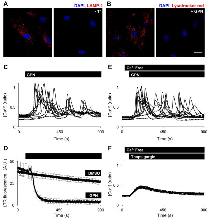
To probe the relationship between lysosomal and ER Ca2+ stores, we used primary cultured human fibroblasts. These cells are large (~120 μm) and flat thus facilitate imaging. As shown in Fig. 1A, immunocytochemical analysis using a primary antibody raised to LAMP-1, revealed well resolved structures that were distributed throughout the cell. A similar distribution was obtained when live cells were incubated with Lysotracker Red, a fluorescent weak base that accumulates in acidic organelles (Fig. 1B). Addition of the lysosomotropic agent GPN resulted in a prompt loss of Lysotracker Red fluorescence consistent with the labelling of lysosomes (Fig. 1B). To quantify lysosomal Ca2+ content, we monitored cytosolic Ca2+ concentration in response to acute addition of GPN using cells loaded with the Ca2+ sensitive indicator fura-2. Surprisingly, despite rapid permeabilisation of lysosomes, GPN evoked long-lasting Ca2+ signals that persisted up to 15 min (Fig. 1C). Parallel measurements of Lysotracker Red fluorescence confirmed that GPN abolished Lysotracker Red fluorescence within ~3 min of addition under these conditions (Fig. 1D). Removal of extracellular Ca2+ had little effect on Ca2+ signals evoked by GPN indicating that Ca2+ influx does not contribute significantly to GPN responses (Fig. 1E). For comparison, the effect of the SERCA pump inhibitor, thapsigargin, was used to estimate ER Ca2+ content (Fig. 1F). GPN thus evokes relatively large cytosolic Ca2+ signals that outlast lysosome integrity.
Fig. 1. Osmotic permeabilisation of lysosomes evokes complex Ca2+ signals.
(A) Confocal fluorescence image (red) of fibroblasts that were fixed and left unlabelled (right) or labelled (left) with a primary antibody raised to LAMP-1 and a far-red Alexa-Fluor-647-conjugated secondary antibody. (B) Confocal fluorescence images (red) of live fibroblasts that were labelled with Lysotracker Red before (left) or 216 seconds after (right) addition of 200 μM GPN. Nuclei were stained using DAPI (blue). Scale bar: 25 μm. (C,D) Single cell fluorescence responses recorded from cells loaded with either the Ca2+ indicator fura-2 (C) or Lysotracker Red (D) and stimulated with 200 μM GPN or the vehicle (DMSO). (E,F) Ca2+ signals in response to 200 μM GPN (E) or 1 μM thapsigargin (F) in the absence of extracellular Ca2+.
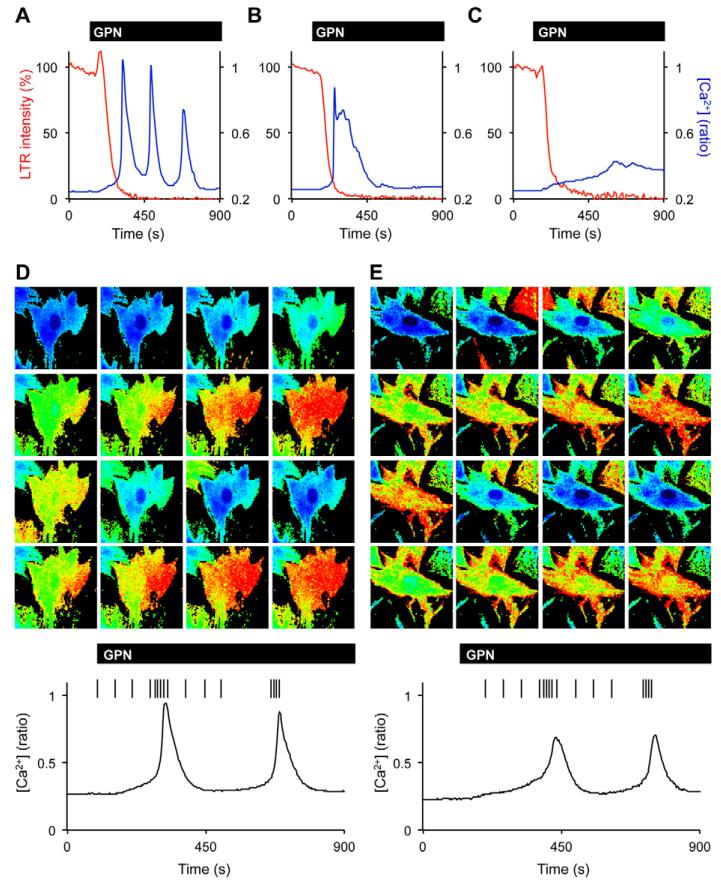
The kinetic mismatch between lysosome permeabilisation and cytosolic Ca2+ responses was further characterised by simultaneous measurements of fura-2 and Lysotracker Red fluorescence at the single cell level (Fig. 2A-C). In all 36 coloaded cells examined (n = 3), we noted a primary linear increase in Ca2+ upon GPN stimulation which correlated with the decrease in Lysotracker Red fluorescence. We term this increase in cytosolic Ca2+ concentration the ‘pacemaker’ response. This was followed by one of three secondary responses. In most cells, the rise in cytosolic Ca2+ accelerated, resulting in a prominent Ca2+ spike. These spikes either repeated resulting in a series of Ca2+ oscillations (Fig. 2A) or were abortive (Fig. 2B). In the remainder of cells, cytosolic Ca2+ levels continued to increase monotonically primarily peaking after complete loss of Lysotracker Red fluorescence (Fig. 2C). Thus, there was marked variation in the time to peak response upon GPN stimulation. Similar results were obtained in the absence of Lysotracker Red. From all 15 experiments analysed (with and without Lysotracker Red), the proportion of oscillatory (Fig. 2A) and abortive cells (Fig. 2B) were 48±5% and 29±5%, respectively (total number of cells = 177). At the spatial level, secondary responses to GPN were non-uniform. In cells displaying repeated Ca2+ spikes, the responses derived from a particular sub-cellular location and spread throughout the cell in wave-like manner. These responses either initiated at one-end of the cell (67±5%, n = 15; Fig. 2D) or in the cell periphery (33±5%, n = 15; Fig. 2E). In the latter case, the wave propagated in a centripetal fashion resembling the closing of an iris. In all 104 oscillatory cells examined, the waves repeatedly initiated from the same sub-cellular locale. Taken together, these data indicate that GPN-evoked Ca2+ signals are biphasic comprising an initial pacemaker response, which is probably lysosome dependent and a secondary response displaying spatial heterogeneity that appears lysosome independent.
Fig. 2. GPN-evoked Ca2+ signals are spatiotemporally complex.
(A-C) Simultaneous measurement of fura-2 (blue line) and Lysotracker Red (LTR; red line) fluorescence from individual fibroblasts stimulated with 200 μM GPN in the absence of extracellular Ca2+. Responses were grouped into three classes and representative examples of each are shown. (D,E) Pseudo-coloured images of GPN-evoked Ca2+ increases (top left to bottom right). Representative responses showing Ca2+ increases that propagated across the cell (D) or in a centripetal manner (E). The bottom traces are the temporal profiles from which spatial information was derived. Each vertical line (left to right) corresponds to an image above (top left to bottom right).
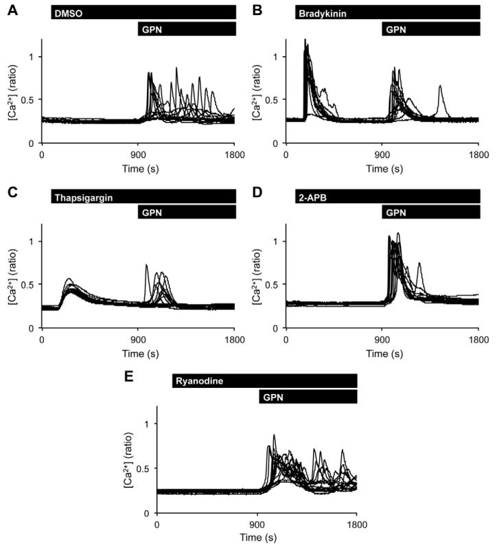
GPN-evoked Ca2+ responses are highly reminiscent of those evoked by cell-surface agonists. We therefore examined the effect of the physiological agonist bradykinin, which mobilises intracellular Ca2+ stores, on GPN responses. Bradykinin generated robust cytosolic Ca2+ signals in Ca2+-free media. Subsequent treatment with GPN induced largely monotonic responses (Fig. 3B). Thus, the proportion of cells displaying oscillations was 57±10% (n = 5, 74 cells) and 10±4% (n = 4, 60 cells) in the absence and presence of bradykinin, respectively (Fig. 3A,B). To probe the contribution of ER Ca2+ stores to GPN-evoked Ca2+ signals, cells were pre-treated with thapsigargin. As shown in Fig. 3C, GPN stimulation evoked only a transient response after thapsigargin treatment similar to bradykinin. Secondary responses were not observed in any of the 279 cells examined (n = 17). To define the role of inositol trisphosphate (InsP3) receptors, cells were pre-treated with the InsP3 receptor antagonist 2-aminoethoxydiphenyl borate (2-APB). 2-APB did not induce a Ca2+ signal (Fig. 3D). However, in its presence, the secondary responses to GPN were again largely eliminated (11±8% of oscillatory cells, n = 3, 45 cells). In contrast, GPN evoked typical complex Ca2+ signals upon blockade of ryanodine receptors by ryanodine pre-treatment (Fig. 3E; 63±13% oscillatory cells, n = 3, 45 cells). Prolonged GPN-evoked Ca2+ signals thus require InsP3 receptors likely on the ER but the possible contribution of Golgi-localised InsP3 receptors (Pizzo et al., 2011) cannot be excluded.
Fig. 3. Prolonged GPN-evoked Ca2+ signals derive from the ER.
Ca2+ signals evoked by GPN (200 μM) in the absence of extracellular Ca2+ following pre-treatment with vehicle (DMSO; A), 10 nM bradykinin (B), 1 μM thapsigargin (C), 100 μM 2-APB (D) or 100 μM ryanodine (E).
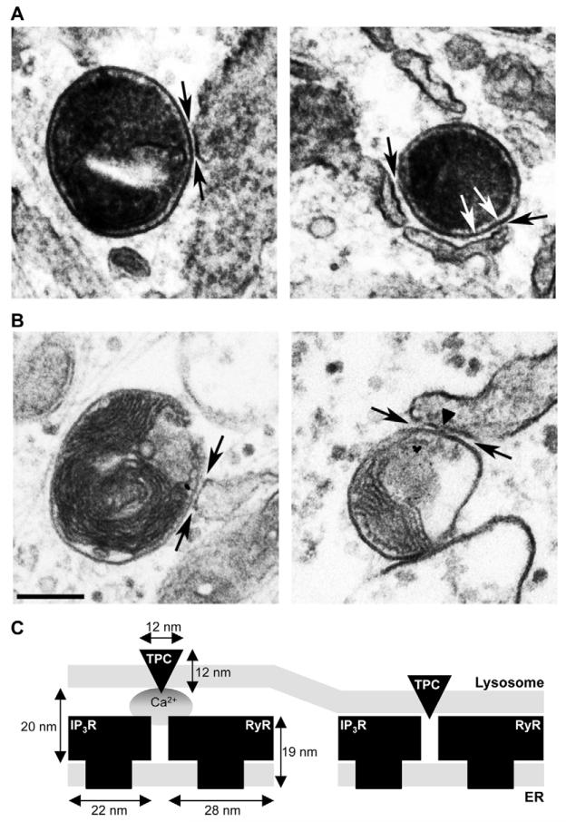
The ER is known to form several inter-organellar contact sites important for both Ca2+ and lipid dialogue (Toulmay and Prinz, 2011). But much less is known concerning contacts between the ER and acidic organelles. We and others recently described contact sites between endosomes/multi-vesicular bodies (MVBs) and the ER (Eden et al., 2010; Rocha et al., 2009). As shown in Fig. 4, membrane contact sites (regions of close apposition of ~20 nm) were clearly visible between lysosomes and the ER. Quantification in random sections revealed that 82±2% of 140 lysosomes analysed form contact sites with the ER (n = 3). This is probably an underestimate, as lysosomes have a diameter of 200–500 nm and thus will extend over a number of experimental sections (70 nm thick) where additional contacts in different planes may have been missed. That such a large proportion of lysosomes display contacts in random sections indicates extensive physical coupling between lysosomes and the ER. Thus, a considerable fraction of the perimeter membrane of each lysosome is coupled. Consistent with our previous observations on ER–MVB contact sites, regions where fibres appear to connect opposing ER and lysosome membranes (Fig. 4A, white arrows) were visible. The fibres resemble the tethers described within ER–mitochondria membrane contact sites (Csordás et al., 2006). Additionally, we identify areas where the apposing membranes appear to make direct contact with no discernible space between them (Fig. 4B, arrowhead).
Fig. 4. Lysosomes and ER form membrane contact sites.
(A,B) Electron micrographs of fibroblasts pulsed with horseradish peroxidase (A) or BSA-gold (B) to label lysosomes. Membrane contact sites between lysosomes and the ER are indicated by the black arrows. Fibres connecting opposing membranes (white arrows) and regions of very close membrane apposition (arrowhead) are marked. Scale bar: 200 nm. (C) Model showing juxtaposition of lysosomal TPCs (triangle) and ER inositol trisphosphate (IP3R)/ryanodine (RyR) receptors (square shapes) at lysosome-ER membrane contact sites. TPCs have been shown to couple to both InsP3 (Calcraft et al., 2009) and RyR (Brailoiu et al., 2009). Lysosomes and ER may communicate through Ca2+ released from TPCs (circle; left) or through more direct interaction (right) where apposing membranes are more closely positioned. Dimensions for the TPC were assumed to be similar to the skeletal muscle L-type Ca2+ channel to which TPCs have sequence and topological similarity (Serysheva et al., 2002). Dimensions for InsP3 and RyR were taken from Hamilton and Serysheva, and Ludtke et al. (Hamilton and Serysheva, 2009; Ludtke et al., 2011).
To conclude, we functionally dissect GPN-evoked Ca2+ signals into lysosomal and ER-derived components. Mobilisation of lysosomal Ca2+ is thus sufficient to evoke complex Ca2+ signals. These data provide direct support for the trigger hypothesis for NAADP action whereby physiological activation of endolysosomal Ca2+ channels is proposed to trigger secondary Ca2+ release via ER Ca2+ channels (Cancela et al., 1999). In our experiments, potential confounding effects of NAADP on ER channels are circumvented. We also demonstrate that the majority of lysosomes make contact with the ER via extensive membrane contact sites which likely represent an important site for the triggering of complex Ca2+ signals through juxtaposition of trigger and amplification Ca2+ channels (Fig. 4C). The tethered contacts we identify would facilitate CICR between TPCs and ER Ca2+ release channels (Fig. 4C, left). This is similar to the physical and functional arrangement of voltage-gated Ca2+ channels and ryanodine receptors at diadic junctions of cardiac muscle (Protasi, 2002). More direct contacts provide a basis for potential protein-protein interactions between Ca2+ release channels (Fig. 4C, right). This echoes the architecture of the excitation-contraction machinery in skeletal muscle (Protasi, 2002). Further ultra-structural and molecular definition of these novel membrane contact sites is warranted.
Materials and Methods
Cell culture
Human skin fibroblasts established from healthy females were maintained in DMEM supplemented with 10% v/v fetal bovine serum, 100 units/ml penicillin and 100 μg/ml streptomycin (all from Invitrogen) at 37 °C in a humidified atmosphere with 5% CO2. Cells were plated on glass coverslips before experimentation.
Immunocytochemistry
Fibroblasts were fixed for 10 min with 4% w/v paraformaldehyde, washed three times in phosphate-buffered saline (PBS) and then permeabilised for 5 min with 40 μM β-escin. Cells were washed again (three times in PBS), and blocked for 1 h with PBS supplemented with 1% w/v BSA and 10% v/v fetal bovine serum. Fibroblasts were sequentially incubated for 1 h at 37 °C with a primary anti-LAMP-1 (lysosomal-associated membrane protein 1) mouse antibody (1:100 dilution; H4A3 clone from Developmental Hybridoma Bank) and a secondary mouse antibody conjugated to Alexa Fluor 647 (1:100 dilution; Invitrogen) in blocking solution. Nuclei were labelled for 5 min with 1 μg/ml DAPI (4′, 6-diamidino-2-phenylindole). Cells were washed three times in PBS containing 0.1% v/v Tween in between incubations and mounted onto microscope slides with DABCO (1,4-diazabicyclo[2,2,2]octane).
Live-cell imaging
Experiments were performed in HEPES-buffered saline (HBS) comprising (in mM) 1.25 KH2PO4, 2 CaCl2, 2 MgSO4, 3 KCl, 156 NaCl, 10 glucose and 10 HEPES (pH 7.4; all from Sigma). For measurement of cytosolic Ca2+ concentration, fibroblasts were incubated with Fura-2AM (2.5 μM) and 0.005% v/v pluronic acid (from Invitrogen) for 1 h. For measurement of lysosome distribution/integrity, fibroblasts were incubated with 100 nM Lysotracker Red (Invitrogen) for 30 min. Cells were stimulated with 200 μM GPN (Santa Cruz Biotech), 10 nM bradykinin (Sigma), 1 μM thapsigargin, 100 μM 2-APB (2-aminoethoxydiphenyl borate) and 100 μM ryanodine (all sourced from Merck). Where indicated, Ca2+ in HBS was replaced with 1 mM EGTA (Sigma).
Epifluorescence microscopy
Epifluorescence images were captured every 3–8 s with a cooled coupled device camera (TILL Photonics) attached to an Olympus IX71 inverted fluorescence microscope fitted with a 20× objective, and a monochromator light source. Fura-2 and Lysotracker Red were excited at 340/380 nm and 560 nm and emitted fluorescence was captured using a 440 nm long pass filter and 590 nm filter, respectively.
Confocal microscopy
Confocal images were captured using an LSM510 confocal scanner (Zeiss) attached to a Zeiss Axiovert 200 M inverted microscope fitted with a 63× Plan Apochromat water-immersion objective. DAPI, Lysotracker Red and Alexa Fluor 647 were excited at 364 nm, 560 nm and 633 nm and emitted fluorescence was captured using long pass 385 nm, 560–615 nm band pass or 655–719 nm band pass filters, respectively.
Electron microscopy
BSA-gold was prepared by coupling 5 nm colloidal gold (BBI) to BSA as described previously (Slot and Geuze, 1985). Fibroblasts cultured on thermanox coverslips (Agar Scientific) were pulsed for 15 min with either BSA-gold or horseradish peroxidase (Sigma) and chased for 3 h in complete medium. This procedure labels exclusively lysosomes, as shown by their degradative capacity and LAMP-1 content (Futter et al., 1996). Cells were fixed and processed for electron microscopy essentially as described (Tomas et al., 2004) except that uranyl acetate (1%, 40 min incubation) was used in place of tannic acid, and samples were embedded in TAAB 812 hard formulation resin. Samples were viewed on a JEOL 1010 TEM and images acquired by a Gatan Orius SC100B charge-coupled device camera. Contact sites between lysosome and ER membranes were defined as regions of close apposition (~20 nm).
Acknowledgements
We thank Tim Levine and Chi Li for useful discussions.
Funding: This was supported by the Biotechnology and Biological Sciences Research Council [grant number BB/K000942/1 to S.P.], Parkinson’s UK [grant number K-1107 to S.P. and A.H.S.] and an IMPACT studentship from University College London [to B.S.K.].
References
- Berridge MJ. The endoplasmic reticulum: a multifunctional signaling organelle. Cell Calcium. 2002;32:235–249. doi: 10.1016/s0143416002001823. [DOI] [PubMed] [Google Scholar]
- Berridge MJ, Lipp P, Bootman MD. The versatility and universality of calcium signalling. Nat. Rev. Mol. Cell Biol. 2000;1:11–21. doi: 10.1038/35036035. [DOI] [PubMed] [Google Scholar]
- Brailoiu E, Churamani D, Pandey V, Brailoiu GC, Tuluc F, Patel S, Dun NJ. Messenger-specific role for nicotinic acid adenine dinucleotide phosphate in neuronal differentiation. J. Biol. Chem. 2006;281:15923–15928. doi: 10.1074/jbc.M602249200. [DOI] [PubMed] [Google Scholar]
- Brailoiu E, Churamani D, Cai X, Schrlau MG, Brailoiu GC, Gao X, Hooper R, Boulware MJ, Dun NJ, Marchant JS, et al. Essential requirement for two-pore channel 1 in NAADP-mediated calcium signaling. J. Cell Biol. 2009;186:201–209. doi: 10.1083/jcb.200904073. [DOI] [PMC free article] [PubMed] [Google Scholar]
- Brailoiu E, Rahman T, Churamani D, Prole DL, Brailoiu GC, Hooper R, Taylor CW, Patel S. An NAADP-gated two-pore channel targeted to the plasma membrane uncouples triggering from amplifying Ca2+ signals. J. Biol. Chem. 2010;285:38511–38516. doi: 10.1074/jbc.M110.162073. [DOI] [PMC free article] [PubMed] [Google Scholar]
- Calcraft PJ, Ruas M, Pan Z, Cheng X, Arredouani A, Hao X, Tang J, Rietdorf K, Teboul L, Chuang KT, et al. NAADP mobilizes calcium from acidic organelles through two-pore channels. Nature. 2009;459:596–600. doi: 10.1038/nature08030. [DOI] [PMC free article] [PubMed] [Google Scholar]
- Cancela JM, Churchill GC, Galione A. Coordination of agonist-induced Ca2+-signalling patterns by NAADP in pancreatic acinar cells. Nature. 1999;398:74–76. doi: 10.1038/18032. [DOI] [PubMed] [Google Scholar]
- Churchill GC, Galione A. NAADP induces Ca2+ oscillations via a two-pool mechanism by priming IP3- and cADPr-sensitive Ca2+ stores. EMBO J. 2001;20:2666–2671. doi: 10.1093/emboj/20.11.2666. [DOI] [PMC free article] [PubMed] [Google Scholar]
- Churchill GC, Okada Y, Thomas JM, Genazzani AA, Patel S, Galione A. NAADP mobilizes Ca2+ from reserve granules, lysosome-related organelles, in sea urchin eggs. Cell. 2002;111:703–708. doi: 10.1016/s0092-8674(02)01082-6. [DOI] [PubMed] [Google Scholar]
- Coen K, Flannagan RS, Baron S, Carraro-Lacroix LR, Wang D, Vermeire W, Michiels C, Munck S, Baert V, Sugita S, et al. Lysosomal calcium homeostasis defects, not proton pump defects, cause endo-lysosomal dysfunction in PSEN-deficient cells. J. Cell Biol. 2012;198:23–35. doi: 10.1083/jcb.201201076. [DOI] [PMC free article] [PubMed] [Google Scholar]
- Csordás G, Renken C, Várnai P, Walter L, Weaver D, Buttle KF, Balla T, Mannella CA, Hajnóczky G. Structural and functional features and significance of the physical linkage between ER and mitochondria. J. Cell Biol. 2006;174:915–921. doi: 10.1083/jcb.200604016. [DOI] [PMC free article] [PubMed] [Google Scholar]
- Dammermann W, Zhang B, Nebel M, Cordiglieri C, Odoardi F, Kirchberger T, Kawakami N, Dowden J, Schmid F, Dornmair K, et al. NAADP-mediated Ca2+ signaling via type 1 ryanodine receptor in T cells revealed by a synthetic NAADP antagonist. Proc. Natl. Acad. Sci. USA. 2009;106:10678–10683. doi: 10.1073/pnas.0809997106. [DOI] [PMC free article] [PubMed] [Google Scholar]
- Eden ER, White IJ, Tsapara A, Futter CE. Membrane contacts between endosomes and ER provide sites for PTP1B-epidermal growth factor receptor interaction. Nat. Cell Biol. 2010;12:267–272. doi: 10.1038/ncb2026. [DOI] [PubMed] [Google Scholar]
- Futter CE, Pearse A, Hewlett LJ, Hopkins CR. Multivesicular endosomes containing internalized EGF-EGF receptor complexes mature and then fuse directly with lysosomes. J. Cell Biol. 1996;132:1011–1023. doi: 10.1083/jcb.132.6.1011. [DOI] [PMC free article] [PubMed] [Google Scholar]
- Galione A, Morgan AJ, Arredouani A, Davis LC, Rietdorf K, Ruas M, Parrington J. NAADP as an intracellular messenger regulating lysosomal calcium-release channels. Biochem. Soc. Trans. 2010;38:1424–1431. doi: 10.1042/BST0381424. [DOI] [PubMed] [Google Scholar]
- Gerasimenko JV, Maruyama Y, Yano K, Dolman NJ, Tepikin AV, Petersen OH, Gerasimenko OV. NAADP mobilizes Ca2+ from a thapsigargin-sensitive store in the nuclear envelope by activating ryanodine receptors. J. Cell Biol. 2003;163:271–282. doi: 10.1083/jcb.200306134. [DOI] [PMC free article] [PubMed] [Google Scholar]
- Gerasimenko JV, Lur G, Sherwood MW, Ebisui E, Tepikin AV, Mikoshiba K, Gerasimenko OV, Petersen OH. Pancreatic protease activation by alcohol metabolite depends on Ca2+ release via acid store IP3 receptors. Proc. Natl. Acad. Sci. USA. 2009;106:10758–10763. doi: 10.1073/pnas.0904818106. [DOI] [PMC free article] [PubMed] [Google Scholar]
- Guse AH. Linking NAADP to ion channel activity: a unifying hypothesis. Sci. Signal. 2012;5:pe18. doi: 10.1126/scisignal.2002890. [DOI] [PubMed] [Google Scholar]
- Guse AH, Lee HC. NAADP: a universal Ca2+ trigger. Sci. Signal. 2008;1:re10. doi: 10.1126/scisignal.144re10. [DOI] [PubMed] [Google Scholar]
- Hamilton SL, Serysheva II. Ryanodine receptor structure: progress and challenges. J. Biol. Chem. 2009;284:4047–4051. doi: 10.1074/jbc.R800054200. [DOI] [PMC free article] [PubMed] [Google Scholar]
- Hohenegger M, Suko J, Gscheidlinger R, Drobny H, Zidar A. Nicotinic acid-adenine dinucleotide phosphate activates the skeletal muscle ryanodine receptor. Biochem. J. 2002;367:423–431. doi: 10.1042/BJ20020584. [DOI] [PMC free article] [PubMed] [Google Scholar]
- Jadot M, Colmant C, Wattiaux-De Coninck S, Wattiaux R. Intralysosomal hydrolysis of glycyl-L-phenylalanine 2-naphthylamide. Biochem. J. 1984;219:965–970. doi: 10.1042/bj2190965. [DOI] [PMC free article] [PubMed] [Google Scholar]
- Kinnear NP, Boittin FX, Thomas JM, Galione A, Evans AM. Lysosome-sarcoplasmic reticulum junctions. A trigger zone for calcium signaling by nicotinic acid adenine dinucleotide phosphate and endothelin-1. J. Biol. Chem. 2004;279:54319–54326. doi: 10.1074/jbc.M406132200. [DOI] [PubMed] [Google Scholar]
- Lin-Moshier Y, Walseth TF, Churamani D, Davidson SM, Slama JT, Hooper R, Brailoiu E, Patel S, Marchant JS. Photoaffinity labeling of nicotinic acid adenine dinucleotide phosphate (NAADP) targets in mammalian cells. J. Biol. Chem. 2012;287:2296–2307. doi: 10.1074/jbc.M111.305813. [DOI] [PMC free article] [PubMed] [Google Scholar]
- Lloyd-Evans E, Morgan AJ, He X, Smith DA, Elliot-Smith E, Sillence DJ, Churchill GC, Schuchman EH, Galione A, Platt FM. Niemann-Pick disease type C1 is a sphingosine storage disease that causes deregulation of lysosomal calcium. Nat. Med. 2008;14:1247–1255. doi: 10.1038/nm.1876. [DOI] [PubMed] [Google Scholar]
- Ludtke SJ, Tran TP, Ngo QT, Moiseenkova-Bell VY, Chiu W, Serysheva II. Flexible architecture of IP3R1 by Cryo-EM. Structure. 2011;19:1192–1199. doi: 10.1016/j.str.2011.05.003. [DOI] [PMC free article] [PubMed] [Google Scholar]
- Luzio JP, Bright NA, Pryor PR. The role of calcium and other ions in sorting and delivery in the late endocytic pathway. Biochem. Soc. Trans. 2007;35:1088–1091. doi: 10.1042/BST0351088. [DOI] [PubMed] [Google Scholar]
- Marchant JS, Lin-Moshier Y, Walseth TF, Patel S. The Molecular Basis for Ca2+ Signalling by NAADP: Two-Pore Channels in a Complex? Messenger. 2012;1:63–76. doi: 10.1166/msr.2012.1003. [DOI] [PMC free article] [PubMed] [Google Scholar]
- Patel S, Brailoiu E. Triggering of Ca2+ signals by NAADP-gated two-pore channels: a role for membrane contact sites? Biochem. Soc. Trans. 2012;40:153–157. doi: 10.1042/BST20110693. [DOI] [PubMed] [Google Scholar]
- Patel S, Docampo R. Acidic calcium stores open for business: expanding the potential for intracellular Ca2+ signaling. Trends Cell Biol. 2010;20:277–286. doi: 10.1016/j.tcb.2010.02.003. [DOI] [PMC free article] [PubMed] [Google Scholar]
- Patel S, Churchill GC, Galione A. Coordination of Ca2+ signalling by NAADP. Trends Biochem. Sci. 2001;26:482–489. doi: 10.1016/s0968-0004(01)01896-5. [DOI] [PubMed] [Google Scholar]
- Patel S, Marchant JS, Brailoiu E. Two-pore channels: Regulation by NAADP and customized roles in triggering calcium signals. Cell Calcium. 2010;47:480–490. doi: 10.1016/j.ceca.2010.05.001. [DOI] [PMC free article] [PubMed] [Google Scholar]
- Pizzo P, Lissandron V, Capitanio P, Pozzan T. Ca2+ signalling in the Golgi apparatus. Cell Calcium. 2011;50:184–192. doi: 10.1016/j.ceca.2011.01.006. [DOI] [PubMed] [Google Scholar]
- Protasi F. Structural interaction between RYRs and DHPRs in calcium release units of cardiac and skeletal muscle cells. Front. Biosci. 2002;7:d650–d658. doi: 10.2741/A801. [DOI] [PubMed] [Google Scholar]
- Pryor PR, Mullock BM, Bright NA, Gray SR, Luzio JP. The role of intraorganellar Ca(2+) in late endosome-lysosome heterotypic fusion and in the reformation of lysosomes from hybrid organelles. J. Cell Biol. 2000;149:1053–1062. doi: 10.1083/jcb.149.5.1053. [DOI] [PMC free article] [PubMed] [Google Scholar]
- Rizzuto R, Duchen MR, Pozzan T. Flirting in little space: the ER/mitochondria Ca2+ liaison. Sci. STKE. 2004;2004:re1. doi: 10.1126/stke.2152004re1. [DOI] [PubMed] [Google Scholar]
- Rocha N, Kuijl C, van der Kant R, Janssen L, Houben D, Janssen H, Zwart W, Neefjes J. Cholesterol sensor ORP1L contacts the ER protein VAP to control Rab7-RILP-p150 Glued and late endosome positioning. J. Cell Biol. 2009;185:1209–1225. doi: 10.1083/jcb.200811005. [DOI] [PMC free article] [PubMed] [Google Scholar]
- Serysheva II, Ludtke SJ, Baker MR, Chiu W, Hamilton SL. Structure of the voltage-gated L-type Ca2+ channel by electron cryomicroscopy. Proc. Natl. Acad. Sci. USA. 2002;99:10370–10375. doi: 10.1073/pnas.162363499. [DOI] [PMC free article] [PubMed] [Google Scholar]
- Shen D, Wang X, Li X, Zhang X, Yao Z, Dibble S, Dong XP, Yu T, Lieberman AP, Showalter HD, et al. Lipid storage disorders block lysosomal trafficking by inhibiting a TRP channel and lysosomal calcium release. Nat. Commun. 2012;3:731. doi: 10.1038/ncomms1735. [DOI] [PMC free article] [PubMed] [Google Scholar]
- Slot JW, Geuze HJ. A new method of preparing gold probes for multiple-labeling cytochemistry. Eur. J. Cell Biol. 1985;38:87–93. [PubMed] [Google Scholar]
- Tomas A, Futter C, Moss SE. Annexin 11 is required for midbody formation and completion of the terminal phase of cytokinesis. J. Cell Biol. 2004;165:813–822. doi: 10.1083/jcb.200311054. [DOI] [PMC free article] [PubMed] [Google Scholar]
- Toulmay A, Prinz WA. Lipid transfer and signaling at organelle contact sites: the tip of the iceberg. Curr. Opin. Cell Biol. 2011;23:458–463. doi: 10.1016/j.ceb.2011.04.006. [DOI] [PMC free article] [PubMed] [Google Scholar]
- Yamaguchi S, Jha A, Li Q, Soyombo AA, Dickinson GD, Churamani D, Brailoiu E, Patel S, Muallem S. Transient receptor potential mucolipin 1 (TRPML1) and two-pore channels are functionally independent organellar ion channels. J. Biol. Chem. 2011;286:22934–22942. doi: 10.1074/jbc.M110.210930. [DOI] [PMC free article] [PubMed] [Google Scholar]
- Yamasaki M, Masgrau R, Morgan AJ, Churchill GC, Patel S, Ashcroft SJH, Galione A. Organelle selection determines agonist-specific Ca2+ signals in pancreatic acinar and beta cells. J. Biol. Chem. 2004;279:7234–7240. doi: 10.1074/jbc.M311088200. [DOI] [PubMed] [Google Scholar]
- Zong X, Schieder M, Cuny H, Fenske S, Gruner C, Rötzer K, Griesbeck O, Harz H, Biel M, Wahl-Schott C. The two-pore channel TPCN2 mediates NAADP-dependent Ca(2+)-release from lysosomal stores. Pflugers Arch. 2009;458:891–899. doi: 10.1007/s00424-009-0690-y. [DOI] [PMC free article] [PubMed] [Google Scholar]


