Figure 5.

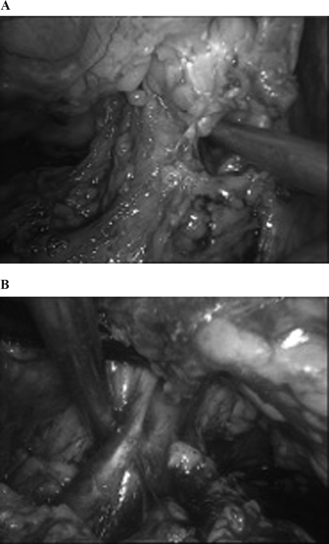
A, Right side; B, left side. A window is created with the suction cannula cranial to the renal vein to allow one of the jaws of the staple. Notice that the wide dissection allows a clear window posterolateral to the renal hilum.

A, Right side; B, left side. A window is created with the suction cannula cranial to the renal vein to allow one of the jaws of the staple. Notice that the wide dissection allows a clear window posterolateral to the renal hilum.