

Abstract
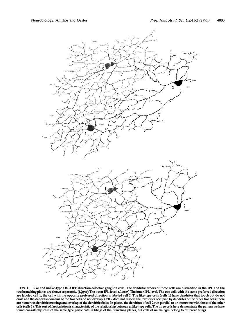
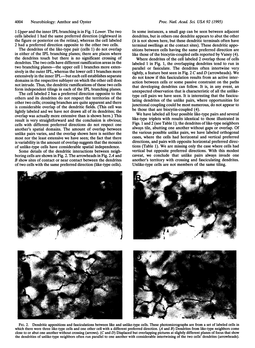
The visual stimuli that elicit neural activity differ for different retinal ganglion cells and these cells have been categorized by the visual information that they transmit. If specific visual information is conveyed exclusively or primarily by a particular set of ganglion cells, one might expect the cells to be organized spatially so that their sampling of information from the visual field is complete but not redundant. In other words, the laterally spreading dendrites of the ganglion cells should completely cover the retinal plane without gaps or significant overlap. The first evidence for this sort of arrangement, which has been called a tiling or tessellation, was for the two types of "alpha" ganglion cells in cat retina. Other reports of tiling by ganglion cells have been made subsequently. We have found evidence of a particularly rigorous tiling for the four types of ganglion cells in rabbit retina that convey information about the direction of retinal image motion (the ON-OFF direction-selective cells). Although individual cells in the four groups are morphologically indistinguishable, they are organized as four overlaid tilings, each tiling consisting of like-type cells that respond preferentially to a particular direction of retinal image motion. These observations lend support to the hypothesis that tiling is a general feature of the organization of information outflow from the retina and clearly implicate mechanisms for recognition of like-type cells and establishment of mutually acceptable territories during retinal development.
Full text
PDF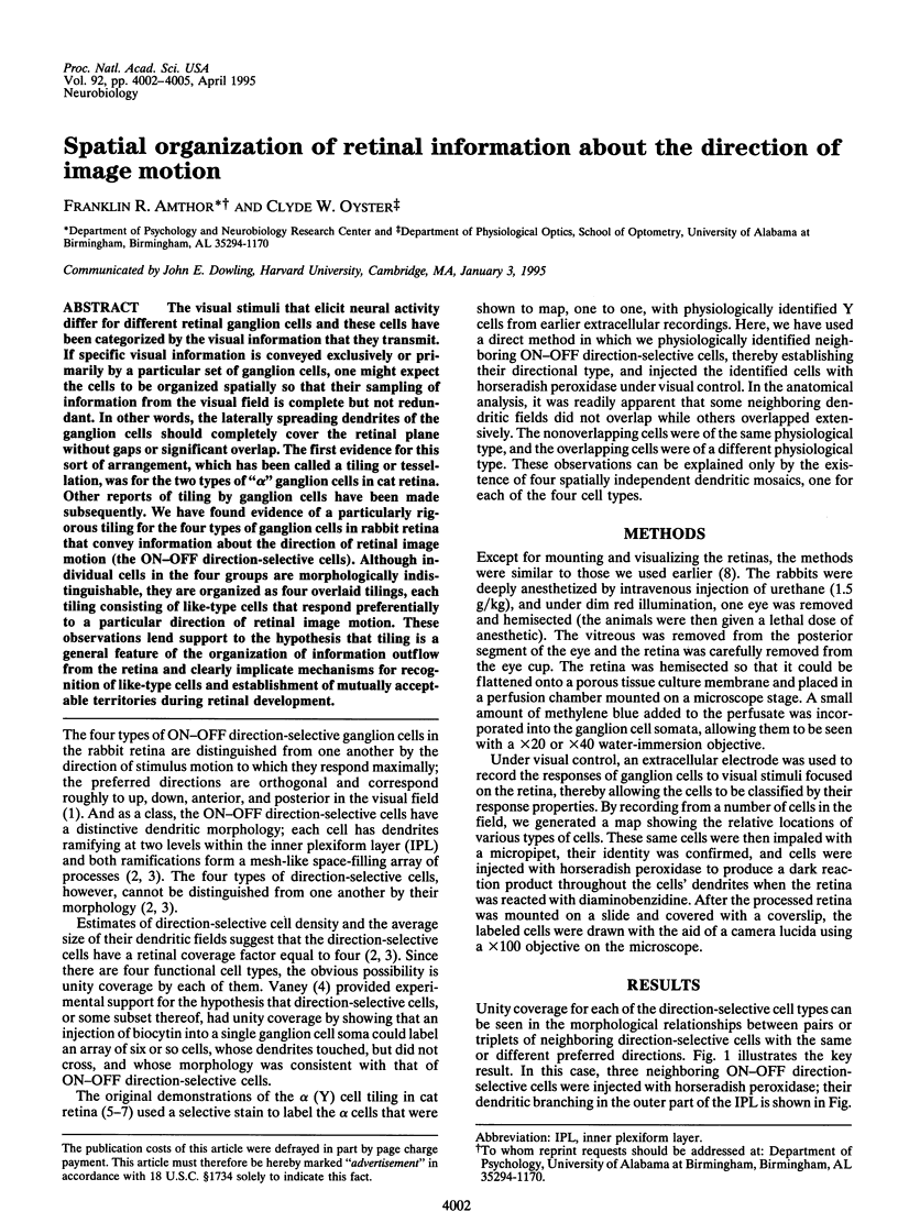
Images in this article
Selected References
These references are in PubMed. This may not be the complete list of references from this article.
- Amthor F. R., Takahashi E. S., Oyster C. W. Morphologies of rabbit retinal ganglion cells with complex receptive fields. J Comp Neurol. 1989 Feb 1;280(1):97–121. doi: 10.1002/cne.902800108. [DOI] [PubMed] [Google Scholar]
- Amthor F. R., Takahashi E. S., Oyster C. W. Morphologies of rabbit retinal ganglion cells with complex receptive fields. J Comp Neurol. 1989 Feb 1;280(1):97–121. doi: 10.1002/cne.902800108. [DOI] [PubMed] [Google Scholar]
- Amthor F. R., Takahashi E. S., Oyster C. W. Morphologies of rabbit retinal ganglion cells with concentric receptive fields. J Comp Neurol. 1989 Feb 1;280(1):72–96. doi: 10.1002/cne.902800107. [DOI] [PubMed] [Google Scholar]
- Amthor F. R., Takahashi E. S., Oyster C. W. Morphologies of rabbit retinal ganglion cells with concentric receptive fields. J Comp Neurol. 1989 Feb 1;280(1):72–96. doi: 10.1002/cne.902800107. [DOI] [PubMed] [Google Scholar]
- Dacey D. M. The mosaic of midget ganglion cells in the human retina. J Neurosci. 1993 Dec;13(12):5334–5355. doi: 10.1523/JNEUROSCI.13-12-05334.1993. [DOI] [PMC free article] [PubMed] [Google Scholar]
- Levick W. R. Receptive fields and trigger features of ganglion cells in the visual streak of the rabbits retina. J Physiol. 1967 Feb;188(3):285–307. doi: 10.1113/jphysiol.1967.sp008140. [DOI] [PMC free article] [PubMed] [Google Scholar]
- Oyster C. W., Amthor F. R., Takahashi E. S. Dendritic architecture of ON-OFF direction-selective ganglion cells in the rabbit retina. Vision Res. 1993 Mar-Apr;33(5-6):579–608. doi: 10.1016/0042-6989(93)90181-u. [DOI] [PubMed] [Google Scholar]
- Oyster C. W., Amthor F. R., Takahashi E. S. Dendritic architecture of ON-OFF direction-selective ganglion cells in the rabbit retina. Vision Res. 1993 Mar-Apr;33(5-6):579–608. doi: 10.1016/0042-6989(93)90181-u. [DOI] [PubMed] [Google Scholar]
- Oyster C. W., Barlow H. B. Direction-selective units in rabbit retina: distribution of preferred directions. Science. 1967 Feb 17;155(3764):841–842. doi: 10.1126/science.155.3764.841. [DOI] [PubMed] [Google Scholar]
- Oyster C. W., Barlow H. B. Direction-selective units in rabbit retina: distribution of preferred directions. Science. 1967 Feb 17;155(3764):841–842. doi: 10.1126/science.155.3764.841. [DOI] [PubMed] [Google Scholar]
- Oyster C. W. The analysis of image motion by the rabbit retina. J Physiol. 1968 Dec;199(3):613–635. doi: 10.1113/jphysiol.1968.sp008671. [DOI] [PMC free article] [PubMed] [Google Scholar]
- Peichl L., Buhl E. H., Boycott B. B. Alpha ganglion cells in the rabbit retina. J Comp Neurol. 1987 Sep 1;263(1):25–41. doi: 10.1002/cne.902630103. [DOI] [PubMed] [Google Scholar]
- Peichl L., Ott H., Boycott B. B. Alpha ganglion cells in mammalian retinae. Proc R Soc Lond B Biol Sci. 1987 Jul 22;231(1263):169–197. doi: 10.1098/rspb.1987.0040. [DOI] [PubMed] [Google Scholar]
- Vaney D. I. Many diverse types of retinal neurons show tracer coupling when injected with biocytin or Neurobiotin. Neurosci Lett. 1991 Apr 29;125(2):187–190. doi: 10.1016/0304-3940(91)90024-n. [DOI] [PubMed] [Google Scholar]
- Wässle H., Boycott B. B., Illing R. B. Morphology and mosaic of on- and off-beta cells in the cat retina and some functional considerations. Proc R Soc Lond B Biol Sci. 1981 May 22;212(1187):177–195. doi: 10.1098/rspb.1981.0033. [DOI] [PubMed] [Google Scholar]
- Wässle H., Peichl L., Boycott B. B. Dendritic territories of cat retinal ganglion cells. Nature. 1981 Jul 23;292(5821):344–345. doi: 10.1038/292344a0. [DOI] [PubMed] [Google Scholar]
- Wässle H., Peichl L., Boycott B. B. Morphology and topography of on- and off-alpha cells in the cat retina. Proc R Soc Lond B Biol Sci. 1981 May 22;212(1187):157–175. doi: 10.1098/rspb.1981.0032. [DOI] [PubMed] [Google Scholar]
- Yang G., Masland R. H. Direct visualization of the dendritic and receptive fields of directionally selective retinal ganglion cells. Science. 1992 Dec 18;258(5090):1949–1952. doi: 10.1126/science.1470920. [DOI] [PubMed] [Google Scholar]