Abstract
Merkel cell carcinoma is recognized by its morphologic features as well as by its classic immunophenotypic properties. Although Merkel cell carcinomas demonstrating non-classic immunoreactivities have been described, a case documenting a change in immunophenotype during the course of disease progression has not been previously reported. We report a case of Merkel cell carcinoma which initially demonstrated cytokeratin 20 positivity but lost expression in subsequent metastases. Likewise, thyroid transcription factor-1 was initially negative in the tumor but expression was present in metastatic lesions.
INTRODUCTION
Merkel cell carcinoma (MCC) is a rare neuroendocrine tumor of the skin with aggressive behavior and a rising incidence. Characteristically, MCC is positive for cytokeratin 20 (CK20) and negative for thyroid transcription factor -1 (TTF-1). Although CK20 negative primary MCCs or TTF-1 positive MCCs have been described, loss of CK20 and simultaneous acquisition of TTF-1 expression in metastatic MCC has not been reported.1,2
CASE REPORT
A 68-year-old Caucasian immunocompetent male with a history of squamous cell carcinoma of the tongue in remission presented with a primary MCC on the left frontal scalp in 2006 which was treated with surgery and radiation therapy at that time. He then developed recurrent MCC on the forehead and scalp in 2007, and subsequent metastasis to a neck lymph node in 2008. Patient underwent multiple surgeries and radiation for these recurrent tumors. In 2012 he was found to have a solitary left frontal brain metastasis and was treated with a gross total surgical resection followed by intensity modulated radiation therapy (IMRT) with 15 Gy delivered to the resection cavity and margin in a single fraction. However, biopsy-proven tumor recurred in the same region of the brain early in 2013, along with an additional new metastatic left frontal dural-based lesion not in continuity with the previous seen lesion or craniotomy site. He was no longer a surgical candidate and elected to be in hospice. Multiple imaging studies failed to demonstrate other primary lesions.
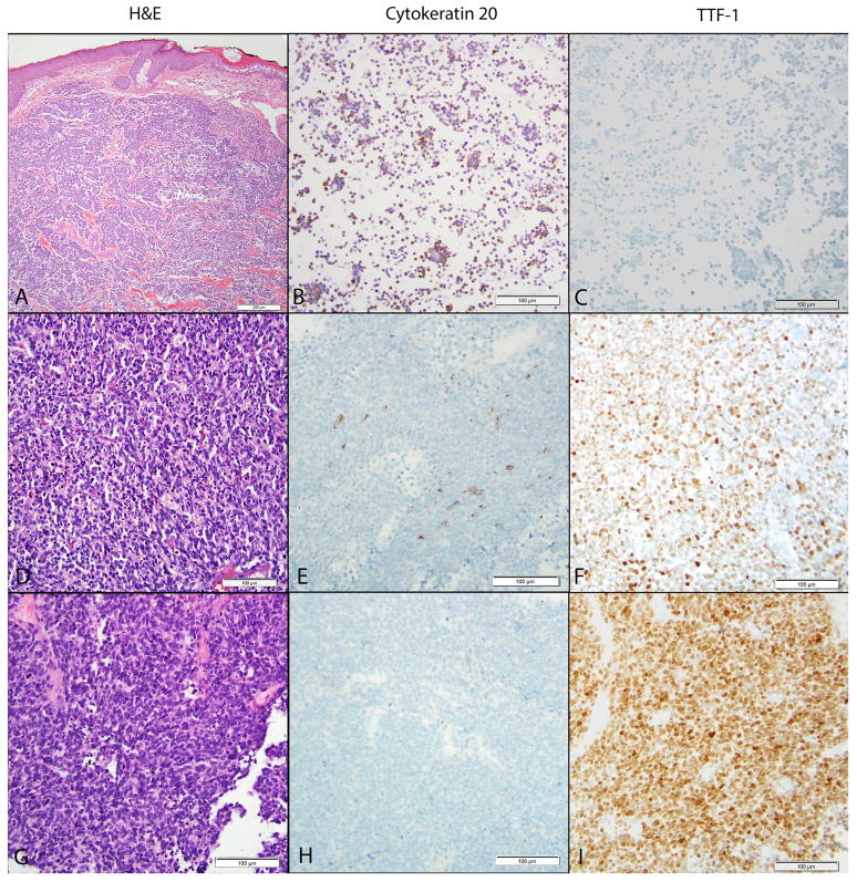
Histologic examination of the primary tumor demonstrated neuroendocrine morphology, as well as characteristic paranuclear accentuation of CK20 immunoreactivity. The tumor cells were negative for TTF-1 (Figure, top panel). Interestingly, in the 2012 brain metastasis, only a few CK20 positive tumor cells were identified while CK20 expression was completely lost in the brain tumor recurrent in 2013 (Figure mid panel). Conversely, TTF-1 positivity was detected in the patient’s 2012 brain metastasis and was diffusely positive in brain tumor biopsied in 2013 (Figure, right panel). CK7 was negative in both the primary and metastatic brain tumors (data not shown). Based on the known history of MCC and the absence of other malignancy, the findings were felt to be compatible with metastatic MCC with an unusual immunohistochemical pattern.
Figure 1.
Evolution of the patient’s tumor immunoreactivities. The patient’s primary tumor (A–C), 2012 brain metastasis (D–F), and 2013 brain metastasis (G–I). The patient’s primary tumor showed cords and nests of tumor cells with a high nuclear:cytoplasm ratio infiltrating through dermal collagen (A). The tumor cells showed CK20 positivity, demonstrating areas with paranuclear accentuation (B). The tumor cells were negative for TTF-1 (C). Examination of the brain metastases in 2012 showed a cellular tumor with neuroendocrine features (D). Focal CK20 expression was detected (E) with patchy areas of strong nuclear TTF-1 expression (F). Recurrence of brain metastases in 2013 showed a cellular tumor (G) with an absence of CK20 expression (H) and diffuse nuclear TTF-1 expression (I).
DISCUSSION
CK20 is an intermediate filament and is expressed in the gastrointestinal epithelial cells and in epidermal Merkel cells. TTF-1 is a nuclear transcriptional factor expressed in epithelial cells of the thyroid and lung. Antibodies to CK20 and TTF-1 are frequently employed in the work-up of cutaneous neuroendocrine carcinomas to help differentiate MCC (CK20+, TTF-1 negative) from metastatic small cell lung carcinoma (CK20 negative, TTF-1 +). CK20 negative primary MCCs have been described, in which case the diagnosis rests on an expanded battery of immunohistochemical markers coupled with clinical and radiographic exclusion of a neuroendocrine carcinoma arising from another organ.3 However, expression patterns of CK20 in metastatic MCCs have not been evaluated. Likewise, negative TTF-1 had been considered to have a high specificity in differentiating MCC from small cell carcinomas of lung origin. However, two recent studies have demonstrated TTF-1 positivity in 4/65 cases of MCCs.4 Similarly, Sierakowski et al describes a case with TTF-1 positivity in both primary and lymph node metastatic tumors.1 Moreover, a CK20 negative primary MCC with TTF-1 positivity is reported.2 Nevertheless, this altered expression pattern of CK20 and TTF-1 in the brain metastases of MCC as seen in our patient has never been reported. The etiology and clinical significance remains to be elucidated. Whether it coincides with epithelial mesenchymal transition or as a result of radiation therapy needs further investigation.
Acknowledgments
Finding sources: The project was supported by the Translational Research Institute (TRI), grants UL1TR000039 and KL2TR000063 through the NIH National Center for Research Resources and the National Center for Advancing Translational Sciences. The content is solely the responsibility of the authors and does not necessarily represent the official views of the NIH. This study was also supported by funds from the Department of Dermatology and the Winthrop P. Rockefeller Cancer Institute, University of Arkansas for Medical Sciences.
Footnotes
The authors state that there are no conflicts of interest.
References
- 1.Sierakowski A, Al-Janabi K, Dam H, et al. Metastatic Merkel cell carcinoma with positive expression of thyroid transcription factor-1--a case report. Am J Dermatopathol. 2009;31:384–6. doi: 10.1097/DAD.0b013e31819821e8. [DOI] [PubMed] [Google Scholar]
- 2.Koba S, Inoue T, Okawa T, et al. Merkel cell carcinoma with cytokeratin 20-negative and thyroid transcription factor-1-positive immunostaining admixed with squamous cell carcinoma. J Dermatol Sci. 2011;64:77–9. doi: 10.1016/j.jdermsci.2011.06.011. [DOI] [PubMed] [Google Scholar]
- 3.Calder KB, Coplowitz S, Schlauder S, et al. A case series and immunophenotypic analysis of CK20−/CK7+ primary neuroendocrine carcinoma of the skin. J Cutan Pathol. 2007;34:918–23. doi: 10.1111/j.1600-0560.2007.00759.x. [DOI] [PubMed] [Google Scholar]
- 4.Ordonez NG. Value of thyroid transcription factor-1 immunostaining in tumor diagnosis: a review and update. Appl Immunohistochem Mol Morphol. 2012;20:429–44. doi: 10.1097/PAI.0b013e31825439bc. [DOI] [PubMed] [Google Scholar]


