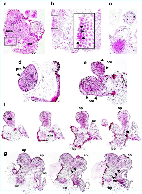
Fig. 3.

Regeneration process in wheat mature embryo tissues: somatic embryogenesis—intermediate steps of somatic embryo development. Initiation of cell division 1 DAC (a), small clusters of a few dividing cells. Cell proliferation, first clues apparent 2 DAC (b) and clumps of highly meristematic cells apparent 6 DAC (c). Globular embryo (pre-embryo); a widened globular structure bordered by a well-developed protoderm (the outer unicellular layer, arrowed), clearly delimited from the callus 8 DAC (d). Polar structure without vascular connection with the original tissue 12 DAC; a polarized embryonic structure next to a globular one (e). Bipolar structure, with root meristem differentiation at the basal pole; the organized formation next to this basal part represents the scutellum, indicating a level of further differentiation (f). Bipolar structures, well-organized somatic embryos; in one of the two shown, the vascular connections are visible between the apical and basal poles (arrowed), with a scutellum next to its basal part (g). Three major embryonic tissue systems—shoot apical meristem, root apical meristem, and the differentiation of procambial strands—are visible. In order to better describe post-globular development of the embryo, we have assembled a series of representative pictures of the same object, sequentially ordered, so that different planes can be observed (the different structures appear in different sections because they have not developed in the same plane). DAC days after culture initiation, mes mesocotyl, sc scutellum, ep scutellar epithelium of the original zygotic embryo, pro protoderm, sm shoot meristem, rm root meristem, ap apical pole, bp basal pole, vc vascular connections
