



Abstract
AIM: To explore the therapeutic role of globular adiponectin (gAd) in high-fat diet/streptozotocin (STZ)-induced type 2 diabetic rats with nonalcoholic fatty liver disease (NAFLD).
METHODS: Seven rats were fed a basic diet (normal control group; NC) during the experiment. Experimental rats (14 rats) were given a high-fat diet for 4 wk and were then injected with STZ to induce type 2 diabetes mellitus (T2DM) and NAFLD. Half of the T2DM/NAFLD rats were randomly injected intraperitoneally with gAd for 7 d (gAd-treated group), while the other 7 rats (T2DM/NAFLD group) received 0.9% saline. Plasma biochemical parameters and insulin concentrations were measured. Liver histopathology was examined by hematoxylin-eosin staining. Insulin receptor expression in the liver was analyzed by immunohistochemical staining, Western blot and quantitative real-time reverse transcription polymerase chain reaction analysis.
RESULTS: Compared to the control group, the T2DM/NAFLD group had increased levels of glucolipid and decreased levels of insulin. Plasma glucose and lipid levels were decreased in the gAd-treated group, while serum insulin levels increased. The expression of insulin receptor in the T2DM/NAFLD group increased compared with the NC group, and gAd downregulated insulin receptor expression in the livers of T2DM/NAFLD rats. Steatosis of the liver was alleviated in the gAd-treated group compared to the T2DM/NAFLD group (NAS 1.39 ± 0.51 vs 1.92 ± 0.51, P < 0.05).
CONCLUSION: Globular adiponectin exerts beneficial effects in T2DM rats with NAFLD by promoting insulin secretion, mediating glucolipid metabolism, regulating insulin receptor expression and alleviating hepatic steatosis.
Keywords: Adiponectin, Insulin secretion, Insulin receptor, Steatosis
Core tip: Nonalcoholic fatty liver disease (NAFLD) is the most common cause of chronic liver disease and is closely associated with obesity and type 2 diabetes mellitus (T2DM). Adiponectin is a fat-derived hormone with anti-diabetic properties, and evidence indicates that adiponectin plays a protective role in NAFLD. Our study focused on the beneficial roles of adiponectin in T2DM rats with NAFLD as a potential therapeutic target. The findings demonstrated that adiponectin exerts its beneficial effects in T2DM rats with NAFLD by promoting insulin secretion, mediating glucolipid metabolism, regulating insulin receptor expression and alleviating hepatic steatosis.
INTRODUCTION
Nonalcoholic fatty liver disease (NAFLD) is a common cause of chronic liver disease and is strongly associated with type 2 diabetes mellitus (T2DM)[1]. The prevalence of NAFLD has been estimated at approximately 70% in people with T2DM by ultrasound techniques, which have a low sensitivity for detecting NAFLD at steatosis levels below 33%[2,3]. NAFLD has been recognized as a common complication in patients with T2DM[4]. Traditionally, patients with NAFLD were treated by hepatologists; however, NAFLD has become a medical concern for endocrinologists because most T2DM patients have NAFLD. Therefore, NAFLD may be a new target for T2DM prevention and treatment[5].
Both T2DM and NAFLD are closely associated with obesity[6,7]. Adiponectin secreted from adipose tissue acts as an anti-diabetic adipocytokine[8], exerting its biological effects by binding to its receptors. Additionally, a recent study showed that plasma adiponectin was significantly lower in NAFLD patients than in controls, which demonstrated a role for low circulating adiponectin in the pathogenesis of NAFLD[9]. Moreover, increased levels of adiponectin are observed when patients are treated for NAFLD through diet interventions and pharmacotherapy[10,11]. These data suggest that adiponectin may contribute to the maintenance of liver integrity and may be a potential therapeutic target in NAFLD.
To explore the therapeutic role of globular adiponectin (gAd) in HFD/STZ-induced type 2 diabetic rats with NAFLD, we examined the biochemical parameters of glucolipid metabolism in the plasma, plasma insulin levels and the expression of insulin receptor in the liver, and we further analyzed liver histopathology upon replenishment of gAd.
MATERIALS AND METHODS
Animals
All the procedures involving animals were conducted in accordance with the ethical principles adopted by the Animal Experimental Center of Xiamen University and were approved by the Ethics Committee on Animal Experiments at Xiamen University. Twenty-one adult male Wistar rats (6-wk-old, body weight 160-210 g) (Shanghai Slac Laboratory Animal Co. Ltd) were transferred to the Animal Experimental Center of Xiamen University, where they were housed in individual cages maintained under ambient temperature (25 °C ± 2 °C) with a 12/12 h light/dark cycle and ad libitum access to water and food. The rats were randomly assigned to three groups: an NC group, comprising normal control rats (n = 7); a T2DM/NAFLD group, comprising type 2 diabetic rats with NAFLD (n = 7); and a gAd-treated group, comprising type 2 diabetic rats with NAFLD that were treated with gAd (n = 7).
After a week of acclimation, the T2DM rats were administered a high-fat diet (containing 10% fat, 10% carbohydrate, 5% cholesterol and 75% basic diet) for 4 wk combined with a single intraperitoneal injection of low-dose STZ (STZ 28 mg/kg; Sigma, St. Louis, MO, United States) in 0.1 mol/L citrate buffer (pH 4.2), while the normal control rats were fed the basic diet and received citrate buffer alone. Seventy-two hours post-injection, random non-fasting blood glucose was measured from tail snips using a portable glucometer. Diabetes was determined by the presence of hyperglycemia (random non-fasting glucose level > 16.7 mmol/L). Low-dose STZ has been known to induce mild impairment in insulin secretion, which is similar to what is observed in the later stages of T2DM. Subsequent liver histological evaluation indicated that the T2DM rats also suffered from NAFLD. Then, seven T2DM rats with NAFLD were randomly selected for the gAd-treatment group, and each rat was injected intraperitoneally with 3.5 μg gAd (BioVision, CA, United States) daily for 7 d, while the NC (n = 7) and T2DM/NAFLD (n = 7) groups received an equal volume of 0.9% saline. During the experimental period, the NC rats were fed the basic diet, while the T2DM/NAFLD group and the gAd-treated group were fed the high-fat diet.
At the end of the experiment, blood samples were collected from the heart and were centrifuged at 3000 g for 15 min to separate the plasma for use in assays. Some liver tissues were harvested, frozen in liquid nitrogen and subsequently stored at -80 °C until required. Other liver tissues were fixed in 10% neutral formaldehyde for paraffin sectioning.
Detection of plasma biochemical parameters and insulin levels
To confirm diabetes, random non-fasting blood glucose levels were measured using a glucometer (Johnson, New Jersey, United States) based on the glucose oxidase method. Fasting plasma glucose, total cholesterol (TC), triglyceride (TG), high-density lipoprotein cholesterol (HDL-C) and low-density lipoprotein cholesterol (LDL-C) were measured using commercial assay kits (Nanjing Jiancheng Bioengineering Institute, Jiangsu, China) according to the manufacturer’s instructions. Fasting plasma insulin levels were measured by ELISA using commercial kits (EMD Millipore Corporation, Billerica, MA, United States).
Histopathological and immunohistochemical analyses
Formalin-fixed paraffin-embedded sections (5 μm) were used for hematoxylin-eosin (HE) staining. Liver tissues from all rats were subjected to routine histological examination. Histopathological grading and staging of the NAFLD biopsies were performed by two liver pathologists using Brunt’s criteria. In this study, 14 rats fed a high-fat diet were diagnosed with simple steatosis by histology.
Immunohistochemical staining for insulin receptor was performed on additional histological sections of liver tissues. Serial sections of 5-μm thickness were cut from paraffin-embedded tissue blocks and floated onto glass slides. After drying overnight at 65 °C, the sections were deparaffinized and hydrated, and endogenous peroxidase activity was inactivated with 0.3% hydrogen peroxide. For antigen retrieval, the slides were immersed in citrate buffer (0.01 mol/L, pH 6.0) at 95 °C for 20 min, cooled to room temperature and blocked with 10% goat serum in PBS for 20 min. The specimens were incubated with primary mouse anti-insulin receptor antibody (1:1000 dilution; Abcam, Cambridge, United States) overnight at 4 °C. On the second day, after washing in PBS (3 min × 5 min), the tissues were incubated with HRP-polymer secondary antibody (Max Vision™ HRP-polymer anti-mouse/rabbit IHC kit) for 20 min at room temperature and were then visualized using diaminobenzidine substrate. A negative control was performed by omitting the primary antibody. The immunostaining results were evaluated and scored independently by two pathologists under double blind conditions.
The intensity of staining was evaluated semi-quantitatively in 10 random microscopic fields with more than 200 cells per field. The scoring criteria were based on the percentage of positive areas (no positive staining was scored as 0 points, less than 25% as 1 point, 25%-50% as 2 points, 51%-75% as 3 points and more than 75% as 4 points) multiplied by the staining intensity (1-4, representing weak, moderate, strong and very strong staining, respectively).
Western blot analysis
Total protein was extracted from liver tissue using a protein extraction kit (Applygen Technologies Inc., Beijing, China) according to the manufacturer’s protocol. Total protein levels were determined by the bicinchoninic acid (BCA) method (Applygen Technologies Inc., Beijing, China). Equal amounts of the protein samples were separated by sodium dodecyl sulfate-polyacrylamide gel electrophoresis (SDS-PAGE) and electro-transferred to PVDF membranes. The membranes were incubated overnight with anti-GAPDH antibody (1:1000 dilution; Good Here, Hangzhou, China) and mouse anti-insulin receptor antibody (1:1000 dilution; Abcam, Cambridge, MA, United States) followed by incubation with horseradish peroxidase HRP)-conjugated rabbit anti-mouse IgG (1:5000 dilution; Multi Sciences Biotech Co., Hangzhou, China) for 1 h. Peroxidase activity was detected with the Super ECL Plus Detection Reagent (Applygen Technologies Inc., Beijing, China).
Quantitative real-time reverse transcription polymerase chain reaction analysis
Total RNA was extracted from liver tissue using TRIzol Reagent (Invitrogen, San Diego, CA, United States). All samples were dissolved in nuclease-free water, and the concentrations of total RNA were measured by absorbance at 260/280 nm. The reverse transcription (RT) reaction for first-strand cDNA synthesis was carried out with reverse transcriptase (Bio-Rad, Hercules, CA, United States) using 2 μg of total RNA. Real-time polymerase chain reaction (PCR) was performed on a 7500 Real-time PCR System (ABI Applied Biosystems) using Power SYBR Green PCR Master Mix. The sequences of the primers used are as follows: insulin receptor, forward 5’- TGAAAAGTCACCTCCGTTCTCT-3’ and reverse 5’-CTCTCGTCATTCCAAAGTCTCC-3’; β-actin, forward 5’-GTAGCCATCCAGGCTGTGTT-3’ and reverse 5’-AACACAGCCTGGATGGCTAC-3’. The cycling conditions were as follows: 12 min at 95 °C followed by 40 cycles of 15 s at 95 °C and 1 min at 60 °C. A melting curve analysis was used to confirm the specificity of the PCR product, which was demonstrated as a single peak (data not shown). The expression of β-actin served as the internal control. Every sample was analyzed in triplicate. A comparative C (T) method reported previously [analyzing real-time PCR data by the comparative C (T) method] was used for analyzing the real-time PCR data.
Statistical analysis
The results are expressed as mean ± SD. Significant differences were determined by one-way ANOVA. P < 0.05 was considered statistically significant. SPSS 13.0 for Windows was used for statistical analyses.
RESULTS
Liver histological evaluation
HE staining of liver specimens from the rats fed a high-fat diet revealed steatosis, which indicated that the T2DM rats also suffered from NAFLD. The NAFLD activity score (NAS) of the T2DM/NAFLD group was 1.92 ± 0.51, which was higher than that of the NC group (0.11 ± 0.33) (P < 0.01). The steatosis in the T2DM/NAFLD group was alleviated by gAd treatment (1.92 ± 0.51 vs 1.39 ± 0.51, P < 0.05) (Figure 1).
Figure 1.

Changes in hepatic histology. A: Normal control group; B: Type 2 diabetes mellitus/nonalcoholic fatty liver disease group; C: Globular adiponectin-treated group (hematoxylin-eosin staining, × 200).
Effects of gAd on biochemical parameters and insulin levels of T2DM rats with NAFLD
The results of fasting plasma glucose, insulin, insulin sensitivity index (ISI), TC, TG, HDL-C and LDL-C are summarized in Table 1. The glucose level of the T2DM/NAFLD group was markedly higher than that of the NC group (P < 0.01). The glucose level of the gAd-treated group was decreased compared with that of the T2DM/NAFLD group (P < 0.01). Fasting insulin and the ISI were significantly decreased in the T2DM/NAFLD group compared with the NC group (P < 0.01). The insulin level of the gAd-treated group was significantly increased compared with that of the T2DM/NAFLD group (P < 0.05). Meanwhile, the ISI of the gAd-treated group showed a slight improvement over that of the T2DM/NAFLD group, although this was not statistically significant (P > 0.05). In addition, rats in the T2DM/NAFLD group had increased levels of TC, TG and LDL-C and decreased levels of HDL-C compared with rats in the NC group (P < 0.01). The concentrations of TC, TG and LDL-C in the gAd-treated group were lower than those in the T2DM/NAFLD group (P < 0.05). In particular, the concentration of TG decreased to nearly normal levels. No change was found in the HDL-C levels between the T2DM/NAFLD and gAd-treated groups (P > 0.05).
Table 1.
Biochemical parameters and insulin levels of rats (mean ± SD)
| NC group | T2DM/NAFLD group | gAd-treated group | |
| (n = 7) | (n = 7) | (n = 7) | |
| Fasting glucose (mmol/L) | 7.1 ± 0.8 | 24.8 ± 1.3b | 20.9 ± 1.9d |
| Fasting insulin (mIU/L) | 26.2 ± 9.5 | 12.8 ± 1.8b | 18.0 ± 3.2c |
| ISI® | -5.0 ± 0.2 | -6.1 ± 0.3b | -5.9 ± 0.2 |
| TC (mmol/L) | 2.6 ± 0.2 | 6.8 ± 0.8b | 6.0 ± 0.8c |
| TG (mmol/L) | 1.3 ± 0.1 | 1.8 ± 0.2b | 1.4 ± 0.2c |
| LDL-C (mmol/L) | 1.0 ± 0.1 | 1.9 ± 0.3b | 1.6 ± 0.2c |
| HDL-C (mmol/L) | 1.8 ± 0.4 | 0.9 ± 0.2b | 0.9 ± 0.2 |
ISI: Insulin sensitivity index®,
indicates the natural logarithm of the original data.
P < 0.01 vs the NC group.
P < 0.05,
P < 0.01 vs the T2DM/NAFLD group. NC: Normal control; T2DM: Type 2 diabetes mellitus; NAFLD: Nonalcoholic fatty liver disease; TC: Total cholesterol; TG: Triglyceride; HDL-C: High-density lipoprotein cholesterol; LDL-C: Low-density lipoprotein cholesterol.
Effects of gAd on insulin receptor protein expression in liver tissue
Immunohistochemical staining revealed that insulin receptor protein was expressed on the cell membrane in liver tissues (Figure 2). The relative expression levels of insulin receptor were evaluated by positive staining. The expression of insulin receptor in the livers of the T2DM/NAFLD group was increased compared to that in the livers of the NC group (8.25 ± 1.5 vs 3.5 ± 1, P < 0.01). Conversely, insulin receptor expression was significantly decreased in the gAd-treated group compared to that in the T2DM/NAFLD group (5.5 ± 1 vs 8.25 ± 1.5, P < 0.05).
Figure 2.

Immunohistochemical staining for insulin receptor in liver tissues. A: Normal control (NC) group; B: Type 2 diabetes mellitus/nonalcoholic fatty liver disease (T2DM/NAFLD) group; C: Globular adiponectin (gAd)-treated group (× 400); D: Bar graph represents the relative expression levels of insulin receptor (IR) in the NC, T2DM/NAFLD and gAd-treated groups based on the intensity and extent of staining. The values are depicted as mean ± SD (n = 7). T2DM/NAFLD vs gAd-treated, aP < 0.05, bP < 0.01 vs T2DM/NAFLD.
In each of the three groups, the results of insulin receptor protein expression obtained by immunohistochemical staining were identical to those obtained by Western blot. The expression level of insulin receptor was 1.5-fold higher in the T2DM/NAFLD group than in the NC group based on Western blot (P < 0.01), and it was reduced by 51% in the gAd-treated group compared with the T2DM/NAFLD group (P < 0.01) (Figure 3).
Figure 3.
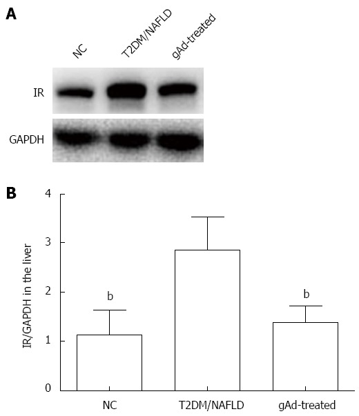
Insulin receptor protein expression in rat liver tissues of each group. A: Insulin receptor (IR) expression was assessed by Western blot analysis; B: The results are shown as the ratio of IR to glyceraldehyde-3-phosphate dehydrogenase (GAPDH) based on densitometry analysis. The values are expressed as mean ± SD (n = 7). bP < 0.01 vs T2DM/NAFLD. NC: Normal control; T2DM: Type 2 diabetes mellitus; NAFLD: Nonalcoholic fatty liver disease; gAd: Globular adiponectin.
Effects of gAd on mRNA expression of insulin receptor in liver tissue
Based on RT-PCR analysis, the expression of insulin receptor mRNA was 2.8-fold higher in the T2DM/NAFLD group than in the NC group (P < 0.01), and it was decreased by 24% in the gAd-treated group compared to the T2DM/NAFLD group (P < 0.01) (Table 2, Figure 4).
Table 2.
Insulin receptor mRNA expression in rat liver tissues of each group (n = 7) (mean ± SD)
P < 0.01 vs the NC group;
P < 0.01 vs the T2DM/NAFLD group. NC: Normal control; T2DM: Type 2 diabetes mellitus; NAFLD: Nonalcoholic fatty liver disease; gAd: Globular adiponectin.
Figure 4.

Insulin receptor mRNA expression in rat liver tissues of each group. Insulin receptor (IR) mRNA expression was assessed by RT-PCR analysis. The results are shown as a ratio of IR to β-actin based on densitometry analysis. The values are expressed as mean ± SD (n = 7). bP < 0.01 vs T2DM/NAFLD. NC: Normal control; T2DM: Type 2 diabetes mellitus; NAFLD: Nonalcoholic fatty liver disease; gAd: Globular adiponectin.
DISCUSSION
The results of many epidemiological and clinical translational studies have led to an increased awareness of the tight link between NAFLD and T2DM. Approximately 70% of patients with T2DM are estimated to have NAFLD based on ultrasound examinations, which have a low sensitivity for detecting NAFLD at steatosis levels below 33%. However, the association of T2DM with NAFLD is likely to be less well known to physicians, and it is therefore important for endocrinologists to be aware of the high likelihood that patients with T2DM will also have NAFLD.
Although the exact mechanism of NAFLD in T2DM remains unclear, evidence indicates that obesity is common in these two diseases. Obesity induces insulin resistance and insulin deficiency, which may be mediated by adipose tissue inflammation and dysregulated adipokine production[12]. Adiponectin, which is secreted from adipose tissue, is an anti-diabetic adipocytokine. Current research shows that plasma adiponectin is decreased in NAFLD patients[9]. Furthermore, increased serum adiponectin was observed in patients with NAFLD after pharmaceutical treatment, which indicated that adiponectin may play a hepatoprotective role in NAFLD[13].
In the present study, a T2DM/NAFLD rat model was developed by the administration of a high-fat diet following a low-dose injection of STZ, which closely mimics the natural history and metabolic characteristics of late stage T2DM with relative insulin deficiency in humans. Subsequent liver histological evaluations revealed steatosis in the rat models, which demonstrated that T2DM rats also had NAFLD.
Targher et al[14] reported that decreased adiponectin levels were closely associated with the degree of hepatic steatosis, necroinflammation and fibrosis in NAFLD patients. Further, increased adiponectin levels following pioglitazone treatment or diet interventions were related to the alleviation of steatosis in NAFLD patients[10,11]. In our study, gAd treatment directly alleviated steatosis in liver tissue.
Factors causing NAFLD in T2DM are still poorly defined; however, they are likely to involve the interplay of dysfunctional lipid metabolism and disordered glucose regulation associated with insulin resistance and relative insulin deficiency. In our study, the ISI in the gAd-treated group showed a tendency toward improvement, although this failed to reach statistical significance, possibly due to the low dose and short course of the gAd treatment. In addition, the levels of TC, TG and LDL-C were dramatically elevated, while HDL-C levels were decreased in the T2DM/NAFLD group compared to the normal control group. Low-dose gAd treatment for 7 d notably decreased the levels of TC, TG and LDL-C. In particular, the levels of TG were restored to nearly normal. Higher fasting glucose concentrations and lower insulin levels were found in the T2DM/NAFLD group compared with the normal control group. gAd increased insulin secretion and decreased glucose levels in the T2DM/NAFLD rats. Lipotoxicity plays an important role in the development of NAFLD. Additionally, hyperglycemia is likely to contribute to the development of NAFLD through the exacerbation of hepatic steatosis by increasing hepatic fat synthesis and reducing fat oxidation[15]. Studies have shown that adiponectin enhances fatty acid oxidation both in skeletal and cardiac muscle as well as in the liver, thus reducing the triglyceride content in these tissues[16]. Yamauchi et al[17] found that both isoforms of the adiponectin receptor could mediate increased AMP-activated protein kinase (AMPK) phosphorylation and PPARa activity by adiponectin binding in vitro, thus activating fatty acid oxidation and glucose uptake. Therefore, gAd may protect the liver from lipid accumulation by increasing insulin secretion and mediating glucolipid metabolism. Rao et al[18] suggested that adiponectin can increase insulin secretion, which was in agreement with our findings. Additionally, evidence has shown that gAd potentiates glucose-induced insulin secretion in both human pancreatic islets and rat beta cells via an AMPK-independent pathway[19]. A recent experiment revealed that adiponectin inhibited apoptosis and stimulated insulin gene expression and secretion in pancreatic beta cells[20]. Furthermore, evidence indicates that both adiponectin receptor 1 (AdipoR1) and adiponectin receptor 2 (AdipoR2) are expressed in human pancreatic beta cells, potentially linking adiponectin to pancreatic function[21].
The liver is one of the most important target organs of insulin. Insulin acts by binding to insulin receptors on the target cell surface. Then, many important cellular signaling cascades are initiated, which finally result in the regulation of blood glucose levels. Thus, defective molecules in the insulin signaling pathway, especially abnormalities in the insulin receptor, may be closely related to the impairment of insulin activity. Hence, the effects of gAd on insulin receptor expression in liver tissue were analyzed. The results showed that the expression of insulin receptor was increased in the T2DM/NAFLD group, and this increase was accompanied by hypoinsulinemia. It was reported that the combination of lipotoxicity and glucotoxicity induced by a high-fat diet and streptozotocin injection led to islet beta-cell failure and apoptosis. This subsequently led to hypoinsulinemia and hyperglycemia[22], which may be the underlying mechanism of hypoinsulinemia in the T2DM/NAFLD group. Indeed, hypoinsulinemia progression can be observed clinically in severe T2DM patients at a late stage of the disease. Thus, we developed T2DM/NAFLD rats with hypoinsulinemia and hyperglycemia, which closely mimics late-stage T2DM. Elevated expression of insulin receptor in the T2DM/NAFLD group may be due to the negative feedback effect caused by decreased insulin levels. After gAd treatment, insulin receptor expression decreased, and this decrease was accompanied by increased insulin levels. Marshall et al[23] showed that high insulin concentrations in the media decreased the number of insulin receptors on cultured human lymphocytes in vitro. Furthermore, exposure of isolated rat adipocytes to continuous hyperinsulinemia led to a dose-dependent loss of insulin receptors. These results suggested that the plasma insulin concentration inversely regulated the number of cellular insulin receptors; therefore, insulin can regulate its own receptor[24]. However, much work remains to be done to delineate the exact mechanisms of gAd in the regulation of insulin function.
In conclusion, gAd augments insulin secretion, mediates the expression of insulin receptor, ameliorates glucolipid metabolism and alleviates hepatic steatosis in HFD/STZ-induced T2DM rats with NAFLD.
COMMENTS
Background
Nonalcoholic fatty liver disease (NAFLD) is the most common cause of chronic liver disease and is closely associated with type 2 diabetes mellitus (T2DM). NAFLD is also associated with an increased risk of all-cause death, which is likely connected to insulin resistance complications in NAFLD patients. Traditionally, NAFLD patients have been treated by hepatologists; however, NAFLD has become a medical concern for endocrinologists because most T2DM patients also suffer from NAFLD. Ongoing research efforts toward understanding the relationship between NAFLD and T2DM have revealed that both T2DM and NAFLD are closely associated with obesity, and evidence indicates that adiponectin plays a crucial role in NAFLD and T2DM.
Research frontiers
Adiponectin is a hormone secreted by adipocytes that acts as an anti-diabetic adipocytokine and exerts its biological effects via binding to its receptors. Additionally, a recent study showed that plasma adiponectin was significantly lower in NAFLD patients than in controls, demonstrating a role for low circulating adiponectin in the pathogenesis of NAFLD. Moreover, increased levels of adiponectin are observed in patients who are treated for NAFLD through diet interventions and pharmacotherapy. These data suggest possible roles for adiponectin in maintaining liver integrity and as a therapeutic target in NAFLD.
Innovations and breakthroughs
Previous studies have shown that the level of adiponectin increases indirectly through diet interventions and pharmacotherapy, which are used in the treatment of NAFLD. In this study, rats were injected intraperitoneally with globular adiponectin (gAd), and the results showed that adiponectin exerted beneficial effects in T2DM rats with NAFLD by promoting insulin secretion, mediating glucolipid metabolism, regulating insulin receptor expression and alleviating hepatic steatosis. Thus, adiponectin may have a potential role in the treatment of diabetes with NAFLD. Additionally, few studies have focused on the relationship between T2DM and NAFLD, and physicians are not likely to be fully aware of the association between T2DM and NAFLD. Therefore, this study urges physicians to be aware of the high likelihood that patients with T2DM also have NAFLD.
Applications
The present study shows that adiponectin has anti-diabetic properties, and evidence indicates that adiponectin plays a protective role in NAFLD. Upon confirmation of these results, future applications of adiponectin as a treatment for T2DM with NAFLD may be merited.
Terminology
NAFLD is the most common hepatic dysregulation associated with the growing epidemic of obesity and is characterized by fat accumulation in the liver. The histologic spectrum of NAFLD ranges from simple fatty liver (i.e., steatosis) to severe steatohepatitis (NASH), which can progress to cirrhosis and liver failure. T2DM is a chronic metabolic disease that affects the ability to convert food into energy. Adiponectin is a recently described adipokine that has been recognized as a key regulator of insulin sensitivity, and evidence indicates that adiponectin has anti-diabetic properties and plays a protective role in NAFLD.
Peer review
In this paper, the authors studied the role of globular adiponectin in an animal model of diabetes/NAFLD, and used a small number of rats to demonstrate that globular adiponectin exerts beneficial effects in T2DM rats with NAFLD. In the study, the authors evaluated the effects of globular adiponectin in diabetic rats with NAFLD. The major result of this study is that gAd promotes insulin secretion, mediates glucolipid metabolism, regulates insulin receptor expression and alleviates hepatic steatosis. The details regarding expression changes in insulin receptor protein and mRNA are very interesting, and the effects of globular adiponectin on fatty acid oxidation should be further discussed.
Footnotes
Supported by Science and Technology Project of Xiamen, China, No. 3502Z20114016
P- Reviewer: Di Minno MND, Grassi A S- Editor: Ma YJ L- Editor: Wang TQ E- Editor: Ma S
References
- 1.Mavrogiannaki AN, Migdalis IN. Nonalcoholic Fatty liver disease, diabetes mellitus and cardiovascular disease: newer data. Int J Endocrinol. 2013;2013:450639. doi: 10.1155/2013/450639. [DOI] [PMC free article] [PubMed] [Google Scholar]
- 2.Musso G, Gambino R, Cassader M, Pagano G. Meta-analysis: natural history of non-alcoholic fatty liver disease (NAFLD) and diagnostic accuracy of non-invasive tests for liver disease severity. Ann Med. 2011;43:617–649. doi: 10.3109/07853890.2010.518623. [DOI] [PubMed] [Google Scholar]
- 3.Saadeh S, Younossi ZM, Remer EM, Gramlich T, Ong JP, Hurley M, Mullen KD, Cooper JN, Sheridan MJ. The utility of radiological imaging in nonalcoholic fatty liver disease. Gastroenterology. 2002;123:745–750. doi: 10.1053/gast.2002.35354. [DOI] [PubMed] [Google Scholar]
- 4.Tolman KG, Fonseca V, Tan MH, Dalpiaz A. Narrative review: hepatobiliary disease in type 2 diabetes mellitus. Ann Intern Med. 2004;141:946–956. doi: 10.7326/0003-4819-141-12-200412210-00011. [DOI] [PubMed] [Google Scholar]
- 5.Fruci B, Giuliano S, Mazza A, Malaguarnera R, Belfiore A. Nonalcoholic Fatty liver: a possible new target for type 2 diabetes prevention and treatment. Int J Mol Sci. 2013;14:22933–22966. doi: 10.3390/ijms141122933. [DOI] [PMC free article] [PubMed] [Google Scholar]
- 6.Tataranni PA. Pathophysiology of obesity-induced insulin resistance and type 2 diabetes mellitus. Eur Rev Med Pharmacol Sci. 2002;6:27–32. [PubMed] [Google Scholar]
- 7.Marchesini G, Bugianesi E, Forlani G, Cerrelli F, Lenzi M, Manini R, Natale S, Vanni E, Villanova N, Melchionda N, et al. Nonalcoholic fatty liver, steatohepatitis, and the metabolic syndrome. Hepatology. 2003;37:917–923. doi: 10.1053/jhep.2003.50161. [DOI] [PubMed] [Google Scholar]
- 8.Böttner A, Kratzsch J, Müller G, Kapellen TM, Blüher S, Keller E, Blüher M, Kiess W. Gender differences of adiponectin levels develop during the progression of puberty and are related to serum androgen levels. J Clin Endocrinol Metab. 2004;89:4053–4061. doi: 10.1210/jc.2004-0303. [DOI] [PubMed] [Google Scholar]
- 9.Pagano C, Soardo G, Esposito W, Fallo F, Basan L, Donnini D, Federspil G, Sechi LA, Vettor R. Plasma adiponectin is decreased in nonalcoholic fatty liver disease. Eur J Endocrinol. 2005;152:113–118. doi: 10.1530/eje.1.01821. [DOI] [PubMed] [Google Scholar]
- 10.Choi S, Choi Y, Choi Y, Kim S, Jang J, Park T. Piperine reverses high fat diet-induced hepatic steatosis and insulin resistance in mice. Food Chem. 2013;141:3627–3635. doi: 10.1016/j.foodchem.2013.06.028. [DOI] [PubMed] [Google Scholar]
- 11.Gastaldelli A, Harrison S, Belfort-Aguiar R, Hardies J, Balas B, Schenker S, Cusi K. Pioglitazone in the treatment of NASH: the role of adiponectin. Aliment Pharmacol Ther. 2010;32:769–775. doi: 10.1111/j.1365-2036.2010.04405.x. [DOI] [PubMed] [Google Scholar]
- 12.Williams KH, Shackel NA, Gorrell MD, McLennan SV, Twigg SM. Diabetes and nonalcoholic Fatty liver disease: a pathogenic duo. Endocr Rev. 2013;34:84–129. doi: 10.1210/er.2012-1009. [DOI] [PubMed] [Google Scholar]
- 13.Shargorodsky M, Omelchenko E, Matas Z, Boaz M, Gavish D. Relation between augmentation index and adiponectin during one-year metformin treatment for nonalcoholic steatohepatosis: effects beyond glucose lowering? Cardiovasc Diabetol. 2012;11:61. doi: 10.1186/1475-2840-11-61. [DOI] [PMC free article] [PubMed] [Google Scholar]
- 14.Targher G, Bertolini L, Scala L, Poli F, Zenari L, Falezza G. Decreased plasma adiponectin concentrations are closely associated with nonalcoholic hepatic steatosis in obese individuals. Clin Endocrinol (Oxf) 2004;61:700–703. doi: 10.1111/j.1365-2265.2004.02151.x. [DOI] [PubMed] [Google Scholar]
- 15.Santos JC, Valentim IB, de Araújo OR, Ataide Tda R, Goulart MO. Development of nonalcoholic hepatopathy: contributions of oxidative stress and advanced glycation end products. Int J Mol Sci. 2013;14:19846–19866. doi: 10.3390/ijms141019846. [DOI] [PMC free article] [PubMed] [Google Scholar]
- 16.Karbowska J, Kochan Z. Role of adiponectin in the regulation of carbohydrate and lipid metabolism. J Physiol Pharmacol. 2006;57 Suppl 6:103–113. [PubMed] [Google Scholar]
- 17.Yamauchi T, Kamon J, Ito Y, Tsuchida A, Yokomizo T, Kita S, Sugiyama T, Miyagishi M, Hara K, Tsunoda M, et al. Cloning of adiponectin receptors that mediate antidiabetic metabolic effects. Nature. 2003;423:762–769. doi: 10.1038/nature01705. [DOI] [PubMed] [Google Scholar]
- 18.Rao JR, Keating DJ, Chen C, Parkington HC. Adiponectin increases insulin content and cell proliferation in MIN6 cells via PPARγ-dependent and PPARγ-independent mechanisms. Diabetes Obes Metab. 2012;14:983–989. doi: 10.1111/j.1463-1326.2012.01626.x. [DOI] [PubMed] [Google Scholar]
- 19.Patané G, Caporarello N, Marchetti P, Parrino C, Sudano D, Marselli L, Vigneri R, Frittitta L. Adiponectin increases glucose-induced insulin secretion through the activation of lipid oxidation. Acta Diabetol. 2013;50:851–857. doi: 10.1007/s00592-013-0458-x. [DOI] [PubMed] [Google Scholar]
- 20.Wijesekara N, Krishnamurthy M, Bhattacharjee A, Suhail A, Sweeney G, Wheeler MB. Adiponectin-induced ERK and Akt phosphorylation protects against pancreatic beta cell apoptosis and increases insulin gene expression and secretion. J Biol Chem. 2010;285:33623–33631. doi: 10.1074/jbc.M109.085084. [DOI] [PMC free article] [PubMed] [Google Scholar]
- 21.Stolzenberg-Solomon RZ, Weinstein S, Pollak M, Tao Y, Taylor PR, Virtamo J, Albanes D. Prediagnostic adiponectin concentrations and pancreatic cancer risk in male smokers. Am J Epidemiol. 2008;168:1047–1055. doi: 10.1093/aje/kwn221. [DOI] [PMC free article] [PubMed] [Google Scholar]
- 22.Nugent DA, Smith DM, Jones HB. A review of islet of Langerhans degeneration in rodent models of type 2 diabetes. Toxicol Pathol. 2008;36:529–551. doi: 10.1177/0192623308318209. [DOI] [PubMed] [Google Scholar]
- 23.Marshall S, Olefsky JM. Effects of insulin incubation on insulin binding, glucose transport, and insulin degradation by isolated rat adipocytes. Evidence for hormone-induced desensitization at the receptor and postreceptor level. J Clin Invest. 1980;66:763–772. doi: 10.1172/JCI109914. [DOI] [PMC free article] [PubMed] [Google Scholar]
- 24.Gavin JR, Roth J, Neville DM, de Meyts P, Buell DN. Insulin-dependent regulation of insulin receptor concentrations: a direct demonstration in cell culture. Proc Natl Acad Sci USA. 1974;71:84–88. doi: 10.1073/pnas.71.1.84. [DOI] [PMC free article] [PubMed] [Google Scholar]