Abstract
Quantitation of individual mAbs within a combined antibody drug product is required for preclinical and clinical drug development. We have developed two antitoxins (XOMA 3B and XOMA 3E) each consisting of three monoclonal antibodies (mAbs) that neutralize type B and type E botulinum neurotoxin (BoNT/B and BoNT/E) to treat serotype B and E botulism. To develop mAb-specific binding assays for each antitoxin, we mapped the epitopes of the six mAbs. Each mAb bound an epitope on either the BoNT light chain (LC) or translocation domain (HN). Epitope mapping data was used to design LC-HN domains with orthogonal mutations to make them specific for only one mAb in either XOMA 3B or 3E. Mutant LC-HN domains were cloned, expressed, and purified from E. coli. Each mAb bound only to its specific domain with affinity comparable to the binding to holotoxin. Further engineering of domains allowed construction of ELISAs that could characterize the integrity, binding affinity, and identity of each of the six mAbs in XOMA 3B, and 3E without interference from the three BoNT/A mAbs in XOMA 3AB. Such antigen engineering is a general method allowing quantitation and characterization of individual mAbs in a mAb cocktail that bind the same protein.
Keywords: botulinum neurotoxin, botulism, oligoclonal antibodies, protein domain, protein characterization, protein purification
Introduction
Monoclonal antibody (mAb)-based drugs have proven successful in the clinic, with more than 27 now approved by the FDA. All of the approved antibody drugs consist of single mAbs (1). In order to enhance efficacy or broaden specificity, combinations of mAbs are now in preclinical and clinical development for a wide range of diseases (2). The most advanced of these is CL184, a combination of two mAbs designed for the post-exposure prophylaxis against rabies (3) now in Phase II clinical trials (clinicaltrials.gov identifier: NCT01228383). Other mAb combinations under investigation include mAbs for influenza (4), for anthrax (5, 6), and for cancer (7).
Regulatory agency requirements for characterization of mAb combinations (oligoclonal antibody) and polyclonal antibodies are based on several existing guidelines (8-10). Because oligoclonal therapies typically have mechanism of actions requiring the presence of all of the mAb components, they are clinically evaluated as the mixture of antibodies. Despite this, analytical methods to characterize and measure the individual components of the mAb cocktails are required for use in product characterization, for production of clinical material, for pharmacokinetic studies, and for monitoring of drug product stability.
We are developing XOMA 3B and 3E, equimolar mixtures of three human mAbs binding non-overlapping epitopes on botulinum neurotoxin type B (BoNT/B) and type E (BoNT/E) respectively. Botulism is characterized by flaccid paralysis, which if not rapidly fatal requires prolonged hospitalization in an intensive care unit and mechanical ventilation. Botulism is an orphan disease, with approximately 100 cases/year in the United States. BoNTs are also classified by the Centers for Disease Control and Prevention as one of the six highest-risk threat agents for bioterrorism, due to the high potency and lethality of the toxin (11). It is estimated that major civilian exposure to BoNT would have catastrophic effects (12).
The mainstay of treatment for adults with botulism is antitoxin produced by hyperimmunizing horses, equine antitoxin (13, 14). Equine antitoxin, however, has a significant incidence of side effects, including hypersensitivity reactions including serum sickness and anaphylaxis (13, 14). An alternative to equine antitoxin is serotype specific recombinant mAb based antitoxin, however, individual mAbs do not have the requisite potency for development (15). Combining the three mAbs in the XOMA 3B and XOMA 3E cocktails increases the potency of BoNT neutralization by at least three orders of magnitude compared to the potency of the individual antibodies (unpublished data).
Oligoclonal antibodies that target the same protein are especially challenging for analytical method development since ELISA assays using the target protein for capture will not distinguish between individual mAbs. One approach is to generate and use anti-idiotype antibodies specific for each mAb (16-18). However, this approach requires generating sets of mAbs or polyclonal antibody binding unique epitopes in the binding site of each of the three mAbs, with high affinity and with specificity only for that mAb and not the other two. Furthermore, the anti-idiotype antibodies would need to have affinities of less than 5 nM, the anticipated effective serum concentration of the mAbs in XOMA 3B and 3E in humans. Frequently, anti-idiotype antibodies do not have this level of sensitivity (16).
Recently, we reported the development of assays that could distinguish the binding of the three component mAbs of XOMA 3AB, a combination of three mAbs binding non-overlapping epitopes on botulinum neurotoxin type A (BoNT/A), using epitope based capture. (19) This proved possible by expressing and purifying three different folded domains of BoNT/A, each recognized by only one of the three component mAbs. This is not possible for XOMA 3B and 3E since at least two of the three mAbs in each antitoxin bind the same BoNT domain. To overcome this limitation, we report here the identification, expression, and purification of BoNT domains consisting of the light chain (LC) and translocation domain (HN) (LC-HN) of BoNT/B and BoNT/E that have been engineered to be specific for only one of the three mAbs in each combination. Using the domains, ELISA assays were developed for characterization of the integrity and purity of each mAb in the three-mAb drug products. We believe this is a general approach that can be applied to mAb combinations binding the same protein target. In the case of BoNTs, the use of mAb specific non-toxic domains also eliminates exposure of laboratory workers to the highly toxic BoNT and obviates the burdensome requirements for handling select agents such as BoNT.
Materials and Methods
Domain cloning
PCR primers were designed to amplify the BoNT/B1 (NCBI accession number YP_001693307) HC (amino acids N862-E1291) HN (amino acids P443-L861), or LC (amino acids P2-A442) gene fragment respectively adding the restriction sites NcoI and NotI. Each gene fragment was amplified by using PCR from a synthetic gene construct (20). Following digestion of the yeast display vector pYD2 and the resulting PCR amplification product with NcoI and NotI, the BoNT/B gene fragments were gel-purified and ligated into pYD2. Ligation mixtures were used to transform E. coli DH5 and correct clones identified by DNA sequencing. Similarly, the BoNT/E (NCBI accession number YP_001920504) HC (amino acids G805-K1248) HN (amino acids S424-L804), or LC (amino acids M1-K423) were cloned into pYD2. LC-HN domains, which are comprised of LC and HN domains, were cloned by the yeast gap repair method inserting HN into a pYD2 plasmid that already had the LC (19). Plasmid DNA was used to transform Lithium Acetate-treated EBY100 cells.
Epitope mapping
The BoNT/B or BoNT/E domain bound by mAbs B-a, B-b and B-c or by mAbs E-a, E-b and E-c were determined by incubating yeast-displayed BoNT/B or BoNT/E HC, HN, LC, or LCHN with the respective mAb followed by goat-anti-human-phycoerythrin with binding detected by flow cytometry as previously described (21). For fine mapping of the mAb epitopes, mutations were randomly introduced into the BoNT/B and BoNT/E LC-HN by using error prone PCR. Mutant LC-HN gene repertoires were then cloned into the pYD2 vector by gap repair and display of the domains on the surface of yeast induced (21). Amino acid residues in the BoNT/B LC-HN critical for the binding of mAbs B-a, B-b, and B-c were identified by incubating the mutant BoNT/B LC-HN library with either mAb B-a, B-b, or B-c followed by goat-anti-human-phycoerythrin and flow sorting yeast that had minimal or no mAb binding as we have previously described (21). The LC-HN genes from yeast clones with reduced or absent mAb binding were sequenced and the location of mutations modeled on the X-ray crystal structure of BoNT/B to identify each of the three putative mAb epitopes as previously described (21). Mutations in the epitopes were then combined until there was no mAb binding to the yeast-displayed BoNT/B domain at a concentration of 1 uM mAb. Amino acid residues in the BoNT/E LC-HN critical for binding of mAbs E-A, E-b, and E-c were similarly identified using the BoNT/E LC-HN random mutant library.
Generation of antibody-specific domains in E. coli for ELISA assays
Wild-type BoNT/B LC-HN domain (amino acids 1-861) and the wild-type BoNT/E LC-HN domain (amino acids 1-834) were both cloned from the pYD2 vector into the pET21d vector in the same way as previously described (19). In this vector, each domain construct has a SV5 epitope tag and a hexa-histidine tag at the C-terminal. Mutations which knocked out individual mAb binding to the yeast-displayed BoNT domains were introduced into the BoNT/B or BoNT/E LC-HN, expression induced at small scale and the domains purified as described in Meng et al, 2012 (19) for BoNT/A domains. The purified mutant domains were tested for binding to mAbs B-a, B-b and B-e (for the BoNT/B LC-HN) or for binding to mAbs E-a, E-b, and E-c (for the BoNT/E LC-HN) using a Attana A100 Quartz Crystal Microbalance (QCM) (Attana AB, Stockholm, Sweden). Once mutations were identified that knocked out binding of a single mAb, a second set of mutations were introduced into each of the six domains to knock out binding of the second of the three mAbs. This work yielded three BoNT/B and three BoNT/E LC-HN domains specific for each of the three mAbs in XOMA 3B and XOMA 3E respectively.
Attana binding assays
Quartz crystal microbalance technology was used for rapid evaluation of antibody binding. Antihuman IgG (Fc) antibody was immobilized on LNB-carboxyl chip (Catalog #: 3623-3033) using the Attana amine coupling kit (Catalog # 3501-3001). Purified domains were injected at 10 μg/ml in HBST buffer (100 mM HEPES, 1.5 M NaCl, 0.05% Tween 20, pH 7.4). Chips were regenerated using HCl (0.1 M) followed by NaOH (0.02 M) solution.
Large scale purification of domains
We developed a scalable purification scheme for the domains to be used for drug characterization. Frozen cell paste from a 20 L fermentation culture (about 120 g of wet cell weight) was resuspended in 10ml of 2-10°C lysis buffer (50 mM Tris-HCl, 500 mM NaCl, 5% Glycerin, 0.5% Triton X-100, pH 8.0, 1% v/w protease inhibitor cocktail. Pastes were dispersed using an Ultra Turax mixer, keeping the paste suspension below 8°C. The suspended cells were lysed by passing through a high pressure homogenizer (Avestin EmulsiFlex-C55), at 18000-22000 psi with cooling. The lysate was cooled and CaCl2 and sodium phosphate buffer and Tris added to achieve pH 8.2-8.5. The lysate was centrifuged and pH of supernatant was 7.3 - 7.7 with acetic acid. Lysate was clarified by ultrafiltration. The clarified lysate was subjected to IMAC chromatography (Ni Sepharose HP resin, GE Healthcare Life Sciences). The column was equilibrated and pH adjusted to 7.5 at 20°C. After loading, the column was washed sequentially with 8 CV of equilibration buffer, 20 CV wash buffer (30 mM Imidazole, 50 mM Tris.HCl, 500 mM NaCl, 0.5% Triton X-100, pH 8.0), and 40 CV Buffer A (25 mM sodium succinate, 25 mM sodium phosphate, 500 mM NaCl, pH 7.5). Elution was carried out with a linear pH gradient to achieve pH 3.5. The IMAC pool was concentrated against formulation buffer (5 mM NaPO4, 4% sucrose, pH 7.0 at 20°C) using ultrafiltration.
Size exclusion chromatography and reverse phase chromatography analysis of domains
All HPLC analysis was performed with the Agilent 1200 Series LC System and analyzed with the Agilent ChemStation Version B.03.02. SE-HPLC was performed with a TosoH TSK gel Super SW2000 column packed with 4μm particles containing 250 Å pores. The injected samples were eluted isocratically with elution buffer (20 mM NaPO4, 0.2 M ammonium sulfate, pH 6.8). Protein was detected by absorbance at 280 nm and 214 nm.
mAb binding by SE-HPLC and concentration measurement: Domains and their corresponding mAb were mixed in a 2:1 mAb:domain molar ratio based on the concentrations determined by A280 nm (Supplemental Table 1 lists extinction coefficients). The domain and the corresponding mAb concentrations during binding assay were in the range of 10-20μM. The binding reaction buffer was essentially the same as that of the individual domain, which was 5mM NaPO4, 120mM sucrose, pH 7. The domain/mAb mixtures were left at ambient temperature for approximately 10 minutes and then kept at 2-8°C auto-injector until HPLC testing. The samples were analyzed using the SE-HPLC method described above. The G3000 column was found to give superior resolution for the binding assay mixture samples.
RP-HPLC: Reversed phase HPLC was conducted with an Agilent Zorbax 300SB-C3 column. Mobile phase A was 0.1% (v/v) TFA in water and mobile phase B was 0.1% TFA in acetonitrile. The domains were eluted with increasing concentration of acetonitrile. The column was washed and re-equilibrated with 62% A for before making a subsequent sample injection. Protein was detected by A214 nm.
Measurement of affinity and binding specificity of mAbs for domains
Binding affinities (KD) were measured using KinExA 3000 flow fluorimeter (Sapidyne Instruments Inc., Boise Idaho) as previously described (22-24). KinExA provides accurate binding constants for high affinity binding interactions. Binding reaction mixtures were performed in PBS (pH 7.4) at room temperature with 1 mg/mL bovine serum albumin (BSA), and 0.02% (w/v) sodium azide added as a preservative. mAb solution was serially diluted into a constant concentration of either BoNT/B and E holotoxin or the corresponding domain sufficient to produce a reasonable signal, where the antibody concentration was varied tenfold above and below the value of the apparent KD and domain concentrations were no more than fourfold above the KD to ensure a KD controlled experiment. Samples were equilibrated up to two days, then each of the 13 dilutions were passed over a flow cell containing a 4 mm column of NHS-activated Sepharose 4 Fast Flow beads (GE Healthcare) covalently coated with the corresponding antibody to capture the free holotoxin or domain. An Alexa-647 labeled antibody was passed over the beads to detect the free antigen by binding and producing a signal relative to the amount of free holotoxin or domain bound to the beads. In the case of the domains, a detection antibody that bound the SV5 tag was used, and in the case of holotoxin, a detection antibody binding a non-overlapping epitope was used. Each dilution was tested in duplicate. Sample volumes varied from 3 to 25 mL depending on antibody affinity. The equilibrium titration data were fit to a 1:1 reversible binding model using KinExA Pro Software (version 3.0.6; Sapidyne Instruments) to determine the KD. Binding curves are provided as Supplementary Figures 1 and 2.
ELISA binding assay
Direct coating plate ELISAs specific for each of the six mAbs were developed using the purified domains. 96 well microtitre plates (Thermo Scientific) were coated overnight at 2-8°C with 100 L/well of purified domains (5 to 15 g/mL). Plates were blocked with BSA, Tween 20 (0.05%) in PBS (PBS-T) for up to 2 hours. Plates were washed with PBS-T by an automated plate washer (Molecular Devices SKAN400). MAb (100 L) was added to each well and incubated at room temperature for 1-2 hrs, washed three times with PBS-T. Horseradish peroxidase conjugated goat anti-human IgG was added for signal detection over 1-2 hours. Plates were washed again as described above and the reaction developed by the addition of 2,2-Azinobis [3-ethylbenzothiazoline-6-sulfonic acid]-diammonium salt (ABTS, 100 L/well) (25-27). After 8-15 min, the reaction was stopped and plates were read in a Molecular Devices M2e instrument, and plotted using a 4-parameter fit algorithm against the mAb concentration to yield a sigmoidal curve. Every sample or standard was measured in duplicate on the same plate.
Indirect capture plate ELISA assays were developed to detect the binding of mAbs A-c and B-b without interference from the binding of other lower affinity mAbs due to avidity resulting from the direct capture format. In this assay, mixtures of eight or nine mAbs were used to demonstrate binding specificity. The nine-mAb mixture used in this assay was prepared by mixing equal molar amounts of each of the nine mAbs (3 mAbs in XOMA 3AB, 3 mAbs in XOMA 3B and 3 mAbs in XOMA 3E); the control eight-mAb mixture was prepared in the same way except one mAb, which was specific for the domain in solution, was not included. In the capture plate, mAbs were coated on the plate at the concentration of 1 g/mL, and then blocked with BSA in PBS-T for 1-2 hours. The plate was washed once with PBS-T by an automated plate washer. In a separate incubation plate, serial dilutions of either a single mAb or mixtures of eight or nine mAbs was mixed with a constant concentration of the domain at 0.4 g/mL. After incubating at room temperature for 60 min, the mixture was transferred to the capture plate and incubated at room temperature for 2 hours to allow mAb capture of free domain. The plates were washed three times with PBS-T before mouse IgG anti-SV5 antibody was added at (100 l/well of 0.2 g/ml) and incubated for 1 hour at room temperature. Plate washing and signal detection was done as for the direct ELISA, except the HRP concentration was reduced by 50%.
Expression and purification of XOMA 3B and XOMA 3E mAbs
BoNT/B (mAbs B-a, B-b, and B-c) and BoNT/E (mAbs E-a, E-b, and E-c) IgG were expressed from stable Chinese Hamster Ovary (CHO) cell lines and purified by Protein A chromatography followed by flow-through anion exchange and hydrophobic interaction chromatographies as described in Meng et al., (19).
Results
Overview of the strategy to construct mAb specific BoNT domains
Three BoNT/B and three BoNT/E LC-HN domains specific for only one of the three mAbs in XOMA 3B or 3E were generated. To accomplish this, we first determined that each of the three mAbs in BoNT/B and BoNT/E bound to the BoNT/B or BoNT/E LC-HN. We then used yeast-displayed libraries of BoNT/B or BoNT/E LC-HN mutants to identify amino acid mutations in the domains that knocked out binding of each of the mAbs. Finally, six recombinant BoNT LC-HN domains each specific for a different mAb in XOMA 3B or 3E were produced from E. coli by combining mutations which eliminated binding of two of the three mAbs in XOMA 3B or XOMA 3E.
Construction of mAb specific domains
The domain recognized by each of the three component mAbs of XOMA 3B and 3E was determined using yeast-displayed BoNT/B and BoNT/E LC, HN, and HC domains as we have previously done for BoNT/A mAbs (21). For XOMA 3B, mAbs B-a and B-c bound the BoNT/B LC and mAb B-b bound the BoNT/B HN (Table 1). For XOMA 3E, mAb E-a bound a complex epitope requiring both the LC and HN, while mAbs E-b and E-c bound the BoNT/E HN. Efforts to express the HN as a separate folded domain in E. coli required detergent solubilization, complicating both purification and use in assays. We therefore used the BoNT/B or BoNT/E LCHN domain for detection of each of the mAbs in XOMA 3B and 3E respectively.
Table 1.
Binding affinities and kinetics of mAbs for BoNT/B and BoNT/E and their mAb-specific LC-HN domains. The equilibrium binding constant (KD) was measured by flow fluorimetry in a KinExA.
| BoNT/B LC-HN mAb specific domain | BoNT/B Holotoxin | ||
|---|---|---|---|
| mAb | Domain | KD (pM) | KD (pM) |
| mAb B-a | B-LC-HN-a | 0.18 | 0.34 |
| mAb B-b | B-LC-HN-b | 60.81 | 56.88 |
| mAb B-c | B-LC-HN-c | 39.08 | 38.07 |
| BoNT/E LC-HN mAb specific domain | BoNT/E Holotoxin | ||
|---|---|---|---|
| mAb | Domain | KD (pM) | KD (pM) |
| mAb E-a | E-LC-HN-a | 22.09 | 11.52 |
| mAb E-b | E-LC-HN-b | 6.71 | 35.11 |
| mAb E-c | E-LC-HN-c | 115.6 | 730.30 |
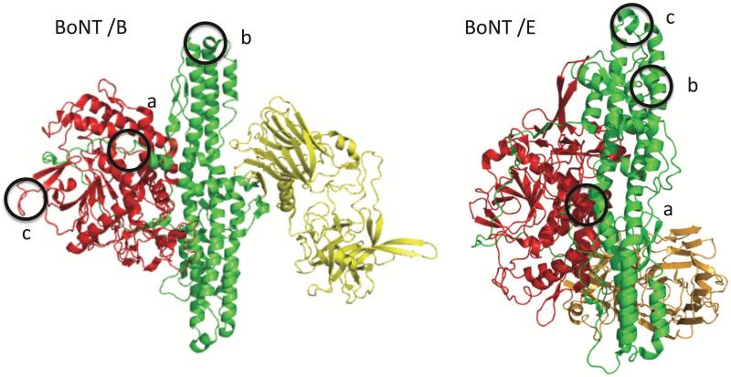
We next used error prone PCR to generate BoNT/B and BoNT/E LC-HN gene repertoires with random mutations and then cloned the mutant repertoires for display on the surface of yeast (21). Mutant yeast clones which had lost binding for each of the mAbs were generated by staining the mutant LC-HN yeast libraries with one of the mAbs and then collecting those yeast which did not bind the mAb by fluorescent activated cell sorting as we have previously described (21). LC-HN genes from yeast clones with reduced or absent mAb binding were sequenced and the location of mutations modeled on the X-ray crystal structure of BoNT/B (PDB accession code 1S0E) (28) or BoNT/E (PDB accession code 3FFZ) (29) to identify putative mAb epitopes. Single alanine mutations were then introduced into the wild type BoNT/B or BoNT/E LC-HN displayed on yeast to identify the amino acids important for binding. Consistent with the domain mapping, all mAbs bound either the BoNT LC, HN or a complex epitope requiring the presence of both the LC and HN (LC-HN) (Figure 1). Based on the knowledge of the fine epitopes, recombinant BoNT/B and BoNT/E LC-HN domains with multiple amino acid substitutions in solvent accessible residues at the identified epitopes were constructed and expressed in E. coli (Table 1). Each mutant LC-HN was purified at small scale to test for binding of each of the three serotype-specific mAbs in XOMA 3B or XOMA 3E by ELISA and Attana (Supplementary Figure 3). Based on the binding results, further mutations were added, or alternative amino acid substitutions were made to eliminate binding of two of the three mAbs before finalizing the construct. Based on these criteria, domains B-LC-HN-a, -b, and -c were specific for mAbs B-a, -b, and –c; and domains E-LC-HN-a, -b -c were specific for mAbs E-a, -b, and -c respectively (Table 1).
Figure 1. Epitopes of BoNT/B and BoNT/E mAbs.
Left panel: Ribbon cartoon of the X-ray crystal structure of BoNT/B secondary structure (PDB accession code 1S0E) showing the catalytic light chain (LC, red), heavy chain binding domain (HC, yellow) and translocation domain (HN, green). The epitopes of the three mAbs comprising XOMA 3B are circled and labeled a, b, c for each mAb respectively. Right panel: Ribbon cartoon of the X-ray crystal structure of BoNT/E secondary structure (PDB accession code 3FFZ) showing catalytic light chain (LC, red), heavy chain binding domain (HC, yellow) and translocation domain (HN, green). The epitopes of the three mAbs comprising XOMA 3E are circled and labeled a, b, c for each mAb respectively.
Characterization of purity of domains
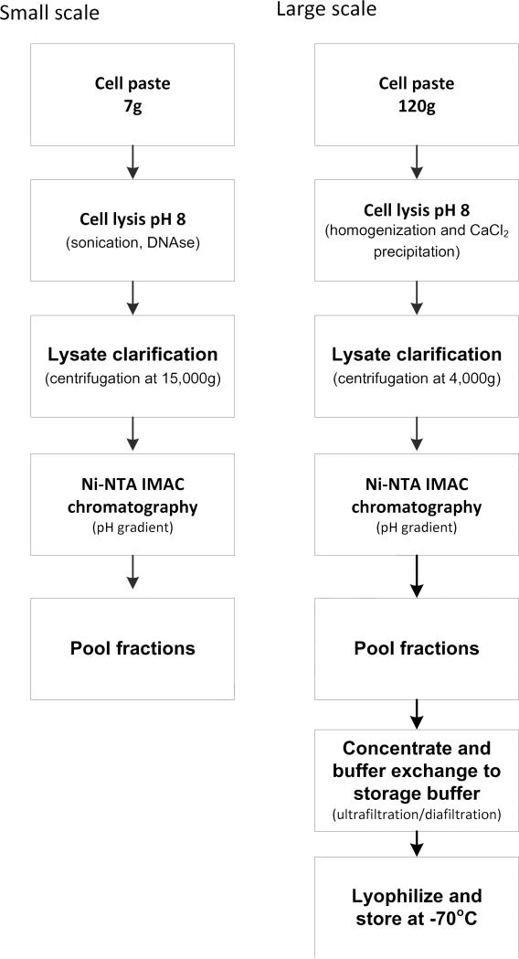
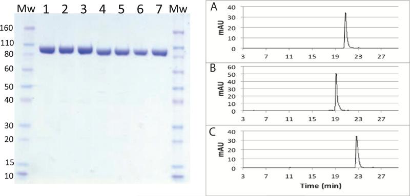
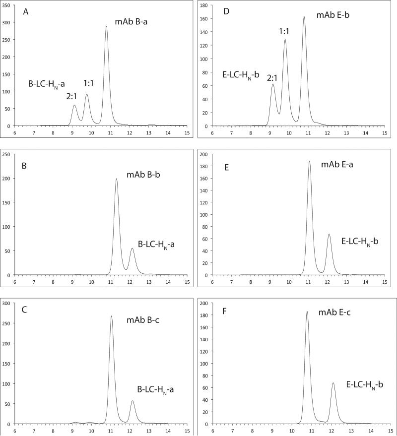
A suite of analytical methods was developed to characterize the purity, specificity, and binding activity and affinity of the domains, and these characteristics were used to define a set of specifications and action limits to ensure consistency across multiple batches. Using the purification scheme in Figure 2, a purity of greater than 90% by SDS-PAGE was achieved for each of the domains with yields of approximately 0.1 mg/gm to 5 mg/gm of E. coli paste for the domains (Figure 3, left panel). Rapid SE-HPLC assays were also developed for each domain that simultaneously determined domain product monomer content and impurities as well as antibody binding activity and specificity. When a single domain was assayed by SE-HPLC, a single peak was seen and the purity of the domain could be determined by the peak area in the elution profile (data not shown). When a single domain (for example domain B-LC-HN-a or domain E-LC-HN-b) was mixed with its corresponding binding mAb at an equimolar ratio, three peaks were seen (Figure 4A and 4D). The size of the first two peaks was determined by light scattering to be consistent with the size of either one IgG complexed with one domain or one IgG complexed with two domains. The third peak was unbound mAb. No free domain was seen suggesting all purified domain were immunoreactive. When the B-LC-HN-a or domain E-LC-HN-b were mixed with the other mAbs (B-b or B-c for domain B-LC-HN-a and mAb E-a or mAb E-c for domain E-LC-HN-b), separate mAb and domain peaks were seen providing evidence of binding specificity in solution (Figure 4B-C and 4E-F). Similar binding results were seen for B-LCHN-b, B-LCHN-c, E-LCHN-a and E-LCHN-c (data not shown). Besides the SE-HPLC, RP-HPLC was developed for each domain to assess purity and presence of breakdown products. RP-HPLC can separate domains that differ in disulfide bonding status that is not separable with SE-HPLC (19), therefore providing additional information to the purity of the domains. RP-HPLC results confirmed that all six domains were over 90% pure. Three examples of this assay for domains E LC-HN-a, -b, -c are shown in Figure 3, right panels A-C. Similar purity was seen for B-LCHN-a, -b, and -c (data not shown).
Fig.2. Scalable domain purification methods.
Small scale (left) and large-scale (right) purification schemes were developed to purify the six BoNT/B and BoNT/E LC-HN domains.
Figure 3. Analysis of the purity of BoNT/B and BoNT/E domains by SDS-PAGE and RP-HPLC.
Left panel; A Coomassie-stained SDS-PAGE gel was run to characterize the purity of domains. Molecular weight (MW) markers are at each side of the gel. Lane 1: B-LC-HN-a, lane 2: B-LC-HN-b, lane 3: B-LC-HN-c, lane 4: E-LC-HN-a, lane 5: E-LC-HN-b, lane 6: E-LC-HN-c, lane 7: A-LC-HN-mut. Right panel: Reverse phase high performance liquid chromatography (RP-HPLC) was used to assess domain purity and presence of aggregation. Shown here are three E-LC-HN domain examples: (A) E-LC-HN-a; (B) E-LC-HN-b; (C) E-LC-HN-c.
Figure 4. Analysis of the purity and immunoreactivity of BoNT/B and E domain by size exclusion high performance liquid chromatography (SE-HPLC).
(A-C). When each domain is mixed with its corresponding mAb, the domain peak disappears, indicating that the domain is near 100% immunoreactive. Both 1:1 complex and 2:1 complexed peaks are present. When the domain is mixed with other non-binding mAbs, no complex formation is observed, indicating binding specificity. Shown here are results for two domains: B-LC-HN-a and E-LC-HN-b. (A) B-LC-HN-a with mAb B-a; (B) B-LC-HN-a with mAb B-b; (C) B-LC-HN-a with mAb B-c; (D) E-LC-HN-b with mAb E-b; (E) E-LC-HN-b with mAb E-a; and (F) E-LC-HN-b with mAb E-c.
Characterization of affinity of domains
Flow fluorimetry in a KinExA (19, 24) was used to measure the affinity and binding kinetics of each of the XOMA 3B and XOMA 3E mAbs for their respective domains and was compared to the affinity of the mAbs for the BoNT B and E holotoxin (Table 1). The affinity of the mAbs to their respective domains was comparable to their binding affinity for the holotoxin. Each of the BoNT/B mAbs bound only to its specific domain and not to the other two domains by flow fluorimetry. Similarly, each of the BoNT/E mAbs bound only to its specific domain and not to the other two domains.
ELISA binding assays to specifically detect each mAb in XOMA 3B and 3E
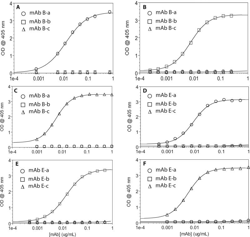
A direct coating ELISA was developed to quantitate each of the mAbs in XOMA 3B and 3E. In this assay, each domain was directly coated onto microtiter plates to measure the concentration of a single mAb in the three-mAb combinations for XOMA 3B and 3E. In this ELISA, each of the individual mAbs in XOMA 3B or 3E bound specifically to only their respective domains and not to the other domains (Figure 5A-F). Binding could be detected at a mAb concentration of 0.1 ng/mL, with saturation occurring at a mAb concentration of 0.1 ug/mL (Figure 5A-F).
Figure 5. Direct-coating ELISAs.
(A-C), Binding of mAbs B-a, B-b and B-c to plates coated with mAb specific domains B-LC-HN-a (A),B-LC-HN-b (B) or B-LC-HN-c (C). (D-F), Binding of mAbs E-a, E-b, and E-c to plates coated with mAb specific domains E-LC-HN-a (D), E-LC-HN-b (E), or E-LC-HN-c (F). Each mAb bound only to its respective domain, with no binding observed to the other domains.
Development of ELISA assays to detect the individual mAbs in a trivalent BoNT/A, B, and E antitoxin
BoNT/A, B, and E cause more than 95% of human botulism. Thus a single antitoxin that could neutralize all three BoNTs would be of high value. We previously reported the development of a combination of three mAbs that potently neutralize BoNT/A (XOMA 3AB) and the development of three domains that could measure the concentration of the three BoNT/A mAbs (19). To support development of the nine-antibody, trivalent BoNT A/B/E antitoxin, which combines the three mAbs in each of XOMA 3AB, 3B and 3E, we tested the binding specificity of the nine domains against the nine component mAbs. Seven out of the nine mAbs showed specific binding to their corresponding domain and no binding to the other eight domains (data not shown). Two domains, A-LC-HN (19) and B-LC-HN-b, bound more than 1 mAb. The domain A-LC-HN binds mAb B-b and mAb E-c in addition to mAb A-c (data not shown) and the domain B-LC-HN-b binds mAb E-c as well as mAb B-b. mAbs B-b and E-c bind an epitope at residues 750–758 of the HN (Figure 1) that is partially homologous in serotypes A, B and E.
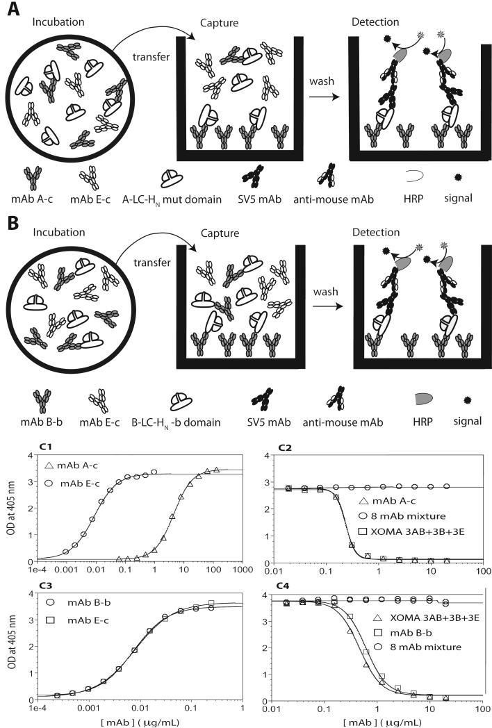
Knocking out the binding of mAbs B-b and E-c to the A-LC-HN domain appeared straightforward; the same mutations that knocked out binding of mAb B-b and E-c to the B-LCHN and E-LC-HN respectively were introduced into the A-LC-HN domain to create the domain A-LC-HN mut. The A-LC-HN mut domain showed no binding to mAb B-b or E-c by flow fluorimetry at concentrations up to 1 M (data not shown). In ELISA with the A-LC-HN mut coated on the plate, there was also no binding of mAb B-b, however mAb E-c did show binding with an approximately 2.5 log lower affinity than mAb A-c (Figure 6C1). Binding of mAb E-c in ELISA but not by flow fluorimetry is likely explained by the avid binding of the IgG to the immobilized domain on the plate, which cannot occur in the solution KD measured by flow fluorimetry. We have seen more than 1000 fold higher affinities for bivalent IgG binding compared to monovalent solution binding (30). Similarly, mAb E-c bound the B-LC-HN-b domain with a binding curve superimposable with mAb B-b (Figure 6C3) despite the fact that in solution mAb B-b binds to domain B-LC-HN-b with a KD of 61 pM while mAb E-c binds to domain B-LC-HN-b with a 500 fold lower KD (31 nM, Table 1).
Figure 6. Specific detection of mAbs A-c and B-b using an indirect capture ELISA.
Top panels (A and B): Cartoon of the indirect capture ELISA.
Incubation: A fixed amount of mAb specific domain and increasing amounts of mAbs are mixed in individual wells and incubated together until binding reaches equilibrium. Amount of mAb bound is a function of the mAb solution KD. Capture: Each reaction mixture is then transferred to a new well on the capture plate which has been coated with the domain specific mAb for capturing of the domain. Domains bound by a mAb binding the same epitope are not captured and are washed away. Detection: Captured domains are detected with anti-SV5 antibody binding the C-terminal SV5 epitope tag followed by anti-mouse Ab-HRP (A) A-LC-HN mut domain binds mAb A-c with high affinity and mAb E-c with low affinity in solution at a non-overlapping epitope. At the mAb concentrations studied, the affinity of mAb E-c for the A-LC-HN domain is too low for binding to occur during the incubation stage. On the capture plate coated with mAb A-c, only free A-LC-HN mut not bound by mAb E-c is captured, allowing quantitation of the mAb E-c concentration. (B). The B-LC-HN-b domain binds both mAb B-b and mAb E-c with overlapping epitopes, and with a 500-fold higher affinity for mAb B-b than to mAb E-c. At the mAb concentrations studied, the affinity of mAb E-c for the B-LC-HN-b domain is too low for binding to occur during the incubation stage. On the capture plate coated with mAb B-b, the free B-LC-HN-b is captured, while B-LC-HN-b bound by mAb B-b in solution is washed away, allowing quantitation of the mAb E-c concentration. HRP = horse radish peroxidase.
Bottom panel (C). Binding specificity of direct vs. indirect capture ELISAs. C1. Direct ELISA with a plate coated with domain A-LC-HN mut shows binding of mAb E-c at higher mAb concentrations than required for mAb A-c. C2 Indirect capture ELISA showed specificity of domain A-LC-HN-mut for only mAb A-c. mAb A-c was coated on the plate as capture antibody, A-LC-HN-mut was incubated with either mAb A-c, or a mixture of eight mAbs (mAbA-a + mAbA-b + XOMA 3B + XOMA 3E) or a mixture of nine mAbs (XOMA 3AB + XOMA 3B + XOMA 3E). C3. Direct ELISA with a plate coated with domain B-LC-HN-b cannot distinguish between mAb B-b and mAb E-c. C4. Indirect capture ELISAs showing specificity of domains B-LC-HN-b for only mAb B-b. mAb B-b was coated on the plate as capture antibody, B-LC-HN-b was incubated with mAb B-b, a mixture of eight mAbs (XOMA 3 AB and mAb B-a, -b and XOMA 3E) or a mixture of nine mAbs (XOMA 3AB + XOMA 3B + XOMA 3E).
To avoid the avidity effect, we developed an indirect capture ELISA assay. In this assay, samples of mAb and domain are incubated in solution to equilibrium, and then the free antigen remaining in solution is captured on an ELISA plate coated with the domain specific mAb (Figure 6A and 6B). Since the initial binding of mAbs takes place in solution, avidity is avoided and the mAb with the significantly lower affinity cannot bind at the concentrations studied.
In this assay format, domains A-LC-HN-mut and LC-HN B-b bound only to their respective mAbs (Figure 6C2 and 6C4). The A-LC-HN mut bound to mAb A-c but no binding was seen with a mixture of the other eight mAbs comprising XOMA 3AB, 3B, and 3E (Figure 6C2). In addition, the binding curve of the nine mAbs in XOMA 3AB, 3B, and 3E was superimposable to the curve from the single mAb A-c (Figure 6C2). For the domain LC-HN B-b, no binding was seen with the mixture of the other eight mAbs comprising XOMA 3AB, 3B, and 3E (Figure 6C4) and the binding curve of the nine mAbs mixture in XOMA 3AB, 3B, and 3E was the same as the curve from the single mAb B-b (Figure 6C4).
Discussion
Antibody combinations are in clinical development for a number of diseases (2-7). Such combinations pose challenges for development compared to individual mAbs, including the need to be able to measure the concentration and activity of each mAb independently when combined, both in vitro and in vivo. We previously showed that individual domains or subdomains of BoNT/A, a multidomain protein, could be independently expressed and purified at scale and used to develop binding assays that can specifically measure the concentration of the three BoNT/A mAbs in XOMA 3AB both in vitro and in vivo. In XOMA 3AB, each mAb bound a different epitope contained within either a unique folded domain of BoNT/A (the LC-HN) or within subdomains of the HC (HCC and HCN), increasing the likelihood that the epitopes could be successfully expressed and purified in a stable form (31). Here we show that even when mAb epitopes are located on the same domain, it is possible to generate mutant domains within the mAb epitopes that knock out binding of several mAbs but retain binding of one of the mAbs. Such mAb-specific domains were developed for the three BoNT/B and three BoNT/E mAbs in XOMA 3B and XOMA 3E respectively. These domains were used to develop highly specific and sensitive ELISAs that could be used for both characterizing the monoclonal and oligoclonal drug products during manufacturing and for monitoring their stability over time to ensure quality of the drug and its component mAbs.
Additional domain engineering and ELISA modification made it possible to develop assays capable of distinguishing and quantitating each of the nine mAbs in a mixture comprising XOMA 3AB, XOMA 3B, and XOMA 3E. For two of these ELISAs, cross reactivity was observed in the direct coated ELISA for one of the mAbs in the nine mAb combination due to avidity effects. By using a capture ELISA, it was possible to eliminate avidity and eliminate the cross reactivity for both of these assays. This work provides the analytical tools that would permit the development of a trivalent human BoNT/A, B, and E antitoxin for treating the three commonest types of human botulism. The type of antigen engineering described here should be broadly applicable for the generation of sensitive assays for quantitation of individual mAbs in an oligoclonal antibody cocktail that bind the same protein.
Supplementary Material
Acknowledgements
This work was partially supported by NIAID contract number HHSN266200600008C and HHSN272200800028C and NIAID Cooperative Agreement U01AI056493. We would also like to acknowledge Anwar Zaman for his help with purifying the domains, and Jacob Kung for help with ELISAs.
Abbreviations used
- A-LC-HN mut
serotype A botulinum neurotoxin LC-HN mutant domains
- ABTS
2,2-Azinobis [3-ethylbenzothiazoline-6-sulfonic acid]-diammonium salt
- KDa
kilodalton
- AUC
area under the concentration-time curve
- BoNT
Botulinum neurotoxin
- BoNT/A
BoNT serotype A
- BoNT/B
BoNT serotype B
- BoNT/B1
BoNT sub-serotype B1
- BoNT/E
BoNT serotype E
- BSA
bovine serum albumin
- Cmax
maximum concentration
- CV
column volumes
- Fab’2
fragment antigen binding
- Fc
fragment crystallizable
- IgG
immunoglobulin G
- IMAC
Ni-NTA immobilized metal affinity chromatography
- LD50
lethal dose 50%
- mAb
monoclonal antibody
- mAbB-a, -b, -c
antibody component of three mAb mixture that binds to BoNT/B
- mAbE-a, -b, -c
antibody component of three mAb mixture that binds to BoNT/E
- MRT
Mean residence time
- scFv
single chain variable fragment
- BoNT/A HC
C-terminal domain of the BoNT/A heavy chain
- BoNT/A HN
N-terminal domain of the BoNT/A heavy chain
- BoNT/A LC
BoNT/A light chain
- BoNT/B HC
C-terminal domain of the BoNT/B heavy chain
- BoNT/B HN
N-terminal domain of the BoNT/B heavy chain
- BoNT/B LC
BoNT/B light chain
- LC-HN
BoNT light chain plus N-terminal domain of the heavy chain
- BLC-HN-a, -b, -c
mutants of the BoNT light chain plus N-terminal domain of the heavy chain that binds specifically to the mAb B-a, -b, -c respectively
- E- LC-HN-a, -b, -c
mutants of the BoNT light chain plus N-terminal domain of the heavy chain that binds specifically to the mAbE-a, -b, -c respectively
- A-LC-HN mut
mutant LC-HN domains
- pM
picomolar
- fM
femtomolar
- ECL
electrochemiluminescence
- ELISA
Enzyme-linked immunosorbent assay
- FACS
fluorescent activated cell sorting
- HBST
HEPES, NaCl, Tween
- HPLC
high pressure liquid chromatography
- IU
International Unit
- IPTG
Isopropyl-β-D-thio-galactoside
- KD
equilibrium dissociation constant
- kon
association rate constant
- koff
dissociation rate constant
- MFI
mean fluorescent intensity
- MW
molecular weight
- PBS
phosphate buffered saline
- PBS-T
0.05% (v/v) Tween 20 in PBS
- RP-HPLC
reverse phase high pressure liquid chromatography
- QCM
Quartz crystal microbalance
- SD
standard deviation
- SDS-PAGE
sodium dodecyl sulfate PAGE
- SEC
Size-exclusion chromatography
- SE-HPLC
Size-exclusion high pressure liquid chromatography
- UV
ultraviolet
- XOMA 3AB
an equimolar mixture of three human/humanized mAbs binding non-overlapping epitopes on botulinum neurotoxin type A (BoNT/A)
- XOMA 3B
an equimolar mixture of three human/humanized mAbs binding non-overlapping epitopes on botulinum neurotoxin type B (BoNT/B)
- XOMA 3E
an equimolar mixture of three human/humanized mAbs binding non-overlapping epitopes on botulinum neurotoxin type E (BoNT/E)
Footnotes
Publisher's Disclaimer: This is a PDF file of an unedited manuscript that has been accepted for publication. As a service to our customers we are providing this early version of the manuscript. The manuscript will undergo copyediting, typesetting, and review of the resulting proof before it is published in its final citable form. Please note that during the production process errors may be discovered which could affect the content, and all legal disclaimers that apply to the journal pertain.
References
- 1.Nelson AL, Dhimolea E, Reichert JM. Development trends for human monoclonal antibody therapeutics. Nat Rev Drug Discov. 2010;9:767–774. doi: 10.1038/nrd3229. [DOI] [PubMed] [Google Scholar]
- 2.Logtenberg T. Antibody cocktails: next-generation biopharmaceuticals with improved potency. Trends Biotechnol. 2007;25:390–394. doi: 10.1016/j.tibtech.2007.07.005. [DOI] [PubMed] [Google Scholar]
- 3.Bakker AB, Python C, Kissling CJ, Pandya P, Marissen WE, Brink MF, Lagerwerf F, Worst S, van Corven E, Kostense S, Hartmann K, Weverling GJ, Uytdehaag F, Herzog C, Briggs DJ, Rupprecht CE, Grimaldi R, Goudsmit J. First administration to humans of a monoclonal antibody cocktail against rabies virus: safety, tolerability, and neutralizing activity. Vaccine. 2008;26:5922–5927. doi: 10.1016/j.vaccine.2008.08.050. [DOI] [PubMed] [Google Scholar]
- 4.Prabakaran M, Prabhu N, He F, Hongliang Q, Ho HT, Qiang J, Meng T, Goutama M, Kwang J. Combination therapy using chimeric monoclonal antibodies protects mice from lethal H5N1 infection and prevents formation of escape mutants. PLoS One. 2009;4:e5672. doi: 10.1371/journal.pone.0005672. [DOI] [PMC free article] [PubMed] [Google Scholar]
- 5.Chen Z, Moayeri M, Crown D, Emerson S, Gorshkova I, Schuck P, Leppla SH, Purcell RH. Novel chimpanzee/human monoclonal antibodies that neutralize anthrax lethal factor, and evidence for possible synergy with anti-protective antigen antibody. Infect Immun. 2009;77:3902–3908. doi: 10.1128/IAI.00200-09. [DOI] [PMC free article] [PubMed] [Google Scholar]
- 6.Mazor O, Ben-Arie E, Mechaly A, Rosenfeld R, Marcus H, Ordentlich A. Combination of Anthrax-Toxin Neutralizing Antibodies: Analysis of Synergism/Antagonism Effect. The Challenge of Highly Pathogenic Microorganisms. 2010:275–285. [Google Scholar]
- 7.Pedersen M, Jacobsen H, Koefoed K, Hey A, Pyke C, Haurum J, Kragh M. Sym004: a novel synergistic anti-epidermal growth factor receptor antibody mixture with superior anticancer efficacy. Cancer Research. 2010;70:588. doi: 10.1158/0008-5472.CAN-09-1417. [DOI] [PubMed] [Google Scholar]
- 8.Production and quality control of monoclonal antibodies. Directive 75/318/EEC3AB4a, COMMITTEE FOR MEDICINAL PRODUCTS FOR HUMAN USE. 1995 [Google Scholar]
- 9.Points to Consider in the Manufacture and Testing of Monoclonal Antibody Products for Human Use. doi: 10.1097/00002371-199705000-00007. [DOI] [PubMed] [Google Scholar]
- 10.Schenerman M, Sunday B, Kozlowski S, Webber K, Gazzano-Santoro H, Mire-Sluis A. CMC Strategy Forum Report: Analysis and structure characterization of monoclonal antibodies. BioProcess Int. 2004;2:42–49. [Google Scholar]
- 11.Arnon SS, Schechter R, Inglesby TV, Henderson DA, Bartlett JG, Ascher MS, Eitzen E, Fine AD, Hauer J, Layton M, Lillibridge S, Osterholm MT, O'Toole T, Parker G, Perl TM, Russell PK, Swerdlow DL, Tonat K. Botulinum toxin as a biological weapon: medical and public health management. JAMA. 2001;285:1059–1070. doi: 10.1001/jama.285.8.1059. [DOI] [PubMed] [Google Scholar]
- 12.St John R, Finlay B, Blair C. Bioterrorism in Canada: An economic assessment of prevention and postattack response. Can J Infect Dis. 2001;12:275–284. doi: 10.1155/2001/904148. [DOI] [PMC free article] [PubMed] [Google Scholar]
- 13.Black RE, Gunn RA. Hypersensitivity reactions associated with botulinal antitoxin. Am J Med. 1980;69:567–570. doi: 10.1016/0002-9343(80)90469-6. [DOI] [PubMed] [Google Scholar]
- 14.Hibbs RG, Weber JT, Corwin A, Allos BM, Abd el Rehim MS, Sharkawy SE, Sarn JE, McKee KT., Jr. Experience with the use of an investigational F(ab')2 heptavalent botulism immune globulin of equine origin during an outbreak of type E botulism in Egypt. Clin Infect Dis. 1996;23:337–340. doi: 10.1093/clinids/23.2.337. [DOI] [PubMed] [Google Scholar]
- 15.Nowakowski A, Wang C, Powers DB, Amersdorfer P, Smith TJ, Montgomery VA, Sheridan R, Blake R, Smith LA, Marks JD. Potent neutralization of botulinum neurotoxin by recombinant oligoclonal antibody. Proc Natl Acad Sci U S A. 2002;99:11346–11350. doi: 10.1073/pnas.172229899. [DOI] [PMC free article] [PubMed] [Google Scholar]
- 16.Blasco H, Lalmanach G, Godat E, Maurel MC, Canepa S, Belghazi M, Paintaud G, Degenne D, Chatelut E, Cartron G, Le Guellec C. Evaluation of a peptide ELISA for the detection of rituximab in serum. J Immunol Methods. 2007;325:127–139. doi: 10.1016/j.jim.2007.06.011. [DOI] [PubMed] [Google Scholar]
- 17.Damen CW, de Groot ER, Heij M, Boss DS, Schellens JH, Rosing H, Beijnen JH, Aarden LA. Development and validation of an enzyme-linked immunosorbent assay for the quantification of trastuzumab in human serum and plasma. Anal Biochem. 2009;391:114–120. doi: 10.1016/j.ab.2009.05.030. [DOI] [PubMed] [Google Scholar]
- 18.Tornetta M, Fisher D, O'Neil K, Geng D, Schantz A, Brigham-Burke M, Lombardo D, Fink D, Knight D, Sweet R, Tsui P. Isolation of human anti-idiotypic antibodies by phage display for clinical immune response assays. J Immunol Methods. 2007;328:34–44. doi: 10.1016/j.jim.2007.08.008. [DOI] [PubMed] [Google Scholar]
- 19.Meng Q, Li M, Silberg MA, Conrad F, Bettencourt J, To R, Huang C, Ma J, Meyer K, Shimizu R, Cao L, Tomic MT, Marks JD. Domain-based assays of individual antibody concentrations in an oligoclonal combination targeting a single protein. Analytical biochemistry. 2012;421:351–361. doi: 10.1016/j.ab.2011.09.030. [DOI] [PMC free article] [PubMed] [Google Scholar]
- 20.Gilsdorf J, Gul N, Smith LA. Expression, purification, and characterization of Clostridium botulinum type B light chain. Protein Expr Purif. 2006;46:256–267. doi: 10.1016/j.pep.2005.09.024. [DOI] [PubMed] [Google Scholar]
- 21.Levy R, Forsyth CM, LaPorte SL, Geren IN, Smith LA, Marks JD. Fine and domain-level epitope mapping of botulinum neurotoxin type A neutralizing antibodies by yeast surface display. J Mol Biol. 2007;365:196–210. doi: 10.1016/j.jmb.2006.09.084. [DOI] [PMC free article] [PubMed] [Google Scholar]
- 22.Blake RC, 2nd, Pavlov AR, Blake DA. Automated kinetic exclusion assays to quantify protein binding interactions in homogeneous solution. Analytical biochemistry. 1999;272:123–134. doi: 10.1006/abio.1999.4176. [DOI] [PubMed] [Google Scholar]
- 23.Lou J, Geren I, Garcia-Rodriguez C, Forsyth CM, Wen W, Knopp K, Brown J, Smith T, Smith LA, Marks JD. Affinity maturation of human botulinum neurotoxin antibodies by light chain shuffling via yeast mating. Protein Eng Des Sel. 2010;23:311–319. doi: 10.1093/protein/gzq001. [DOI] [PMC free article] [PubMed] [Google Scholar]
- 24.Razai A, Garcia-Rodriguez C, Lou J, Geren I, Forsyth CM, Robles Y, Tsai R, Smith TJ, Amith LA, Siegel RW, Feldhaus M, MArks JD. Molecular evolution of antibody affinity for sensitive detection of botulinum neurotoxin type A. J. Mol. Biol. 2005 doi: 10.1016/j.jmb.2005.06.003. [DOI] [PubMed] [Google Scholar]
- 25.Szutowicz A, Kobes RD, Orsulak PJ. Colorimetric assay for monoamine oxidase in tissues using peroxidase and 2,2′-azinodi(3-ethylbenzthiazoline-6-sulfonic acid) as chromogen. Anal Biochem. 1984;138:86–94. doi: 10.1016/0003-2697(84)90773-5. [DOI] [PubMed] [Google Scholar]
- 26.Engvall E. Enzyme immunoassay ELISA and EMIT. Methods Enzymol. 1980;70:419–439. doi: 10.1016/s0076-6879(80)70067-8. [DOI] [PubMed] [Google Scholar]
- 27.Al-Kaissi E, Mostratos A. Assessment of substrates for horseradish peroxidase in enzyme immunoassay. J Immunol Methods. 1983;58:127–132. doi: 10.1016/0022-1759(83)90269-7. [DOI] [PubMed] [Google Scholar]
- 28.Eswaramoorthy S, Kumaran D, Keller J, Swaminathan S. Role of metals in the biological activity of Clostridium botulinum neurotoxins. Biochemistry. 2004;43:2209–2216. doi: 10.1021/bi035844k. [DOI] [PubMed] [Google Scholar]
- 29.Kumaran D, Eswaramoorthy S, Furey W, Navaza J, Sax M, Swaminathan S. Domain organization in Clostridium botulinum neurotoxin type E is unique: its implication in faster translocation. J Mol Biol. 2009;386:233–245. doi: 10.1016/j.jmb.2008.12.027. [DOI] [PubMed] [Google Scholar]
- 30.Tang Y, Lou J, Alpaugh RK, Robinson MK, Marks JD, Weiner LM. Regulation of antibody-dependent cellular cytotoxicity by IgG intrinsic and apparent affinity for target antigen. J Immunol. 2007;179:2815–2823. doi: 10.4049/jimmunol.179.5.2815. [DOI] [PubMed] [Google Scholar]
- 31.Lacy DB, Tepp W, Cohen AC, DasGupta BR, Stevens RC. Crystal structure of botulinum neurotoxin type A and implications for toxicity. Nature Struct. Biol. 1998;5:898–902. doi: 10.1038/2338. [DOI] [PubMed] [Google Scholar]
Associated Data
This section collects any data citations, data availability statements, or supplementary materials included in this article.


