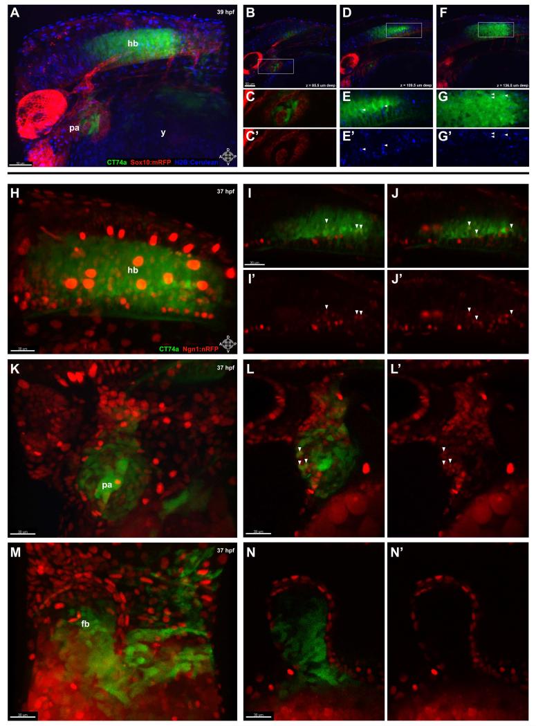
FIG. 2.
CT74a-Citrine is excluded from neural crest-derived tissue and is present in both neuronal and non-neuronal cells. A: In a confocal z-stack projection of a live CT74a-Citrine; Sox10:mRFP; H2B-Cerulean embryo at 39 hpf, CT74a-Citrine (green) is predominantly expressed in the posterior pharyngeal arches and hindbrain, with Sox10:mRFP+ cells (red) and H2B-Cerulean+ nuclei (blue) visible in multiple structures (z = 195 μm). B-D,F: Single 3 μm thick z-plane slices at 85.5 μm (B), 109.5 μm (D), and 136.5 μm (F) depth (0 μm = left side of embryo) through (A) illustrate the exclusionary expression of CT74a-Citrine and Sox10:mRFP, shown at higher zoom in (C,C’) in the pharyngeal arches (B, boxed area). E,E’,G,G’: Boxed areas from (D,F) show nuclear localization of CT74a-Citrine protein in a subset of hindbrain cells (arrowheads). H-N’: Confocal z-stack projections of a live CT74a-Citrine; Ngn1:nRFP embryo at 37 hpf illustrate neurons (red) expressing CT74a-Citrine in the hindbrain (H) and pharyngeal arches (K). Single 2 μm thick z-plane slices at two levels through the hindbrain (I-J’) and one level through the pharyngeal arches (L,L’) confirm the presence of CT74a-Citrine in neuronal nuclei (arrowheads) as well as in non-neuronal cells in both structures. In addition, non-neuronal expression is found in the fin bud mesenchyme as shown in a z-stack projection (M) and a single 2 μm thick z-plane slice (N,N’). CT74a-Citrine, green; Sox10:mRFP, red (A-C’,D,F); Ngn1:nRFP, red (H-L’); H2B:Cerulean, blue (A,B,D-G’). fb, fin bud; hb, hindbrain; pa, pharyngeal arches; y, yolk. Orientation arrows: A, anterior; P, posterior; D, dorsal; V, ventral. Scale bars: 50 μm (A,B,D,F); 30 μm (H-N’).

