Abstract
Macrophages are instrumental in the pathophysiology of liver ischemia/reperfusion injury (IRI). Although Nrf2 regulates macrophage-specific heme oxygenase-1 (HO-1) antioxidant defense, it remains unknown whether HO-1 induction might rescue macrophage Nrf2-dependent antiinflammatory functions. This study explores the mechanisms by which the Nrf2–HO-1 axis regulates sterile hepatic inflammation responses after adoptive transfer of ex vivo modified HO-1 overexpressing bone marrow–derived macrophages (BMMs). Livers in Nrf2-deficient mice preconditioned with Ad-HO-1 BMMs, but not Ad-β-Gal-BMMs, ameliorated liver IRI (at 6 h of reperfusion after 90 min of warm ischemia), evidenced by improved hepatocellular function (serum alanine aminotransferase [sALT] levels) and preserved hepatic architecture (Suzuki histological score). Treatment with Ad-HO-1 BMMs decreased neutrophil accumulation, proinflammatory mediators and hepatocellular necrosis/apoptosis in ischemic livers. Moreover, Ad-HO-1 transfection of Nrf2-deficient BMMs suppressed M1 (Nos2+) while promoting the M2 (Mrc-1/Arg-1+) phenotype. Unlike in controls, Ad-HO-1 BMMs increased the expression of Notch1, Hes1, phosphorylation of Stat3 and Akt in IR-stressed Nrf2-deficient livers as well as in lipopolysaccharide (LPS)-stimulated BMMs. Thus, adoptive transfer of ex vivo generated Ad-HO-1 BMMs rescued Nrf2-dependent antiinflammatory phenotype by promoting Notch1/Hes1/Stat3 signaling and reprogramming macrophages toward the M2 phenotype. These findings provide the rationale for a novel clinically attractive strategy to manage IR liver inflammation/damage.
INTRODUCTION
Ischemia/reperfusion injury (IRI) has been recognized as a major cause of organ dysfunction and failure after liver transplantation. Hepatic IR activates Kupffer cells and triggers reactive oxygen species (ROS) production, leading to the release of proinflammatory cytokines/chemokines and cell apoptosis (1). Macrophages are key mediators, which initiate toll-like receptor-4 (TLR4) response during the inflammatory phase of hepatic IRI (2). We have shown that disruption of TLR4 signaling regulated macrophage inflammation (2) and ameliorated IR-induced liver damage in vivo (3). Activation of HO-1 exerts potent antiinflammatory and antiapoptotic functions in transplant models of liver allograft rejection (4) and IRI (5–7). We and others have shown that HO-1 is expressed primarily by liver macrophages and Kupffer cells (8,9), whereas depletion of Kupffer cells diminished HO-1 expression and increased IR hepatic damage (10). HO-1 is one of the antioxidant response element (ARE)-dependent phase II detoxifying enzymes and antioxidants (11), which are regulated by the redox-sensitive transcription factor nuclear factor erythroid 2–related factor (Nrf2) (12). Activation of Nrf2 induces HO-1 expression (13), suggesting Nrf2 is essential for HO-1–mediated cytoprotection against IR liver injury.
Nrf2, a “master regulator” of cellular resistance to oxidative stress (14), controls both basal and induced expression of ARE-dependent genes to regulate cell defense systems (15). Under normal conditions, Nrf2 is kept in the cytoplasm by Keap1 (Kelch-like ECH-associated protein 1) and Cullin 3, which degrade Nrf2 by ubiquitination (16). Activation of Nrf2 leads to Nrf2 translocation into the nucleus and binding to the ARE, which initiates transcription of Nrf2 target genes (17). Increased Nrf2 activity promotes cell survival and mitigates oxidative stress–induced damage (18,19), whereas disruption of Nrf2 enhances hypoxia-induced acute lung and oxidative stress–mediated inflammation (20). Thus, Nrf2 might be functioning as a key regulator of inflammatory responses against oxidative stress–induced organ injury.
The present study was designed to explore the mechanisms by which the Nrf2–HO-1 axis regulates inflammation response in a mouse model of liver IRI. Our results show that adoptive transfer of ex vivo generated Ad-HO-1–modified bone marrow–derived macrophages (BMMs) ameliorated liver IRI in Nrf2-deficient mice by enhancing the Notch1/Hes1/Stat3 signaling and promoting the M2 phenotype. Thus, HO-1–modified macrophages can be applied to rescue Nrf2-dependent antiinflammatory functions.
MATERIALS AND METHODS
Animals
The Nrf2-deficient (Nrf2−/−; KO) male mice (C57BL/6) at 6–8 wks of age were used (provided by T Kensler, Johns Hopkins University, Baltimore, MD, USA). Animals were housed in the UCLA animal facility under specific pathogen-free conditions and received humane care according to the criteria outlined in the Guide for the Care and Use of Laboratory Animals (NIH publication 86-23, revised 1985) (21).
Mouse Liver IRI Model/Treatment
We used a mouse model of warm hepatic ischemia followed by reperfusion (22). The Nrf2 KO mice were injected with heparin (100 U/kg), and an atraumatic clip was used to interrupt the arterial/portal venous blood supply to the cephalad liver lobes. After 90 min, the clip was removed, and mice were sacrificed at 6 h of reperfusion. In the treatment groups, each mouse was injected via a portal vein with 5 × 10⁶ of transfected BMMs with Ad-HO-1/Ad-β-gal (2.5 × 10⁹ plaque-forming units [pfu]) or untreated BMMs 24 h before the onset of warm ischemia. After adoptive transfer of Ad-β-gal–transfected BMMs, the X-Gal staining in the ischemic liver lobes at 6 h of reperfusion was 40–50%, as compared with control livers (23).
Hepatocellular Function Assay
Serum alanine aminotransferase (sALT) levels, an indicator of hepatocellular injury, were measured by IDEXX Laboratories (Westbrook, ME, USA).
Histology/Immunohistochemistry
Liver sections (5 μm) were stained with hematoxylin and eosin (H&E). The severity of IRI was graded by using Suzuki criteria on a scale from 0 to 4 (24). Liver neutrophils were detected by using primary rat anti-mouse Ly6G monoclonal antibody (BD Biosciences, San Jose, CA, USA). The secondary, biotinylated goat anti-rat IgG (Vector, Burlingame, CA, USA) was incubated with immunoperoxidase (ABC Kit; Vector), according to the manufacturer’s instructions. Positive cells were counted blindly in 10 fields per section.
Myeloperoxidase Activity Assay
The presence of myeloperoxidase (MPO) was used as an index of hepatic neutrophil accumulation (25). The change in absorbance was measured spectrophotometrically at 655 nm. One unit of MPO activity was defined as the quantity of enzyme degrading 1 μmol peroxide per minute at 25°C per gram of tissue.
Terminal Deoxynucleotidyl Transferase dUTP Nick End Labeling (TUNEL) Assay
The Klenow-FragEL DNA Detection Kit (EMD Chemicals, Gibbstown, NJ, USA) was used to detect the DNA fragmentation characteristic of apoptosis in formalin-fixed paraffin-embedded liver sections (2). The results were scored semiquantitatively by averaging the number of apoptotic cells/field. Ten fields were evaluated per tissue sample.
Quantitative Real-Time Polymerase Chain Reaction Analysis
Quantitative real-time polymerase chain reaction (RT-PCR) was performed by using the DNA Engine with Chromo 4 Detector (MJ Research, Waltham, MA, USA). In a final reaction volume of 25 μL, the following were added: 1× SuperMix (Platinum SYBR Green qPCR Kit; Invitrogen/Life Technologies [Thermo Fisher Scientific Inc., Waltham, MA, USA]), cDNA and 10 μmol/L of each primer. Amplification conditions were as follows: 50°C (2 min) and 95°C (5 min), followed by 40 cycles of 95°C (15 s) and 60°C (30 s). Primers used to amplify specific gene fragments are shown in Supplementary Table S1.
Western Blot Assay
Proteins (30 μg/sample) from cell cultures or liver samples were subjected to 12% sodium dodecyl sulfate–polyacrylamide gel electrophoresis and transferred to nitrocellulose membrane (Bio-Rad, Hercules, CA, USA). Polyclonal rabbit anti-mouse HO-1 (StressGen Biotech, Victoria, BC, Canada) and monoclonal rabbit anti-mouse Notch1, Hes1, phos-Akt, Akt, phos-Stat3, Stat3, NF-κB, Bcl-2, Bcl-xL, cleaved caspase-3 and β-actin antibodies were used (Cell Signaling Technology, Danvers, MA, USA).
Cell Isolation/In Vitro Cultures
Murine BMMs were generated, as described (23). Bone marrow cells were removed from the femurs and tibias of Nrf2 KO mice and cultured in Dulbecco’s modified Eagle medium (DMEM) supplemented with 10% fetal calf serum (FCS) and 15% L929-conditioned medium. Cells (5 × 10⁶/well) cultured for 7 d were added with Ad-HO-1 or Ad-β-gal (at a multiplicity of infection [MOI] of 10) and incubated for 1 h. The medium was removed and changed to DMEM + 2% FCS. After 36–48 h, cells were harvested for in vivo adoptive transfer. The X-Gal staining by 48 h of transfection was >90% in BMMs, compared with control BMMs (23). For the in vitro assay, these transfected cells were incubated with lipopolysaccharide (LPS; 100 ng/mL) for an additional 6 h.
Statistical Analysis
Data are expressed as mean ± SD. Statistical comparisons were analyzed by the Student t test. Differences were statistically significant at p < 0.05.
All supplementary materials are available online at www.molmed.org.
RESULTS
Ad-HO-1 BMMs Ameliorate Liver IRI in Nrf2-Deficient Mice
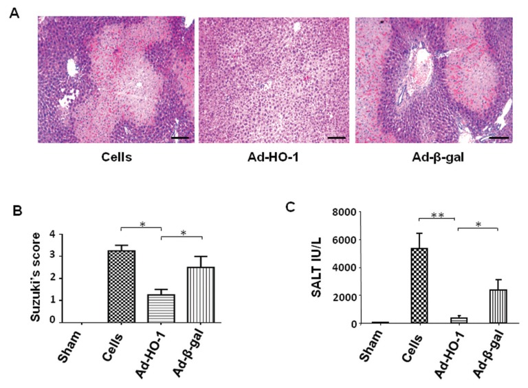
We used a mouse model of hepatic warm ischemia (90 min) followed by reperfusion (22). By 6 h of reperfusion, livers in the control Nrf2-deficient mice preconditioned (at d –1) with Ad-β-gal BMMs, or untreated BMMs, revealed significant edema, severe sinusoidal congestion, cytoplasmic vacuolization and extensive hepatocellular necrosis (30–50%; Figures 1A, B; Suzuki score 2.5 ± 0.45 and 3.15 ± 0.25, respectively). In contrast, livers in Nrf2-deficient hosts that received Ad-HO-1 BMMs had well-preserved hepatic architecture, with minimal sinusoidal congestion, without edema, vacuolization or necrosis (Figures 1A, B; Suzuki score 1.33 ± 0.29; p < 0.05).
Figure 1.
Adoptive transfer of Ad-HO-1 BMMs prevents IR-induced hepatocellular damage in Nrf2-deficient mice. BMMs transfected (5 × 10⁶) with Ad-HO-1 or Ad-β-gal (2.5 × 10⁹ pfu) or untreated BMMs were infused into Nrf2 KO mice (via portal vein) at 24 h before 90 min of ischemia. Animals were killed at 6 h of reperfusion. (A) Representative histological staining (H&E) of ischemic liver tissue. Scale bars indicate 50 μm. (B) Liver damage, evaluated by Suzuki histological score. *p < 0.05. (C) Hepatocellular function, assessed by sALT levels (IU/L). *p < 0.005; **p < 0.0005. Mean ± SD are shown (n = 4–6/group).
The hepatocellular function in Nrf2 KO mice, assessed by sALT levels (IU/L), improved after adoptive transfer of Ad-HO-1 BMMs, as compared with Ad-β-gal BMMs or untreated BMM groups (Figure 1C; 378.8 ± 84.8 versus 2,358 ± 366.9 and 5,321 ± 558.3, respectively; p < 0.005).
Ad-HO-1 BMMs Inhibit Neutrophil Activation in Nrf2-Deficient IR-Stressed Livers
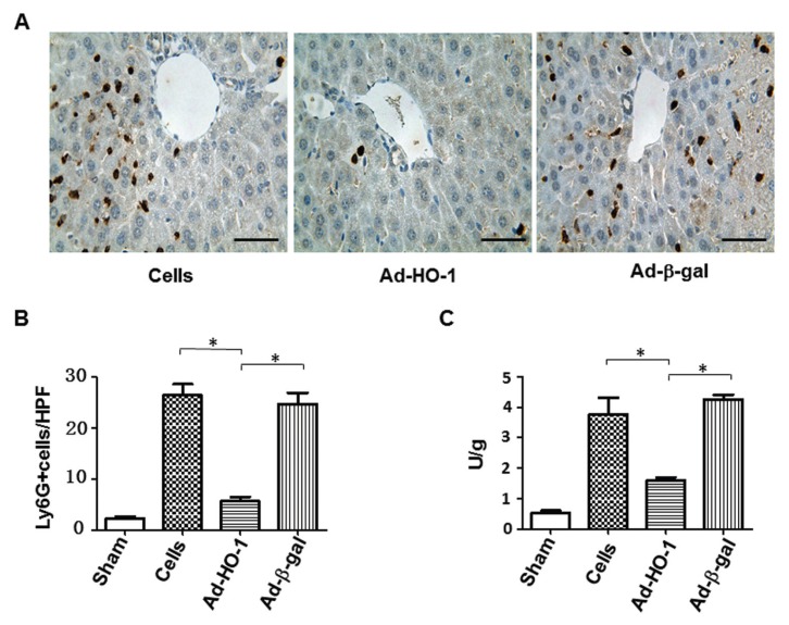
By 6 h of reperfusion after 90 min of ischemia, livers in Nrf2 KO mice pre-treated with Ad-HO-1 BMMs showed reduced infiltration by Ly6G+ neutrophils (Figures 2A, B; 5 ± 1.2) compared with Ad-β-gal (24.33 ± 1.39, p < 0.05) or BMMs (26.33 ± 1.53, p < 0.05). Furthermore, the MPO assay (Figure 2C), reflecting hepatic neutrophil activity (U/g), was decreased from 4.26 ± 0.37 in Ad-β-gal BMMs and 3.78 ± 0.3 in BMMs, to 1.45 ± 0.06 in the Ad-HO-1 BMMs group (p < 0.05).
Figure 2.
Adoptive transfer of Ad-HO-1 BMMs inhibits hepatic neutrophil activation in Nrf2-deficient mice (6 h of reperfusion after 90 min of ischemia). (A, B) Liver neutrophil accumulation by immuohistochemistry. Scale bars indicate 50 μm. (C) Hepatic neutrophil activity analyzed by MPO assay (U/g). *p < 0.05. Mean ± SD are shown (n = 4–6/group).
Ad-HO-1 BMMs Upregulate Notch Signaling in Nrf2-Deficient IR-Stressed Livers
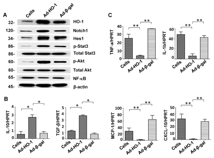
Notch signaling has been shown to regulate macrophage inflammatory responses (26). In our liver IRI model in Nrf2-deficient mice, infusion of Ad-HO-1 BMMs upregulated Western-assisted hepatic expression of Notch1, Hes1, p-Stat3 and p-Akt, yet downregulated NF-κB, as compared with Ad-β-gal or BMMs groups (Figure 3A). Furthermore, unlike in controls, Ad-HO-1 BMMs increased mRNA levels coding for interleukin (IL)-10 and transforming growth factor (TGF)-β (Figure 3B, p < 0.05), while decreasing tumor necrosis factor (TNF)-α, IL-1β, monocyte chemoattractant protein (MCP)-1 and C-X-C motif chemokine 10 (CXCL10) expression (Figure 3C, p < 0.05).
Figure 3.
Adoptive transfer of Ad-HO-1 BMMs upregulates Notch signaling and regulates IR inflammation in Nrf2-deficient livers (6 h of reperfusion after 90 min of ischemia). (A) Western blot analysis of hepatic HO-1, Notch1, Hes1, p-Stat3, total Stat3, p-Akt, total Akt and NF-κB expression in Nrf2 KO mice pretreated with Ad-HO-1/Ad-β-gal–transfected BMMs or untreated BMMs. Anti–β-actin antibody was used to assure equal protein amounts between samples. Data are representative of three experiments. (B, C) Quantitative real-time PCR-assisted detection of cytokine/chemokine programs in ischemic livers. *p < 0.05; **p < 0.005. Mean ± SD; n = 3–4/group.
Ad-HO-1 BMMs Prevent Apoptosis in Nrf2-Deficient IR-Stressed Livers
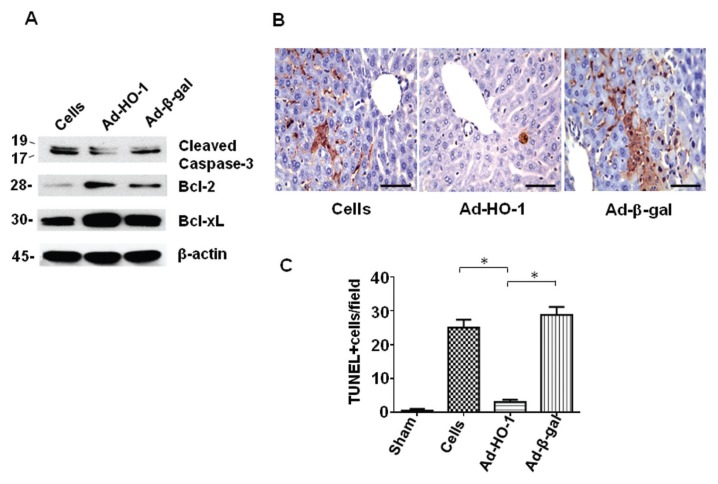
Western-assisted analysis of ischemic tissue has shown that unlike in controls, Ad-HO-1 BMMs decreased the expression of cleaved caspase-3 while increasing Bcl-2 and Bcl-xL levels (Figure 4A). Furthermore, while Nrf2-deficient livers pretreated with Ad-β-gal BMMs or untreated BMMs showed increased frequency of TUNEL+ cells (Figures 4B, C; 28.33 ± 2.1 and 24.67 ± 2.1), the number of hepatic TUNEL+ cells markedly dropped after infusion of Ad-HO-1 BMMs (2 ± 0.4, p < 0.001).
Figure 4.
Adoptive transfer of Ad-HO-1 BMMs ameliorates hepatocellular apoptosis in Nrf2-deficient mice (6 h of reperfusion after 90 min of ischemia). (A) Representative (n = 3) Western blot analysis of hepatic cleaved caspase-3, Bcl-2 and Bcl-xL after infusion of Ad-HO-1– or Ad-β-gal–transfected BMMs or untreated BMMs. (B) Representative (n = 3) TUNEL-assisted detection of apoptosis in ischemic liver tissue. Scale bars indicate 50 μm. (C) TUNEL+ cells were scored semiquantitatively by averaging the number of apoptotic cells (mean ± SD) per field. A minimum of 10 fields was evaluated/sample. *p < 0.001. There were 4–6 mice/group.
Nrf2–HO-1 Axis Promotes Notch1/Hes1/Stat3 Signaling to Regulate Inflammatory Responses In Vitro
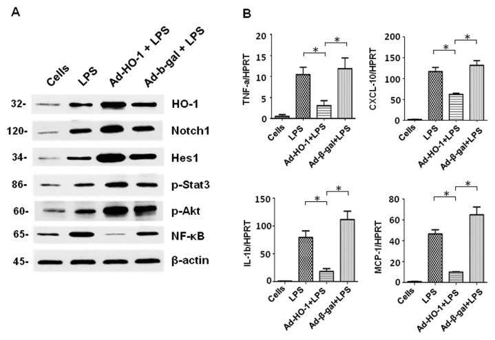
Our in vivo data suggest that HO-1 overexpression may rescue Nrf2-dependent antiinflammatory functions, which in turn diminishes liver IR inflammation. We then used an LPS-stimulated BMM culture system to test a hypothesis that the Nrf2–HO-1 axis regulates inflammatory response through the Notch1/Hes1/Stat3 signaling pathway. Indeed, Ad-HO-1 transfection upregulated HO-1, Notch1, Hes1, p-Stat3 and p-Akt but depressed NF-κB in Nrf2-deficient BMMs (Figure 5A), compared with controls. Consistent with these data, Ad-HO-1 transfection consistently reduced mRNA levels coding for TNF-α/IL-1β and MCP-1/CXCL10 in Nrf2-deficient BMMs, compared with Ad-β-gal BMMs or untreated BMMs (Figure 5B).
Figure 5.
Nrf2–HO-1 axis promotes Notch1/Hes1/Stat3 signaling to regulate inflammatory response in vitro. Nrf2-deficient BMMs were transfected with Ad-HO-1 or Ad-β-gal (at an MOI of 10). After 36–48 h, cells were incubated with LPS (100 ng/mL) for 6 h. (A) Proteins isolated from Ad-HO-1/Ad-β-gal transfected or untreated BMMs were stained for HO-1, Notch1, Hes1, phos-Akt, phos-Stat3 and NF-κB. Anti–β-actin antibody was used to assure equal protein amounts between samples. Data are representative of three experiments. (B) Cytokine/chemokine programs analyzed by quantitative real-time PCR. *p < 0.05. Mean ± SD; n = 3–4/group.
Nrf2–HO-1 Axis–Mediated Notch1/Hes1/Stat3 Signaling Regulates Macrophage Differentiation and Function In Vitro
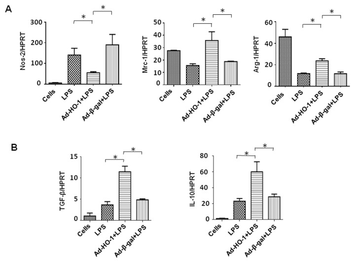
To test whether HO-1 might regulate alternative macrophage activation/function in Nrf2-deficient BMMs, we focused on the M1 (Nos2+) and M2 (Mrc-1+ and Arg-1+) macrophage phenotype. Indeed, Ad-HO-1 transfection in Nrf2-deficient BMMs inhibited Nos2+ while enhancing Mrc-1 and Arg-1+ expression (Figure 6A). Consistent with these data, the expression of TGF-β and IL-10 was consistently increased in Ad-HO-1–transfected BMMs compared with controls (Figure 6B).
Figure 6.
Nrf2–HO-1 axis regulates macrophage differentiation and function in vitro. Nrf2-deficient BMMs were transfected with Ad-HO-1 or Ad-β-gal (at an MOI of 10). After 36–48 h, cells were incubated with LPS (100 ng/mL) for an additional 6 h. Quantitative RT-PCR assisted analysis of macrophage: (A) phenotype (Nos2+, Mrc-1/Arg-1+); (B) TGF-β/IL-10 levels. *p < 0.05. Mean ± SD; n = 3–4/group.
DISCUSSION
Nrf2 is a key transcription factor that regulates oxidative stress and inflammation through activation of its targeting genes encoding antioxidant and detoxifying molecules (27). In this study, adoptive transfer of ex vivo genetically modified HO-1–overexpressing BMMs ameliorated liver IRI in Nrf2 KO mice, suggesting HO-1 can successfully impose an antiinflammatory phenotype in Nrf2-deficient IR-stressed livers.
The major mechanism of Nrf2-dependent cellular defense against oxidative stress is activation of Nrf2-ARE signaling (28). Many antioxidative genes involved in cellular protection are under the transcriptional control of Nrf2-ARE (29). Nrf2 promotes cell survival by binding ARE in the promoter of downstream target genes that induce antioxidant molecules and scavenging of ROS. Indeed, activation of Nrf2 induces HO-1 expression to regulate oxidative stress while inactivating the transcriptional repressor Bach1 (13,30). Increasing cellular levels of HO-1 enhances its antiinflammatory effects in macrophages (31). The ability of Ad-HO-1 BMMs to impose cytoprotection in IRI-susceptible Nrf2-deficient livers, as shown in the current study, underlines the regulatory function of the Nrf2–HO-1 axis in sterile-type inflammation response, consistent with our previous findings that Nrf2 mediates its cytoprotection by inducing HO-1 (32).
Because Nrf2 activity is required to counteract the oxidative stress via induction of HO-1 in the cell defense system (33), the regulatory mechanism of Nrf2–HO-1 axis may involve multiple intercellular signaling pathways. We found that adoptive transfer of Ad-HO-1 BMMs into Nrf2 KO mice activated Notch signaling, a highly conserved pathway that controls cell fate, including cell growth, differentiation and survival (34). Although IR-triggered injury upregulated Notch1 expression in our study, HO-1 induction further enhanced Notch signaling after hepatic IR (Supplementary Figure S1). Indeed, activation of Notch signaling has been shown to increase hepatocyte DNA synthesis and cell survival, whereas disruption of Notch suppressed hepatocyte proliferation during liver regeneration after partial hepatectomy (35). Moreover, blocking Notch increased ROS production and aggravated IR-liver damage. In contrast, enhancing Notch activation reduced intracellular ROS and protected hepatocytes from IR-triggered apoptosis through the activation of STAT3 (36). Consistent with Notch-mediated cytoprotection, our study has revealed that Nrf2 deficiency increased IR-triggered liver damage (Supplementary Figure S2). Overexpression of HO-1 after adoptive transfer of genetically modified BMMs into Nrf2-deficient livers upregulated the expression of Hes1, a Notch downstream effector molecule crucial for the regulation of cell cycle and survival (37,38). Indeed, as a transcriptional regulator, Hes1 has been shown to control differentiation/proliferation of neuronal, endocrine and T-lymphocyte progenitors during cell development (39–41). Induction of Hes1 increases cell survival by directly controlling the activity of transcriptional repression of a cyclin-dependent kinase inhibitor p27Kip1 (42). Thus, Nrf2–HO-1 axis promotes Notch1/Hes1 signaling, which contributes to IR-hepatocellular protection.
Adoptive transfer of Ad-HO-1 BMMs induced Akt activation and enhanced antiapoptotic gene expression in Nrf2-deficient livers, accompanied by decreased frequency of apoptotic cells. In marked contrast, Nrf2 deficiency itself without infusion of Ad-HO-1 BMMs increased hepatocyte apoptosis in parallel with depressed Akt phosphorylation. Indeed, Akt promotes cell survival by phosphorylating Bcl-2/Bcl-xL–associated death promoter (BAD), a pro-apoptotic protein of the Bcl-2 family, and inhibiting caspase-mediated cell death (43). Moreover, Akt promotes cell survival in a variety of apoptotic paradigms (44,45). Activation of Akt inhibits cell apoptosis by reducing apoptosis signal-regulating kinase 1 (ASK1) activity (46). Consistent with our previous findings that Nrf2 activation induces HO-1 to promote phosphatidylinositol 3-kinase (PI3K)/Akt in hepatocyte protection (32), our current results support the cytoprotective role of the Nrf2–HO-1 axis in IR-induced liver apoptosis.
We have shown that macrophages may serve as a cellular delivery system for HO-1 gene transfer in the ischemic livers (23). Indeed, adoptive transfer of HO-1–modified BMMs markedly increased HO-1 mRNA and protein expression levels in ischemic livers (23), suggesting the macrophage–HO-1 delivery system functions as a therapeutic targeting gene in IR-stressed liver. However, because macrophages are pivotal for IR-liver inflammatory injury, the question arises as to whether the Nrf2–HO-1 axis can regulate macrophage activation and induce an alternative macro phage polarization in our model? We analyzed the molecular mechanisms by which Nrf2–HO-1 axis may regulate Notch1/Hes1/Stat3 signaling upon macrophage polarization during inflammatory response in vitro. First, we found that Nrf2-deficient BMMs transfected with Ad-HO-1 showed increased expression of HO-1, Notch1 and Hes1, as well as enhanced phosphorylation of Stat3, critical for macrophage differentiation and function (47). Indeed, activation of Notch signaling, which protects hepatocytes against hepatic IR, is required for Hes-dependent Stat3 activation (36), whereas Hes can bind Stat3 to regulate cell differentiation (48). Here, we show that induction of Hes1 enhanced Stat3 phosphorylation, leading to selective inhibition of the M1 phenotype (Nos2+) while simultaneously increasing the M2 phenotype (Mrc-1/Arg-1+), along with the antiinflammatory (TGF-β/IL-10) cytokine program in Ad-HO-1–transfected BMMs. In contrast, Nrf2 deficiency in LPS-stimulated control BMMs increased Nos2+ while decreasing Mrc-1+/Arg-1+ and the antiinflammatory cytokine program. Consistent with our in vivo findings that infusion of Ad-HO-1 BMMs increased antiinflammatory phenotype to limit IR-induced inflammation in Nrf2-deficient livers, our results are consistent with the key regulatory role of Nrf2–HO-1 axis in Notch1/Hes1/Stat3 signaling to promote alternative macrophage activation toward an antiinflammatory M2 phenotype during hepatic IRI.
CONCLUSION
Adoptive transfer of Ad-HO-1 BMMs rescued Nrf2-dependent antiinflammatory phenotype in vivo and in vitro. The Nrf2–HO-1 axis promoted Notch1/Hes1/Stat3 signaling to regulate macrophage activation, enhance antiinflammatory cytokine program and inhibit liver IRI. The present data, in agreement with our recent report (23), confirm the validity of a novel investigative tool in which native macrophages can be transfected ex vivo with cytoprotective HO-1 and then infused, if needed, to prospective recipients to mitigate IR-mediated inflammation, such as during liver transplantation, resection or trauma. Moreover, as decreasing donor organ quality represents one of the most challenging problems, our findings provide the rationale for a new and clinically attractive gene targeted strategy to “rejuvenate” extended criteria suboptimal donor livers and thus improve their function in transplant recipients.
Supplemental Data
Figure 7.
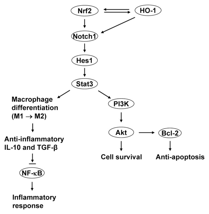
Scheme of signaling pathways by which the Nrf2–HO-1 axis may regulate inflammatory responses in liver IRI. HO-1 rescues Nrf2-dependent antiinflammatory functions by activating Notch1/Hes1/Stat3 and reprogramming macrophages toward the M2 phenotype. This step, in turn, upregulates antiinflammatory IL-10/TGF-β, leading to inhibition of NF-κB activation and inflammatory responses in IR-stressed livers. Furthermore, HO-1–mediated Stat3 activation promotes PI3K/Akt signaling to increase cell survival and enhance antiapoptotic activity, which may also exert hepatoprotection in IR-stressed livers.
ACKNOWLEDGMENTS
This work was supported by National Institutes of Health Grant DK 062357 and the W.M. Keck, Diann Kim and Dumont Research Foundations.
Footnotes
Online address: http://www.molmed.org
DISCLOSURE
The authors declare that they have no competing interests as defined by Molecular Medicine, or other interests that might be perceived to influence the results and discussion reported in this paper.
REFERENCES
- 1.Zhai Y, Busuttil RW, Kupiec-Weglinski JW. Liver ischemia and reperfusion injury: new insights into mechanisms of innate-adaptive immune-mediated tissue inflammation. Am J Transplant. 2011;11:1563–9. doi: 10.1111/j.1600-6143.2011.03579.x. [DOI] [PMC free article] [PubMed] [Google Scholar]
- 2.Ke B, et al. HO-1-STAT3 axis in mouse liver ischemia/reperfusion injury: regulation of TLR4 innate responses through PI3K/PTEN signaling. J Hepatol. 2012;56:359–66. doi: 10.1016/j.jhep.2011.05.023. [DOI] [PMC free article] [PubMed] [Google Scholar]
- 3.Zhai Y, et al. Cutting edge: TLR4 activation mediates liver ischemia/reperfusion inflammatory response via IFN regulatory factor 3-dependent MyD88-independent pathway. J Immunol. 2004;173:7115–9. doi: 10.4049/jimmunol.173.12.7115. [DOI] [PubMed] [Google Scholar]
- 4.Ke B, et al. Heme oxygenase 1 gene transfer prevents CD95/Fas ligand-mediated apoptosis and improves liver allograft survival via carbon monoxide signaling pathway. Hum Gene Ther. 2002;13:1189–99. doi: 10.1089/104303402320138970. [DOI] [PubMed] [Google Scholar]
- 5.Amersi F, et al. Upregulation of heme oxygenase-1 protects genetically fat Zucker rat livers from ischemia/reperfusion injury. J Clin Invest. 1999;104:1631–9. doi: 10.1172/JCI7903. [DOI] [PMC free article] [PubMed] [Google Scholar]
- 6.Ke B, et al. Cytoprotective and antiapoptotic effects of IL-13 in hepatic cold ischemia/reperfusion injury are heme oxygenase-1 dependent. Am J Transplant. 2003;3:1076–82. doi: 10.1034/j.1600-6143.2003.00147.x. [DOI] [PubMed] [Google Scholar]
- 7.Ke B, et al. Viral interleukin-10 gene transfer prevents liver ischemia-reperfusion injury: Toll-like receptor-4 and heme oxygenase-1 signaling in innate and adaptive immunity. Hum Gene Ther. 2007;18:355–66. doi: 10.1089/hum.2007.181. [DOI] [PubMed] [Google Scholar]
- 8.Kato H, et al. Heme oxygenase-1 overexpression protects rat livers from ischemia/reperfusion injury with extended cold preservation. Am J Transplant. 2001;1:121–8. [PubMed] [Google Scholar]
- 9.Patel A, et al. Early stress protein gene expression in a human model of ischemic preconditioning. Transplantation. 2004;78:1479–87. doi: 10.1097/01.tp.0000144182.27897.1e. [DOI] [PubMed] [Google Scholar]
- 10.Devey L, et al. Tissue-resident macrophages protect the liver from ischemia reperfusion injury via a heme oxygenase-1-dependent mechanism. Mol Ther. 2009;17:65–72. doi: 10.1038/mt.2008.237. [DOI] [PMC free article] [PubMed] [Google Scholar]
- 11.Li N, et al. Induction of heme oxygenase-1 expression in macrophages by diesel exhaust particle chemicals and quinones via the antioxidant-responsive element. J Immunol. 2000;165:3393–401. doi: 10.4049/jimmunol.165.6.3393. [DOI] [PubMed] [Google Scholar]
- 12.Itoh K, et al. An Nrf2/small Maf heterodimer mediates the induction of phase II detoxifying enzyme genes through antioxidant response elements. Biochem Biophys Res Commun. 1997;236:313–22. doi: 10.1006/bbrc.1997.6943. [DOI] [PubMed] [Google Scholar]
- 13.Reichard JF, Motz GT, Puga A. Heme oxygenase-1 induction by NRF2 requires inactivation of the transcriptional repressor BACH1. Nucleic Acids Res. 2007;35:7074–86. doi: 10.1093/nar/gkm638. [DOI] [PMC free article] [PubMed] [Google Scholar]
- 14.Ma Q. Role of nrf2 in oxidative stress and toxicity. Annu Rev Pharmacol Toxicol. 2013;53:401–26. doi: 10.1146/annurev-pharmtox-011112-140320. [DOI] [PMC free article] [PubMed] [Google Scholar]
- 15.Nioi P, McMahon M, Itoh K, Yamamoto M, Hayes JD. Identification of a novel Nrf2-regulated antioxidant response element (ARE) in the mouse NAD(P)H:quinone oxidoreductase 1 gene: reassessment of the ARE consensus sequence. Biochem J. 2003;374:337–48. doi: 10.1042/BJ20030754. [DOI] [PMC free article] [PubMed] [Google Scholar]
- 16.McMahon M, Itoh K, Yamamoto M, Hayes JD. Keap1-dependent proteasomal degradation of transcription factor Nrf2 contributes to the negative regulation of antioxidant response element-driven gene expression. J Biol Chem. 2003;278:21592–600. doi: 10.1074/jbc.M300931200. [DOI] [PubMed] [Google Scholar]
- 17.Nguyen T, Nioi P, Pickett CB. The Nrf2-antioxidant response element signaling pathway and its activation by oxidative stress. J Biol Chem. 2009;284:13291–5. doi: 10.1074/jbc.R900010200. [DOI] [PMC free article] [PubMed] [Google Scholar]
- 18.Hagemann JH, Thomasova D, Mulay SR, Anders HJ. Nrf2 signalling promotes ex vivo tubular epithelial cell survival and regeneration via murine double minute (MDM)-2. Nephrol Dial Transplant. 2013;28:2028–37. doi: 10.1093/ndt/gft037. [DOI] [PubMed] [Google Scholar]
- 19.Cho HY, et al. Role of NRF2 in protection against hyperoxic lung injury in mice. Am J Respir Cell Mol Biol. 2002;26:175–82. doi: 10.1165/ajrcmb.26.2.4501. [DOI] [PubMed] [Google Scholar]
- 20.Jin W, et al. Genetic ablation of Nrf2 enhances susceptibility to acute lung injury after traumatic brain injury in mice. Exp Biol Med (Maywood) 2009;234:181–9. doi: 10.3181/0807-RM-232. [DOI] [PubMed] [Google Scholar]
- 21.Institute of Laboratory Animal Resources (U.S.), Committee on Care and Use of Laboratory Animals. Guide for the Care and Use of Laboratory Animals. Bethesda (MD): NIH; 1985. p. 83. Rev 1985. (NIH publication; no. 85-23) [Google Scholar]
- 22.Shen XD, et al. CD154-CD40 T-cell costimulation pathway is required in the mechanism of hepatic ischemia/reperfusion injury, and its blockade facilitates and depends on heme oxygenase-1 mediated cytoprotection. Transplantation. 2002;74:315–9. doi: 10.1097/00007890-200208150-00005. [DOI] [PubMed] [Google Scholar]
- 23.Ke B, et al. Adoptive transfer of ex vivo HO-1 modified bone marrow-derived macrophages prevents liver ischemia and reperfusion injury. Mol Ther. 2010;18:1019–25. doi: 10.1038/mt.2009.285. [DOI] [PMC free article] [PubMed] [Google Scholar]
- 24.Suzuki S, Toledo-Pereyra LH, Rodriguez FJ, Cejalvo D. Neutrophil infiltration as an important factor in liver ischemia and reperfusion injury: modulating effects of FK506 and cyclosporine. Transplantation. 1993;55:1265–72. doi: 10.1097/00007890-199306000-00011. [DOI] [PubMed] [Google Scholar]
- 25.Mullane KM, Kraemer R, Smith B. Myeloperoxidase activity as a quantitative assessment of neutrophil infiltration into ischemic myocardium. J Pharmacol Methods. 1985;14:157–67. doi: 10.1016/0160-5402(85)90029-4. [DOI] [PubMed] [Google Scholar]
- 26.Zhang Q, et al. Notch signal suppresses Toll-like receptor-triggered inflammatory responses in macrophages by inhibiting extracellular signal-egulated kinase 1/2-mediated nuclear factor kappaB activation. J Biol Chem. 2012;287:6208–17. doi: 10.1074/jbc.M111.310375. [DOI] [PMC free article] [PubMed] [Google Scholar]
- 27.Klaassen CD, Reisman SA. Nrf2 the rescue: effects of the antioxidative/electrophilic response on the liver. Toxicol Appl Pharmacol. 2010;244:57–65. doi: 10.1016/j.taap.2010.01.013. [DOI] [PMC free article] [PubMed] [Google Scholar]
- 28.Johnson JA, et al. The Nrf2-ARE pathway: an indicator and modulator of oxidative stress in neurodegeneration. Ann N Y Acad Sci. 2008;1147:61–9. doi: 10.1196/annals.1427.036. [DOI] [PMC free article] [PubMed] [Google Scholar]
- 29.Zhang Y, Gordon GB. A strategy for cancer prevention: stimulation of the Nrf2-ARE signaling pathway. Mol Cancer Ther. 2004;3:885–93. [PubMed] [Google Scholar]
- 30.Shan Y, Lambrecht RW, Donohue SE, Bonkovsky HL. Role of Bach1 and Nrf2 in up-regulation of the heme oxygenase-1 gene by cobalt protoporphyrin. FASEB J. 2006;20:2651–3. doi: 10.1096/fj.06-6346fje. [DOI] [PubMed] [Google Scholar]
- 31.Lee TS, Chau LY. Heme oxygenase-1 mediates the anti-inflammatory effect of interleukin-10 in mice. Nat Med. 2002;8:240–6. doi: 10.1038/nm0302-240. [DOI] [PubMed] [Google Scholar]
- 32.Ke B, et al. KEAP1-NRF2 complex in ischemia-induced hepatocellular damage of mouse liver transplants. J Hepatol. 2013;59:1200–7. doi: 10.1016/j.jhep.2013.07.016. [DOI] [PMC free article] [PubMed] [Google Scholar]
- 33.Rushworth SA, MacEwan DJ, O’Connell MA. Lipopolysaccharide-induced expression of NAD(P)H:quinone oxidoreductase 1 and heme oxygenase-1 protects against excessive inflammatory responses in human monocytes. J Immunol. 2008;181:6730–7. doi: 10.4049/jimmunol.181.10.6730. [DOI] [PMC free article] [PubMed] [Google Scholar]
- 34.Artavanis-Tsakonas S, Rand MD, Lake RJ. Notch signaling: cell fate control and signal integration in development. Science. 1999;284:770–6. doi: 10.1126/science.284.5415.770. [DOI] [PubMed] [Google Scholar]
- 35.Kohler C, et al. Expression of Notch-1 and its ligand Jagged-1 in rat liver during liver regeneration. Hepatology. 2004;39:1056–65. doi: 10.1002/hep.20156. [DOI] [PMC free article] [PubMed] [Google Scholar]
- 36.Yu HC, et al. Canonical notch pathway protects hepatocytes from ischemia/reperfusion injury in mice by repressing reactive oxygen species production through JAK2/STAT3 signaling. Hepatology. 2011;54:979–88. doi: 10.1002/hep.24469. [DOI] [PubMed] [Google Scholar]
- 37.Monahan P, Rybak S, Raetzman LT. The notch target gene HES1 regulates cell cycle inhibitor expression in the developing pituitary. Endocrinology. 2009;150:4386–94. doi: 10.1210/en.2009-0206. [DOI] [PMC free article] [PubMed] [Google Scholar]
- 38.Moriyama M, et al. Notch signaling via Hes1 transcription factor maintains survival of melanoblasts and melanocyte stem cells. J Cell Biol. 2006;173:333–9. doi: 10.1083/jcb.200509084. [DOI] [PMC free article] [PubMed] [Google Scholar]
- 39.Radtke F, et al. Deficient T cell fate specification in mice with an induced inactivation of Notch1. Immunity. 1999;10:547–58. doi: 10.1016/s1074-7613(00)80054-0. [DOI] [PubMed] [Google Scholar]
- 40.Strom A, Castella P, Rockwood J, Wagner J, Caudy M. Mediation of NGF signaling by post-translational inhibition of HES-1, a basic helix-loop-helix repressor of neuronal differentiation. Genes Dev. 1997;11:3168–81. doi: 10.1101/gad.11.23.3168. [DOI] [PMC free article] [PubMed] [Google Scholar]
- 41.Weng AP, et al. Growth suppression of pre-T acute lymphoblastic leukemia cells by inhibition of notch signaling. Mol Cell Biol. 2003;23:655–64. doi: 10.1128/MCB.23.2.655-664.2003. [DOI] [PMC free article] [PubMed] [Google Scholar]
- 42.Murata K, et al. Hes1 directly controls cell proliferation through the transcriptional repression of p27Kip1. Mol Cell Biol. 2005;25:4262–71. doi: 10.1128/MCB.25.10.4262-4271.2005. [DOI] [PMC free article] [PubMed] [Google Scholar]
- 43.Datta SR, et al. Akt phosphorylation of BAD couples survival signals to the cell-intrinsic death machinery. Cell. 1997;91:231–41. doi: 10.1016/s0092-8674(00)80405-5. [DOI] [PubMed] [Google Scholar]
- 44.Scheid MP, Woodgett JR. PKB/AKT: functional insights from genetic models. Nat Rev Mol Cell Biol. 2001;2:760–8. doi: 10.1038/35096067. [DOI] [PubMed] [Google Scholar]
- 45.Nicholson KM, Anderson NG. The protein kinase B/Akt signalling pathway in human malignancy. Cell Signal. 2002;14:381–95. doi: 10.1016/s0898-6568(01)00271-6. [DOI] [PubMed] [Google Scholar]
- 46.Kim AH, Khursigara G, Sun X, Franke TF, Chao MV. Akt phosphorylates and negatively regulates apoptosis signal-regulating kinase 1. Mol Cell Biol. 2001;21:893–901. doi: 10.1128/MCB.21.3.893-901.2001. [DOI] [PMC free article] [PubMed] [Google Scholar]
- 47.Sica A, Bronte V. Altered macrophage differentiation and immune dysfunction in tumor development. J Clin Invest. 2007;117:1155–66. doi: 10.1172/JCI31422. [DOI] [PMC free article] [PubMed] [Google Scholar]
- 48.Kamakura S, et al. Hes binding to STAT3 mediates crosstalk between Notch and JAK-STAT signalling. Nat Cell Biol. 2004;6:547–54. doi: 10.1038/ncb1138. [DOI] [PubMed] [Google Scholar]
Associated Data
This section collects any data citations, data availability statements, or supplementary materials included in this article.


