
Figure 3.
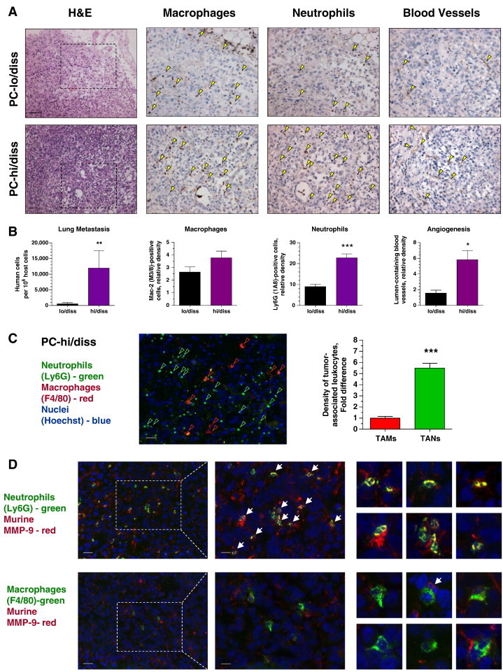
Tumor dissemination and development of angiogenic vasculature in human PC-3 prostate cancer xenografts correlates with recruitment of MMP-9–bearing neutrophils. (A) PC-lo/diss (top) and PC-hi/diss (bottom) xenografts were examined for overall histology (left), infiltration by macrophages and neutrophils (middle), and tumor vasculature (right). Some of IHC-stained (brown) leukocytes and blood vessels are indicated by yellow arrowheads. Bars, 200 μm. (B) Levels of lung metastasis and relative density of TAMs, TANs, and lumen-containing blood vessels in PC-lo/diss versus PC-hi/diss tumors. Pooled data (means ± SEM) from two independent experiments, each employing six mice per variant, are presented. *P < .05, **P < .005, ***P < .0001. (C) PC-hi/diss xenografts were double-stained for neutrophil-specific (Ly6G; green) and macrophage-specific (F4/80; red) markers. Bar, 50 μm. Graph: Relative density of TAMs and TANs, quantified in a total of 17 to 30 tumor sections from four tumors harvested in two independent experiments, is presented as fold difference over TAM density (1.0). (D) Left: PC3-hi/diss tumor sections were double-stained for murine MMP-9 (red) and either neutrophil-specific (Ly6G; green) or macrophage-specific (F4/80; green) markers. Bar, 50 μm. Middle sections depict boxed areas imaged at higher magnification. Bar, 25 μm. Small panels on the right depict examples of double-stained TANs (two top rows) and TAMs (two bottom rows). White arrows point to MMP-9–positive leukocytes with multi-lobed nuclei characteristic of mature neutrophils.