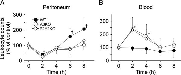
Fig 1. Leukocytes in the peritoneum and peripheral blood after CLP.
The WT, A3KO, and P2Y2KO mice were subjected to intraperitoneal sepsis by CLP with a 22-gauge needle, and the time course of leukocyte recruitment in the peritoneal cavity (A) and the peripheral blood (B) was measured after CLP. Data are expressed relative to leukocyte counts of controls without CLP. Data points represent mean ± SD of 5 animals per group. Statistical analyses were performed with ANOVA followed by Bonferroni test, *P < 0.01, † P < 0.05 compared with WT control.

