Abstract
OBJECTIVE
To explore the advantages of using artificial neural networks (ANNs) to recognize patterns in colposcopy to classify images in colposcopy.
PURPOSE
Transversal, descriptive, and analytical study of a quantitative approach with an emphasis on diagnosis. The training test e validation set was composed of images collected from patients who underwent colposcopy. These images were provided by a gynecology clinic located in the city of Criciúma (Brazil). The image database (n = 170) was divided; 48 images were used for the training process, 58 images were used for the tests, and 64 images were used for the validation. A hybrid neural network based on Kohonen self-organizing maps and multilayer perceptron (MLP) networks was used.
RESULTS
After 126 cycles, the validation was performed. The best results reached an accuracy of 72.15%, a sensibility of 69.78%, and a specificity of 68%.
CONCLUSION
Although the preliminary results still exhibit an average efficiency, the present approach is an innovative and promising technique that should be deeply explored in the context of the present study.
Keywords: colposcopy, artificial intelligence, neural networks, medical informatics, computer-assisted diagnosis
Introduction
A colposcopy is a test that allows the cervix to be viewed with up to 10 times magnification,1,2 which is essential for the evaluation of abnormalities or lesions (which are highly suggestive of cervical neoplasia)3,4 during cytopathological examinations and to assist in biopsies (which are essential for definitive diagnoses).5,6
Infection with human papillomavirus (HPV) is a sexually transmitted disease and is considered a preneoplastic lesion. The incidence of HPV infection has increased in recent decades.7–10 The most common manifestation of HPV infection is subclinical, and the estimated prevalence of HPV infection is greater than 50% in sexually active women.11–14
Therefore, seeking a diagnostic imaging technique, such as colposcopy, medical informatics is proving increasingly helpful for certain clinical tasks, such as data storage, information retrieval, indication of warning signs, critical therapy, and image recognition interpretation.15–18
The artificial neural networks (ANNs) used in medical informatics seek to provide methods for the classification of images from mathematical algorithms. ANNs are systems that are able to acquire, store, and use experiential knowledge to mimic the abilities of the biological nervous system.19 Among the applications of ANNs, we highlight pattern recognition, which can greatly aid in the interpretation of imaging tests such as colposcopy and radiographs. Currently, ANNs have been applied in several areas of medicine and have obtained approximately 80–90% accuracy in areas such as oncology and mastology.20
In this context, the objective of this study is to use pattern recognition via ANNs for the automated classification of colposcopic images on dot pattern.
Methods
A cross-sectional, descriptive, analytical, quantitative approach, with a diagnostic emphasis, approved by the Ethics and Human Research Board, was conducted. The research was performed under protocol 176/2009. This research complied with the principles of the Declaration of Helsinki.
A hybrid neural network based on Kohonen self-organizing maps and multilayer perceptron (MLP) network was used, and this model was chosen because it is a reasonably small net and as a result, it learns faster and reaches good generalization ability with a reasonably small sized training dataset.21
The training set consisted of images of patients submitted to colposcopy from January 2009 to May 2010 from a gynecology service in the city of Criciúma (Santa Catarina, Brazil). A convenience sample was estimated by combining 179 images taken directly in the digital JPEG format (thus negating a need to scan the images) and authorized by informed consent for the original images. The database of images (n = 170) was subdivided into 48 images for training, 58 images for tests, and 64 images for validation, proportions suggested in previous studies.22
The neural network training was performed after the data collection and was divided into two parts. First, the images were processed without a filter, thereby resulting in 66 trials, with an accuracy rate of 65%. In the second step, each image was processed by the user with contrast enhancement, and then the Euclidean distance of each image was found.
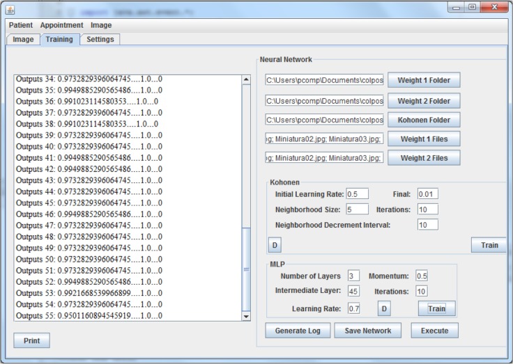
The step after processing is divided into training, testing, and validation. At this stage, we used the application developed by the Research Group on Applied Computational Intelligence and by the Research Group on Information Technology and Communication in Health of the Universidade do Extremo Sul Catarinense (Santa Catarina, Brazil), whose interface is shown in Figure 1. This software offers a hybrid neural network based on Kohonen self-organizing maps and MLP networks.23
Figure 1.
Training of the ANN.
In the Kohonen network training, 48 images were used. Each of these images contained a sub-region of 64 × 64 pixels selected from the original image (Fig. 1) by the software (this was the size that best characterized the dotted pattern, despite tests with 32 × 32 and 128 × 128 images, which resulted in lower accuracy values). The colposcopic image is read into the application, and then the user selects a region of interest with or without the dotted pattern, which will be handled by the library Java Advanced Imaging (JAI) and used in the neural network. The pixels are displayed and stored by the application that used the library JAI, which makes the imaging. When selecting a region on the image to be used, the application stores the 64 × 64 pixels selected by the library JAI area, and for the classification of a new image, the JAI library is used again and each pixel is read to create a neuron network for each image pixel.
The dot pattern was present in 20 of the images and absent in 28 of the images. Such a ratio of images was also used for testing and validation (Fig. 2).
Figure 2.
Selecting subregions of size 64 × 64 pixels.
The architecture of the Kohonen network used for training was 64 neurons in the input layer and a two-dimensional lattice of 8 × 8 neurons in the Kohonen layer. The output of this training was then used as the input for the MLP network trained with a backpropagation algorithm that classified the resulting feature map as belonging to either the group of images that show the dot pattern (class 1) or the group of images without this feature (class 2).
The MLP network architecture used for training and classifying these patterns was defined with 3 layers, 64 neurons in the input layer, 45 neurons in the hidden layer, and 1 neuron in the output layer, and started with 10 iterations and ended in 400 iterations; the learning rate started at 0.5 and ended at 0.6, a momentum of 0.4 with 500 iterations.
The Kohonen network structure was defined with two layers accomplished with an initial learning rate of 0.5 ending at 0.30 (although the final value was set at 0.01), a neighborhood size of 5 and an interval decrement in the neighborhood of 10, and the number of iterations starting at 10 and ending at 300.
In the statistical analysis, we calculated the sensitivity, specificity, positive predictive value (PPV), negative predictive value (NPV), positive likelihood ratio (LR+), negative likelihood ratio (LR−), receiver operating characteristic (ROC) curve, area under curve (AUC) and accuracy of the validation of the ANN, using the Microsoft Excel® 2010 and IBM Statistical Package for the Social Sciences (SPSS) 21.0 software.
Results
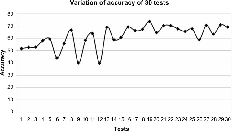
The database of 170 images was used in training, testing, and validation. To reach the final state of ANN, 126 cycles were performed in the training. The accuracy values obtained in the training ranged from 41.78% to 71.5%, and in the test, from 52.5% to 72.15%. The validation was performed with the highest accuracy of 72.15% for the neural network whose parameters are mentioned in the “Methods” section. Figure 3 illustrates the variation of accuracy in the top 30 tests.
Figure 3.
Variation of accuracy.
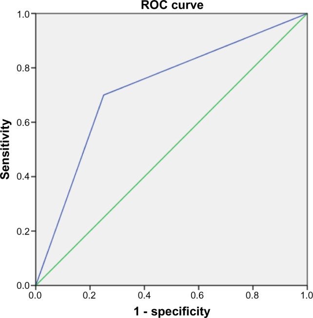
The sensitivity of the ANN was 69.78%, and the specificity was 68.0%; the PPV was 69.47%, and the NPV was 68.29%; and the calculations revealed an LR+ of 1.58 and an LR− of 1.92. Figure 4 illustrates the ROC curve, and the AUC was 0.73.
Figure 4.

ROC curve.
Discussion
Colposcopy is an important imaging method for the diagnosis and classification of cervical lesions.2 In our study, the accuracy demonstrated by the ANN was 72.15%, which is below the average found in the literature of approximately 85%;24–29 as an example, the study conducted in Recife (Pernambuco, Brazil),24 which aimed to develop an intelligent system composed of ANNs and applied to the diagnosis of breast cancer in 2009, exhibited an accuracy of 95%.
Claude and colleagues30 developed a study for colposcopic image classification based on contour parameters used in a comparison study of different ANNs and the k-nearest neighbor reference method. They used 283 samples, and revealed that 95.8% of contour image set has been correctly classified.30 Our findings revealed a lower accuracy, which can be explained by the neural network model applied, and the type of contour identified in our study (dotted), hardly characterized.
Researchers from Alicante (Spain) evaluated the accuracy of using three ANNs in the diagnosis of urological dysfunctions25 with the objective of assisting the diagnosis of varied and complex pathologies and helping to identify sick patients while reducing the cost and wear caused by medical treatment. The accuracy obtained was approximately 83%, thereby demonstrating the method’s effectiveness in helping the clinician make more accurate diagnoses.25 Such an accuracy was superior to that of our study, possibly because three ANNs were used instead of a single network.
The standard colposcopic dot type consists of a focal point in which the capillaries form a dot image.1,2 Speckles that are finer and smoother in appearance indicate a greater likeliness of classification of low-grade injury or metaplasia, especially if the intercapillary distance is small. The probability of high-grade injury increases when the dithering is coarser and less regular.1,2
Researchers at the Albert Einstein College of Medicine in the United States evaluated the reduction in the rates of false-negative cervical smears with the help of ANN, and 487 images of smears of 228 women with documented biopsies of high-grade precursor lesions or invasive carcinoma, which were obtained from 10 different laboratories, were analyzed.26 The instrument was used to detect cytological abnormalities such as atypical squamous or glandular cells of undetermined significance, low- or high-grade squamous intraepithelial lesions, and carcinoma. The results indicate a significant reduction in the rate of false negatives, thereby facilitating early detection of premalignant lesions of the cervix and contributing to prevention of invasive carcinoma of the cervix.26 Similar to our work, this study contributed to explaining new methods to aid in the early identification of changes in the uterine cervix.
In Brazil, a survey by the Federal University of Rio de Janeiro employed ANNs for the classification of breast lesions through the analysis of samples based on digital images of a small section of examinations via fine-needle aspiration.27 For the classification of lesions, backpropagation using nine inputs to generate a logic output was used with the trained neural network algorithm, thereby indicating benignity or malignancy. The accuracy was 97%,27 which is superior to the accuracy observed in our study.
Another Brazilian study performed in São Paulo in 2007 used ANN to classify postural patterns in children with mouth-breathing,28 and included 84 children, of whom 52 were mouth-breathing and 32 were nasal-breathing. The analysis contained anthropometric variables and measured diaphragm excursion and body posture. In the classification of posture, a sensitivity of 0.98 and a specificity of 0.97 were obtained. These results are also superior to the results obtained in this study.28
ANNs have also been used for the prediction of infectious diseases, such as hepatitis A, as demonstrated in a study conducted in 2005 in Rio de Janeiro, which achieved a specificity of 99%, a sensitivity of 70%, and an error rate of 12%.29 A study in Santa Catarina, Brazil, that sought to aid in the diagnostic imaging of primary osteoarthritis of the lumbar spine obtained an accuracy of 62.85% in the use of an ANN for the recognition of image patterns of osteophytes of the lumbar spine in the lateral view.23
As demonstrated above, the number of applications of ANNs is increasing, and they are increasingly used in various areas of medicine.19,20 In gynecology, ANNs were explored in an Italian study that sought to help classify ovarian masses as malignant or non-malignant using images obtained by transvaginal ultrasound Doppler flowmetry. The tests revealed a sensitivity of 96% and a specificity of 97.7%, which are greater than those in our study, perhaps because a different imaging method was used.31
Several other studies have been conducted to evaluate the accuracy of neural networks in the classification of mammography images, and all obtained results demonstrate that the method would be effective in aiding in the early detection of suspicious lesions in the breast.20 Our study also aimed to classify images, but the results were inferior.
Our research considered the use of a hybrid model based on Kohonen self-organizing maps and MLP networks, which was successfully used in previous studies.21,32 Thus, our study presented the first use of hybrid ANNs for pattern classification of dot images obtained via colposcopy. The sensitivity and specificity of the method were approximately 69%. We recommend that future studies conduct tests with more images of patients that present or do not present dot patterns in the colposcopic examination, improve the methodology used in image processing to seek greater system accuracy, and we also recomends to refine the hybrid model used, the use of methods such as random forest and boosting and using other architectures of neural networks. Although the neural network has exhibited moderate results because of the innovative nature of this study, its values demonstrate the potential of ANNs for helping the healthcare professional.
The use of computers to aid in the analysis, interpretation, and classification of images has proven effective in the diagnoses of many medical specialties. The initial goal was not to provide high accuracy but to obtain a performance that is close to that of the specialist, thereby serving as a support tool and not as a substitute method and assisting in the learning process.
Footnotes
Author Contributions
Conceived and designed the experiments: PWS, NBI, RSC, RV, CDV, GPM, ELR, MLS, SAM. Analyzed the data: PWS, NBI. Contributed to the writing of the manuscript: CC, LBC, EC, PJM, RAC. Agree with manuscript results and conclusions: PWS, NBI, RSC, RV, CDV, GPM, ELR, CC, LBC, EC, PJM, RAC, MLS, SAM. Jointly developed the structure and arguments for the paper: PWS, NBI, RSC, RV, MLS, SAM. Made critical revisions and approved final version: PWS, NBI, RSC, RV, CDV, GPM, ELR, CC, LBC, EC, PJM, RAC, MLS, SAM. All authors reviewed and approved of the final manuscript.
ACADEMIC EDITOR: JT Efird, Editor in Chief
FUNDING: This research was supported by Financiadora de Estudos e Projetos (FINEP), Fundação de Amparo à Pesquisa e Inovação do Estado de Santa Catarina (FAPESC), and Universidade do Extremo Sul Catarinense (UNESC). The authors confirm that the funder had no influence over the study design, content of the article, or selection of this journal.
COMPETING INTERESTS: Authors disclose no potential conflicts of interest.
Paper subject to independent expert blind peer review by minimum of two reviewers. All editorial decisions made by independent academic editor. Prior to publication all authors have given signed confirmation of agreement to article publication and compliance with all applicable ethical and legal requirements, including the accuracy of author and contributor information, disclosure of competing interests and funding sources, compliance with ethical requirements relating to human and animal study participants, and compliance with any copyright requirements of third parties.
REFERENCES
- 1.Kyrgiou M, Tsoumpou I, Vrekoussis T, et al. The up-to-date evidence on colposcopy practice and treatment of cervical intraepithelial neoplasia: the Cochrane colposcopy and cervical cytopathology collaborative group (C5 group) approach. Cancer Treat Rev. 2006;32:516–23. doi: 10.1016/j.ctrv.2006.07.008. [DOI] [PubMed] [Google Scholar]
- 2.O’Neill E, Reeves MF, Creinin MD. Baseline colposcopic findings in women entering studies on female vaginal products. Contraception. 2008;78:162–6. doi: 10.1016/j.contraception.2008.04.002. [DOI] [PubMed] [Google Scholar]
- 3.Valentim L. Imaging in gynecology. Best Pract Res Clin Obstet Gynaecol. 2006;20:881–906. doi: 10.1016/j.bpobgyn.2006.06.001. [DOI] [PubMed] [Google Scholar]
- 4.Alvarez RD, Wright TC. Increased detection of high grade cervical intraepithelial neoplasia utilizing an optical detection system as an adjunct to colposcopy. Gynecol Oncol. 2007;106:23–8. doi: 10.1016/j.ygyno.2007.02.028. [DOI] [PubMed] [Google Scholar]
- 5.Manopunya M, Suprasert P, Srisomboon J, Kietpeerakool C. Colposcopy audit for improving quality of services in areas with a high incidence of cervical cancer. Int J Gynaecol Obstet. 2010;108:4–6. doi: 10.1016/j.ijgo.2009.07.042. [DOI] [PubMed] [Google Scholar]
- 6.Bekkers RL, van de Nieuwenhof HP, Neesham DE, Hendriks JH, Tan J, Quinn MA. Does experience in colposcopy improve identification of high grade abnormalities. Eur J Obstet Gynecol Reprod Biol. 2008;141:75–8. doi: 10.1016/j.ejogrb.2008.07.007. [DOI] [PubMed] [Google Scholar]
- 7.Anttila A, Ronco G. Description of the national situation of cervical cancer screening in the member states of European Union. Eur J Cancer. 2009;45:2685–708. doi: 10.1016/j.ejca.2009.07.017. [DOI] [PubMed] [Google Scholar]
- 8.Cain JM, Ngan H, Garland S, Wright T. Control of cervical cancer: women’s options and rights. Int J Gynaecol Obstet. 2009;106:141–3. doi: 10.1016/j.ijgo.2009.03.027. [DOI] [PubMed] [Google Scholar]
- 9.Murillo R, Luna J, Gamboa O, Osório E, Bonilla J, Cendales R. Cervical cancer screening with naked-eye visual inspection in Colombia. Int J Gynaecol Obstet. 2010;109:230–4. doi: 10.1016/j.ijgo.2010.01.019. [DOI] [PubMed] [Google Scholar]
- 10.Elit L, Fyles AW, Devries MC, Oliver TK, Fung-Kee-Fung M. Follow-up for women after treatment for cervical cancer: a sistematic review. Gynecol Oncol. 2009;114:528–35. doi: 10.1016/j.ygyno.2009.06.001. [DOI] [PubMed] [Google Scholar]
- 11.Nicula FA, Anttila A, Neamtiu L, et al. Challenges in starting organised screening programmes for cervical cancer in the new member states of the European Union. Eur J Cancer. 2009;45:2679–84. doi: 10.1016/j.ejca.2009.07.025. [DOI] [PubMed] [Google Scholar]
- 12.Kotaniemi-Talonen L, Nieminen P, Hakama M, et al. Significant variation in performance does not reflect the effectiveness of the cervical cancer screening programme in Finland. Eur J Cancer. 2007;43:169–74. doi: 10.1016/j.ejca.2006.08.026. [DOI] [PubMed] [Google Scholar]
- 13.Llorca J, Rodriguez-Cundin P, Dierssen-Sotos T, Prieto-Salceda D. Cervical cancer mortality is increasing in Spanish women younger than 50. Cancer Lett. 2006;240:36–40. doi: 10.1016/j.canlet.2005.08.021. [DOI] [PubMed] [Google Scholar]
- 14.Bornstein J, Yaakov Z, Pascal B, et al. Decision-making in the colposcopy clinic – a critical analysis. Eur J Obstet Gynecol Reprod Biol. 1999;85:219–24. doi: 10.1016/s0301-2115(99)00026-3. [DOI] [PubMed] [Google Scholar]
- 15.Abbass HA. An evolutionary artificial neural networks approach for breast cancer diagnosis. Artif Intell Med. 2002;25:265–81. doi: 10.1016/s0933-3657(02)00028-3. [DOI] [PubMed] [Google Scholar]
- 16.Ozbay Y, Kara S, Latifoglu F, Ceylan R, Ceylan M. Complex-valued wavelet artificial neural network for Doppler signals classifying. Artif Intell Med. 2007;40:143–56. doi: 10.1016/j.artmed.2007.02.001. [DOI] [PubMed] [Google Scholar]
- 17.Biganzoli EM, Boracchi P, Ambrogi F, Marubini E. Artificial neural network for the joint modelling of discrete cause-specific hazards. Artif Intell Med. 2006;37:119–30. doi: 10.1016/j.artmed.2006.01.004. [DOI] [PubMed] [Google Scholar]
- 18.Smith AE, Nugent CD, McClean SI. Evaluation of inherent performance of intelligent medical decision support systems: utilizing neural networks as an example. Artif Intell Med. 2003;27:1–27. doi: 10.1016/s0933-3657(02)00088-x. [DOI] [PubMed] [Google Scholar]
- 19.Abbod MF, Catto JWF, Linkens DA, Hamdy FC. Application of artificial intelligence to the management of urological câncer. J Urol. 2007;178:1150–6. doi: 10.1016/j.juro.2007.05.122. [DOI] [PubMed] [Google Scholar]
- 20.Glingani FA, Ambrósio PE. Sistema de Análise Computadorizada para Auxílio à Detecção de Lesões de Mama Baseado em Redes Neurais Artificiais; XXIX Congresso da Sociedade Brasileira de Computação; 2009. [Google Scholar]
- 21.Brudzewski K, Osowski S. Gas analysis system composed of a solid-state sensor array and hybrid neural network structure. Sens Actuators B Chem. 1999;55(1):38–46. [Google Scholar]
- 22.San PP, Ling SH, Nguyen HT. Block based neural network for hypoglycemia detection. Conf Proc IEEE Eng Med Biol Soc. 2011;2011:5666–9. doi: 10.1109/IEMBS.2011.6091371. [DOI] [PubMed] [Google Scholar]
- 23.Veronezi CCD, Simões PWTA, Santos RL, et al. Análise computacional para auxílio ao diagnóstico de osteoartrite de coluna lombar baseado em redes neurais artificiais. Rev Bras Ortop. 2011;46:195–9. [Google Scholar]
- 24.Silva NC, Salvador VDN, Cordeiro JR, Araújo RA. Desenvolvimento de um sistema inteligente aplicado ao diagnostico de câncer de mama via redes neurais artificiais [MSc dissertation] Recife: Universidade Federal de Pernambuco; 2007. [Google Scholar]
- 25.Gil D, Johnsson M, Chamizo JMG, Paya AS, Fernandez DR. Application of artificial neural networks in the diagnosis of urological disfunctions. Expert Syst Appl. 2009;36:5754–60. [Google Scholar]
- 26.Koss LG, Sherman ME, Cohen MB, et al. Significant reduction in the rate of false-negative cervical smears with neural network based technology (PAPNET testing system) Hum Pathol. 1997;28:1196–203. doi: 10.1016/s0046-8177(97)90258-6. [DOI] [PubMed] [Google Scholar]
- 27.Leite L, Garcia LGU, Caloba LP. Classificação de lesões da mama usando parametrização e redes neurais [MSc dissertation] Rio de Janeiro: Universidade Federal do Rio de Janeiro; 2004. [Google Scholar]
- 28.Mancini F. Aplicação de redes neurais artificiais na classificação de padrões posturais em crianças respiradoras bucais [PhD thesis] São Paulo: Escola Paulista de Medicina; 2007. [Google Scholar]
- 29.Santos MA, Seixas JM, Pereira BB, Medronho RA. Usando redes neurais artificiais e regressão logística na predição da hepatite A. Res Bras Epidemiol. 2005;2:117–26. [Google Scholar]
- 30.Claude I, Winzenrieth R, Pouletaut P, Boulanger JC. Contour features for colposcopic image classification by artificial neural networks; Proceedings of the 16th International Conference on Pattern Recognition (ICPR’02); Washington, DC, U S A. 2002. pp. 771–4. [Google Scholar]
- 31.Biagiotti R, Desii C, Vanzi E, Gacci G. Predicting ovarian malignancy: application of artificial neural networks to transvaginal and color Doppler flow US. Radiology. 1999;210:399–403. doi: 10.1148/radiology.210.2.r99fe18399. [DOI] [PubMed] [Google Scholar]
- 32.Pal NR, Pal S, Das J, Majumdar K. SOFM–MLP: a hybrid neural network for atmospheric temperature prediction. IEEE Trans Geosci Remote Sens. 2004;41(12):2783–91. [Google Scholar]


