

Abstract
An efficient in vitro protocol has been established for somatic embryogenesis and plantlet conversion of Korean wild ginseng (Panax ginseng Meyer). Wild-type and mutant adventitious roots derived from the ginseng produced calluses on Murashige and Skoog (MS) medium supplemented with 0.5 mg/L 2,4-dichlorophenoxyacetic acid and 0.3 mg/L kinetin; 53.3% of the explants formed callus. Embryogenic callus proliferation and somatic embryo induction occurred on MS medium containing 0.5 mg/L 2,4-dichlorophenoxyacetic acid. The induced somatic embryos further developed to maturity on MS medium with 5 mg/L gibberellic acid, and 85% of them germinated. The germinated embryos were developed to shoots and elongated on MS medium with 5 mg/L gibberellic acid. The shoots developed into plants with well-developed taproots on one-third strength Schenk and Hildebrandt basal medium supplemented with 0.25 mg/L 1-naphthaleneacetic acid. When the plants were transferred to soil, about 30% of the regenerated plants developed into normal plants.
Keywords: mutant adventitious roots, Panax ginseng Meyer, somatic embryo, somatic embryogenesis
1. Introduction
Panax ginseng Meyer is an important medicinal herb that is widely cultivated in Korea, China, and Japan. The root has been used as a drug for over 2000 years in oriental countries. Its use is rapidly expanding in Western countries as complementary and alternative medicine [1]. Ginsenosides are the major pharmacologically active components in P. ginseng. More than 30 types of ginsenosides have been identified from the genus [2,3].
Ginseng is a perennial plant that grows slowly and has a long production cycle (4–6 years). And > 3 years of juvenile period are required for producing seeds [4,5]. This has made the generation of superior genotypes by conventional breeding difficult. Therefore, attempts have been made to achieve a more rapid and increased production of the ginsenosides using other methods such as classical tissue culture [6], bioreactor culture [7], Agrobacterium-mediated hairy root production [8,9], using elicitors in cell cultures [10–12], and mutation breeding by γ-irradiation [13,14]. The last method has been used in many other plant species and has provided a large number of variants useful for plant breeding [15–17]. Mutagenesis by γ-irradiation has been shown to enhance ginsenoside production in P. ginseng [13,14]. Recently, we have also generated mutant cell lines by applying γ-irradiation on P. ginseng adventitious roots which were derived from Korean wild ginseng root [18]. Among the selected mutant cell lines, line 1 showed the highest total ginsenoside content of seven major ginsenosides (Rg1, Re, Rb1, Rb2, Rc, Rf, and Rd). The total ginsenoside content of the mutant line was 2.3 times higher than in the wild-type line [18]. Using γ-irradiation, we have created a useful mutant line for breeding of the ginseng plant. However, there are no reports on in vitro plant regeneration with mutant lines of ginseng adventitious root.
Plant tissue culture system is considered a valuable tool in the plant improvement program. Somatic embryogenesis has been used as a preferred method for rapid in vitro propagation of many plant species [19–21]. P. ginseng is a difficult species to manipulate in vitro; however, its regeneration has generally been accomplished using somatic embryogenesis in callus derived from mature root tissues [22–24], callus derived from zygotic embryo [25,26], protoplast derived from callus [27], and cotyledons [4,28–30]. The development of efficient in vitro culture methods has facilitated the use of mutation technique for improvement of vegetative propagation of ginseng adventitious roots [13,14,18]. At present no information is available on the regeneration of a mutant adventitious root line that has been selected from γ-irradiated P. ginseng adventitious roots.
In this paper, we report on an efficient procedure for the regeneration of wild-type and mutant cell lines of P. ginseng adventitious roots through somatic embryogenesis.
2. Materials and methods
2.1. Callus induction and proliferation
Adventitious roots derived from Korean wild ginseng were provided by Sunchon National University, Sunchon, Korea. The adventitious roots were generated as described previously [7,31,32] and have been maintained in our laboratory for over 10 years. A mutant adventitious root line has been generated from the wild-type adventitious roots by γ-irradiation [18]. For embryogenic callus induction, wild-type and mutant adventitious roots were sectioned into 10 mm in length and were placed on Murashige and Skoog (MS) solid medium supplemented with 2,4-dichlorophenoxyacetic acid (2,4-D), kinetin, and 3% sucrose. The media were solidified with 0.3% Gelite. Callus induction frequency was tested on MS solid medium supplemented with various concentrations of 2,4-D (0.5 mg/L, 1 mg/L, 1.5 mg/L, 2 mg/L) and kinetin (0 mg/L, 0.3 mg/L, 0.5 mg/L). All media were adjusted to pH 5.8 prior to autoclaving. Thirty pieces of adventitious roots were placed on each petri dish. Three replicates were prepared for each treatment. All cultures were incubated at 25°C in the dark. Callus formation was observed after 4 wk of culture. After 6 wk of culture, the frequency of callus induction was estimated. The induced callus was subcultured at 3-wk intervals on the same medium for induction of embryogenic callus and maintenance.
2.2. Induction of somatic embryos
Embryogenic callus induced from the segments of adventitious roots was used for induction of somatic embryos. A 10 g piece of embryogenic callus was incubated in a 15 L airlift bioreactor containing 5 L MS liquid medium with 0.5 mg/L 2,4-D and 3% sucrose for proliferation. After 3 wk, the proliferated embryogenic callus was used as explants for induction of somatic embryogenesis.
To examine the effect of 2,4-D on somatic embryo induction, proliferated callus was placed on a solid MS medium supplemented with different concentrations of 2,4-D (0 mg/L, 0.5 mg/L, 1 mg/L). Ten clumps of embryogenic callus (about 5 mm in diameter) were cultured on petri dishes containing 40 mL of medium and the experiment repeated three times. All cultures were incubated at 25°C in the dark. The frequency of somatic embryo production was examined after 6 wk of culture by counting cultured embryogenic calluses that formed somatic embryos.
When the callus produced globular-stage embryos on MS solid medium with 2,4-D and 3% sucrose, the globular embryos were removed and transferred to 500 mL-Erlenmeyer flasks containing 200 mL of liquid MS medium supplemented with 2,4-D and 3% sucrose for further growth. The liquid cultures were agitated at 100 rpm on a gyratory shaker in the dark. After 1 mo of culture, the proliferated globular embryos in flasks were transferred to individual petri dishes containing solid MS medium with gibberellic acid (GA3) and 3% sucrose for maturation and germination of embryos.
2.3. Maturation and germination of somatic embryos
The proliferated globular embryos in flasks were transferred to 40 mL MS solid medium supplemented with GA3 and 3% sucrose in 100 mm × 20 mm plastic petri dishes for maturation and germination. To investigate the effect of GA3 concentration on maturation and germination of somatic embryos, 150 globular embryos were transferred to germination medium containing 0 mg/L, 1 mg/L, 3 mg/L, 5 mg/L, 7 mg/L, or 10 mg/L of GA3. Cultures were maintained at 23 ± 2°C under dim light illumination (12 μmol/m2/s) with a 16/8 h (light/dark) photoperiod. After 6 wk of culture, maturation and germination of embryos were examined. The experiment was repeated three times.
2.4. Development of plantlets and acclimatization
When shoots reached 0.5–1.0 cm in height, the plantlets were transferred from germination medium to elongation medium, 50 mL MS solid medium supplemented with 5 mg/L GA3 in 100 mm × 40 mm plastic petri dishes, for shoot elongation. When shoots grew 3.0–4.0 cm in height, they were transferred to rooting medium, half or one-third strength MS, or Schenk and Hildebrandt (SH) basal medium supplemented with 0.25 mg/L 1-naphthaleneacetic acid (NAA) or with 0.5% activated charcoal, in 75 mm × 130 mm glass bottles, one shoot per bottle. Cultures were conducted in a culture room and maintained in a 16/8 h (light/dark) photoperiod with white fluorescent light (30 μmol/m2/s) at 23 ± 2°C. After 4 wk, the results of rooting were examined.
Plantlets with both shoots and roots were transferred to plastic pots (10 cm × 18 cm) containing an artificial soil mixture of peat moss, vermiculite, and perlite (2:3:1 v/v) and covered with a transparent polyvinyl film. The potted plants were cultivated in a growth room (40 μmol/m2/s, 16 h photoperiod, 25 ± 1°C). After 3 wk, the plants were hardened by removing the polyvinyl film gradually on a daily basis for 1 wk, and then the film was removed. After 3 mo of culture, the survived plants without wilting were counted. The acclimated plants were transplanted to glasshouse conditions or kept in the growth room for another 4–6 mo.
2.5. Data analysis
Each of the treatments was performed three times. Statistical analyses were performed according to the one-way analysis of variance (ANOVA) using SPSS software version 17.0 (SPSS Inc., Chicago, IL, USA) to assess significant differences in the mean values of different treatments. Comparisons between the mean values were assessed using Duncan's multiple-range test (p < 0.05).
3. Results and discussion
3.1. Induction of callus from adventitious roots
Initiation of callus from adventitious root explants generally occurred after 3 wk on media supplemented with different combinations of growth regulators. The highest frequency of callus induction was observed on the medium containing 0.5 mg/L 2,4-D and 0.3 mg/L kinetin. The frequency of callus induction reduced dramatically as the concentration of 2,4-D increased. Callus was not induced in the presence of 2 mg/L 2,4-D (Table 1). Similar results were reported with the cultures of hairy roots of P. ginseng that 2,4-D at > 3 mg/L strongly suppressed callus induction [33]. When the segments of adventitious roots (Fig. 1A) of P. ginseng were incubated in MS solid medium with 0.5 mg/L 2,4-D and 0.3 mg/L kinetin, callus was induced from the cut sides of the adventitious roots after 6 wk of culture (Fig. 1B). The callus was subcultured on the same medium at 3-wk subculture intervals. After 3 mo, embryogenic callus was induced (Fig. 1C) and the embryogenic callus showed high regenerative capacity and differentiated into somatic embryos and plantlets. Callus induction and growth from adventitious root explants was dependent upon 2,4-D as previously reported [22,24,27]. When embryogenic callus was transferred to MS medium lacking kinetin, a small number of globular embryos formed after 3 wk of culture (Fig. 1D and E). Thus, it is essential to induce and maintain the embryogenic callus in the medium supplemented with 2,4-D in combination with kinetin. Embryogenic callus has been maintained in the dark for > 2 yr through 3-wk subculture intervals on MS solid medium with 0.5 mg/L 2,4-D and 0.3 mg/L kinetin.
Table 1.
Effects of 2,4-Dichlorophenoxyacetic Acid (2,4-D) and Kinetin on the Frequency of Callus Formation from Ginseng Adventitious Roots1)
| 2,4-D (mg/L) | Kinetin (mg/L) | Number of root explants | Number of root explants forming callus |
|---|---|---|---|
| 0.5 | 0 | 30 | 4.3 ± 1.0de |
| 0.3 | 30 | 16.2 ± 1.8a | |
| 0.5 | 30 | 9.1 ± 1.7de | |
| 1 | 0 | 30 | 7.2 ± 1.2bc |
| 0.3 | 30 | 5.4 ± 1.1cd | |
| 0.5 | 30 | 2.3 ± 0.8ef | |
| 2 | 0 | 30 | 0.0 ± 0f |
| 0.3 | 30 | 0.0 ± 0f | |
| 0.5 | 30 | 0.0 ± 0f |
Data were collected after 6 wk of culture. The results represent the means ± standard error of the mean of values obtained from three experiments. Different corresponding letters within a column are significant different at p < 0.05 by Duncan's multiple range test
Fig. 1.

Somatic embryogenesis and regeneration of plantlet from adventitious roots of Panax ginseng. (A) Adventitious roots derived from Korean wild ginseng root. (B) Callus induction from adventitious root explants. (C) Embryogenic callus derived from adventitious roots. (D) Proliferation of embryogenic callus in an airlift bioreactor. (E) Proliferated embryogenic cell clumps from bioreactor culture. (F) Somatic embryos formed on embryogenic callus. (G) Magnified image from (F; scale bar = 2 mm). (H) Proliferation of somatic embryos in conical flasks. (I) Maturation and germination of somatic embryos on MS medium supplemented with 5 mg/L GA3. (J) Well-developed plantlet derived from somatic embryo (scale bar = 0.8 cm).
3.2. Induction of somatic embryos
The embryogenic callus grows better in a liquid medium than a solid medium (data not shown). Therefore, we propagated the embryogenic callus in a bioreactor to assess somatic embryo development and plantlet conversion. When embryogenic callus was inoculated into a 15 L airlift bioreactor containing 5 L MS liquid medium with 0.5 mg/L 2,4-D, the embryogenic callus was propagated and a small number of globular shaped embryos also formed after 3 wk of culture (Fig. 1D and E). The growth rate (final explant fresh weight/initial explant fresh weight) was about 2.1. Embryogenic cell clumps proliferated in bioreactor were transferred onto MS solid medium with different concentrations of 2,4-D (0 mg/L, 0.5 mg/L, 1.0 mg/L) for embryogenesis. The frequency of somatic embryo formation was significantly depended on the concentrations of 2,4-D (Table 2). The highest induction frequency of somatic embryos was observed on the medium supplemented with 0.5 mg/L 2,4-D. The frequency of somatic embryo formation in wild-type and mutant cell line was 15.3% and 14.7%, respectively. The number of somatic embryos per callus was 25.6 and 23.7 in wild-type and mutant cell line, respectively. There was no significant difference in somatic embryo formation frequency between wild-type and mutant cell line (Table 2). Globular shaped somatic embryos formed on the surfaces of embryogenic callus (Fig. 1F and G).
Table 2.
Effects of 2,4-Dichlorophenoxyacetic Acid (2,4-D) on Somatic Embryo Formation from Embryogenic Callus of Wild-type and Mutant Adventitious Roots1)
| Cell line | 2,4-D (mg/L) | Frequency of somatic embryo formation (%) | Number of somatic embryos per callus |
|---|---|---|---|
| Wild-type | 0 | 5.3 ± 0.73b | 10.0 ± 1.2b |
| 0.5 | 15.3 ± 1.21a | 25.6 ± 2.3a | |
| 1 | 2.5 ± 0.46c | 4.7 ± 0.3c | |
| Mutant | 0 | 5.8 ± 0.28b | 12.3 ± 1.9b |
| 0.5 | 14.7 ± 0.45a | 23.7 ± 0.6a | |
| 1 | 2.2 ± 0.27c | 6.0 ± 1.4c |
Data were collected after 6 wk of culture. The results represent the means ± standard error of the mean of values obtained from three experiments. Different corresponding letters within a column are significant different at p < 0.05 by Duncan's multiple range test
3.3. Maturation and germination of somatic embryos
These somatic embryos were transferred into 500 mL-Erlenmeyer flasks containing 200 mL of liquid MS medium supplemented with 0.5 mg/L 2,4-D and 3% sucrose (Fig. 1H) for proliferation. The growth rate (final explant fresh weight/initial explant fresh weight) was about 1.7. After 4 wk of culture, the proliferated globular embryos were transferred to petri dishes containing solid MS medium with various concentrations of GA3 and 3% sucrose. At 5 mg/L GA3, most of the globular embryos turned green and increased in size and developed into torpedo- and cotyledonary-stage embryos within 1 mo. When the mature somatic embryos were transferred to a fresh medium with the same composition, most of the embryos germinated within 2 wk of culture (Fig. 1I). Adventitious shoots were induced from the mature somatic embryos. The optimal concentration of GA3 in germination medium was 5 mg/L, yielding the highest germination frequency of 85%. Without GA3 treatment, the germination frequency was lowest at 36%. Maturation and germination of embryos were strongly influenced by the GA3 concentration (Table 3). This result suggests that GA3 is required for maturation and germination of somatic embryos. Similar results were observed in Eleutherococcus senticosus, that GA3 treatment was necessary to induce germination from somatic embryos [34]. GA3 treatment is also commonly used for maturation and germination of somatic embryos from P. ginseng [22,26,28,29], from Panax quinquefolius [35] and from Panax japonicus [36].
Table 3.
Effects of Gibberellic Acid (GA3) on Germination of Somatic Embryos1)
| Concentration of GA3 (mg/L) | Number of somatic embryos inoculated | Number of somatic embryos germinated | Germination frequency (%) |
|---|---|---|---|
| 0 | 150 | 53 ± 6c | 36 ± 4c |
| 1 | 150 | 60 ± 9c | 40 ± 6c |
| 3 | 150 | 66 ± 10bc | 45 ± 7bc |
| 5 | 150 | 127 ± 7a | 85 ± 5a |
| 7 | 150 | 75 ± 6b | 50 ± 4b |
| 10 | 150 | 65 ± 5bc | 44 ± 3bc |
Data were collected after 6 wk of culture on MS medium with 3% sucrose. The results represent the means ± standard error of the mean of values obtained from three experiments. Different corresponding letters within a column are significant different at p < 0.05 by Duncan's multiple range test
3.4. Development of plantlets and transplantation
When shoots reached 0.5–1.0 cm in height on germination medium, the shoots were transferred to elongation medium, 50 mL MS solid medium supplemented with 5 mg/L GA3 in 100 mm × 40 mm plastic petri dishes, for further growth of shoots. After about 1 mo of culture, the shoots developed to 3.0–4.0 cm in height, but most of the shoots had no visible roots. The shoots without roots were excised and transferred to different rooting media, half or one-third strength MS, or SH basal medium supplemented with 0.25 mg/L NAA or with 0.5% activated charcoal, in 75 mm × 130 mm glass bottles, one shoot per bottle. Adventitious roots formed from the excised regions of the shoots. After 1 mo, the rate of root formation from the shoots was examined (Table 4). As far as root quality is concerned, one-third SH medium with 0.25 mg/L NAA and 1% sucrose showed the best result among the tested rooting media; the roots grew fast and thickened on the medium (Fig. 1J, 2A, 2B; Table 4). Although one-third SH medium with 2% sucrose and 0.5% activated charcoal was most effective in inducing roots, the roots grew well but was weak (Table 4). The optimal medium for rooting is therefore one-third SH medium supplemented with 0.25 mg/L NAA and 1% sucrose among the tested rooting media in this study. In our comparative studies, SH medium was more effective than MS medium in root induction and proliferation. A very similar result was reported in American [35] and Korean ginsengs [30]. It was reported that the high level of ammonium nitrate in MS medium highly suppressed root development in carrot [37]. Choi et al [5] reported that when the ammonium nitrate was omitted in MS medium, root growth of regenerated ginseng plants was enhanced. The concentration of ammonium nitrate in SH medium was about eight times lower than in MS medium. It seems that the different concentrations of ammonium nitrate in SH and MS medium may result in the different root induction efficiency between the two basal medium. From these observations, we suggest that SH medium, especially one-third strength SH medium is suitable for root induction and growth of regenerated ginseng plants.
Table 4.
Comparison of Rooting Media for Ginseng Root Development
| Media | No. of shoots | Root frequency (%) | No. of roots per plant | Description on root quality |
|---|---|---|---|---|
| 1/2 MS 3% sucrose | 30 | 36 | 1.6 | Grows slow, calluses |
| 1/2 MS + 3% sucrose + 0.5% charcoal | 30 | 45 | 1.0 | Grows slow |
| 1/3 MS + 1% sucrose + 0.25 mg/L NAA | 30 | 58 | 1.0 | Grows fast, thin |
| 1/2 SH + 3% sucrose | 30 | 71 | 1.0 | Grows slow |
| 1/2 SH + 2% sucrose + 0.5% charcoal | 30 | 62 | 1.0 | Grows slow, calluses |
| 1/3 SH + 2% sucrose + 0.5% charcoal | 30 | 80 | 1.0 | Grows fast, thin |
| 1/3 SH + 1% sucrose + 0.25 mg/L NAA | 30 | 76 | 1.2 | Grows fast, strong |
MS, Murashige and Skoog medium; NAA, 1-naphthaleneacetic acid; SH, Schenk and Hildebrandt basal medium
Fig. 2.
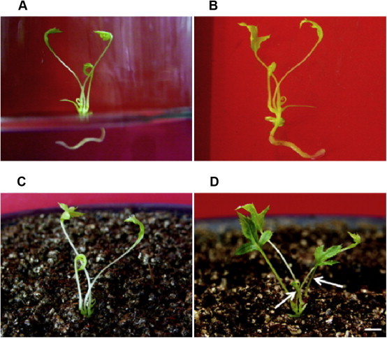
Development of ginseng plant and transplantation into soil. (A) Well-developed plantlet on rooting medium, one-third Schenk and Hildebrandt basal medium supplemented with 0.25 mg/L 1-naphthaleneacetic acid. (B) Plant with a taproot just prior to transplantation into soil. (C) Regenerated plant hardened in soil. (D) New leaves (indicated by arrows) were produced from 3-mo-old potted plant (scale bar = 0.8 cm).
Well-developed plantlets with both shoots and roots derived from adventitious roots were transferred to plastic pots (10 cm × 18 cm) containing an artificial soil mixture of peat moss, vermiculite and perlite (2:3:1 v/v) in a growth room (Fig. 2C). The survival rate of the plantlets was about 30% after 3 mo of culture and new leaf began growing (Fig. 2D). The plants regenerated from both wild-type and mutant cell line acclimatized in the growth room (Fig. 3).
Fig. 3.
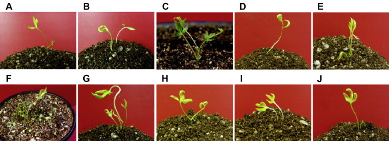
Transplantation of regenerated ginseng plants into soil. (A–E) Plants derived from wild-type adventitious roots. (F–J) Plants derived from mutant adventitious roots.
In conclusion, we have developed an efficient in vitro regeneration protocol for an important medicinal plant of P. ginseng. The protocol described here will allow a relatively rapid mass production of Korean wild ginseng plants. It takes 6–8 mo from the callus induction of adventitious roots to the plantation of plants. In the present study, we also produced the regenerated plants from the mutant adventitious roots which were obtained by γ-irradiation. The combination of mutation technique by γ-irradiation and plant regeneration by tissue cultures may be an effective way to ginseng improvement. The protocol established in this study is currently being used for the genetic transformation of this species.
Conflicts of interest
All contributing authors declare no conflicts of interest.
Acknowledgments
This research was supported by Basic Science Research Program through the National Research Foundation of Korea (NRF) funded by the Ministry of Education (2009-0094059).
Footnotes
This is an Open Access article distributed under the terms of the Creative Commons Attribution Non-Commercial License (http://creativecommons.org/licenses/by-nc/3.0) which permits unrestricted non-commercial use, distribution, and reproduction in any medium, provided the original work is properly cited.
Contributor Information
Yong Hwan Kim, Email: dragon@ipet.re.kr.
Hyo-Yeon Lee, Email: hyoyeon@jejunu.ac.kr.
References
- 1.Shim M., Lee Y.J. Ginseng as a complementary and alternative medicine for postmenopausal symptoms. J Ginseng Res. 2009;33:89–92. [Google Scholar]
- 2.Harrison D.M. The biosynthesis of triterpenoids, steroids, and carotenoids. Nat Prod Rep. 1990;7:459–484. doi: 10.1039/np9900700459. [DOI] [PubMed] [Google Scholar]
- 3.Nah S.Y. Ginseng; recent advances and trends. Korean J Ginseng Sci. 1997;21:1–12. [Google Scholar]
- 4.Ahn I.O., Le B.V., Gendy C., Van K.T.T. Direct somatic embryogenesis through thin layer culture in Panax ginseng. Plant Cell Tiss Organ Cult. 1996;45:237–243. [Google Scholar]
- 5.Choi Y.E., Yang D.C., Park J.C., Soh W.Y., Choi K.T. Regenerative ability of somatic single and multiple embryos from cotyledons of Korean ginseng on hormone-free medium. Plant Cell Rep. 1998;17:544–551. doi: 10.1007/s002990050439. [DOI] [PubMed] [Google Scholar]
- 6.Wu J., Zhong J.J. Production of ginseng and its bioactive components in plant cell culture: current technological and applied aspects. J Biotechnol. 1999;68:89–99. doi: 10.1016/s0168-1656(98)00195-3. [DOI] [PubMed] [Google Scholar]
- 7.Sivakumar G., Yu K.W., Paek K.Y. Production of biomass and ginsenosides from adventitious roots of Panax ginseng in bioreactor cultures. Eng Life Sci. 2005;5:333–342. [Google Scholar]
- 8.Yoshikawa T., Furuya T. Saponin production by cultures of Panax ginseng transformed with Agrobacterium rhizogenes. Plant Cell Rep. 1987;6:449–453. doi: 10.1007/BF00272780. [DOI] [PubMed] [Google Scholar]
- 9.Mallol A., Cusidó R.M., Palazón J., Bonfill M., Morales C., Piñol M.T. Ginsenoside production in different phenotypes of Panax ginseng transformed roots. Phytochemistry. 2001;57:365–371. doi: 10.1016/s0031-9422(01)00062-0. [DOI] [PubMed] [Google Scholar]
- 10.Lu M.B., Wong H.L., Teng W.L. Effects of elicitation on the production of saponin in cell culture of Panax ginseng. Plant Cell Rep. 2001;20:674–677. [Google Scholar]
- 11.Palazón J., Cusidó R.M., Bonfill M., Mallol A., Moyano E., Morales C., Piñol M.T. Elicitation of different Panax ginseng transformed root phenotypes for an improved ginsenoside production. Plant Physiol Biochem. 2003;41:1019–1025. [Google Scholar]
- 12.Bae K.H., Choi Y.E., Shin C.G., Kim Y.Y., Kim Y.S. Enhanced ginsenoside productivity by combination of ethephon and methyl jasmonate in ginseng (Panax ginseng C.A. Meyer) adventitious root cultures. Biotechnol Lett. 2006;28:1163–1166. doi: 10.1007/s10529-006-9071-1. [DOI] [PubMed] [Google Scholar]
- 13.Kim D.S., Kim S.Y., Jeong I.Y., Kim J.B., Lee G.J., Kang S.Y., Kim W. Improvement of ginsenoside production by Panax ginseng adventitious roots induced by γ-irradiation. Biol Plant. 2009;53:408–414. [Google Scholar]
- 14.Kim D.S., Song M., Kim S.H., Jang D.S., Kim J.B., Ha B.K., Kim S.H., Lee K.J., Kang S.Y., Jeong I.Y. The improvement of ginsenoside accumulation on Panax ginseng as a result of γ-irradiation. J Ginseng Res. 2013;37:332–340. doi: 10.5142/jgr.2013.37.332. [DOI] [PMC free article] [PubMed] [Google Scholar]
- 15.Subhan F., Anwar M., Ahmad N., Gulzar A., Siddiq A.M., Rahman S., Ahmad I., Rauf A. Effect of gamma radiation on growth and yield of barley under different nitrogen levels. Pak J Biol Sci. 2004;7:981–983. [Google Scholar]
- 16.Mokobia C.E., Okpakorese E.M., Analogbei C., Agbonwanegbe J. Effect of gamma irradiation on the grain yield of Nigerian Zea mays and Arachis hypogaea. J Radiol Prot. 2006;26:423–427. doi: 10.1088/0952-4746/26/4/N01. [DOI] [PubMed] [Google Scholar]
- 17.Chung B.Y., Lee Y.B., Baek M.H., Kim J.H., Wi S.G., Kim J.S. Effects of low-dose gamma-irradiation on production of shikonin derivatives in callus cultures of Lithospermum erythrorhizon S. Radiat Phys Chem. 2006;75:1018–1023. [Google Scholar]
- 18.Zhang J.Y., Bae T.W., Boo K.H., Sun H.J., Song I.J., Pham C.H., Ganesan M., Yang D.H., Kang H.K., Ko S.M. Ginsenoside production and morphological characterization of wild ginseng (Panax ginseng Meyer) mutant lines induced by γ-irradiation (60Co) of adventitious roots. J Ginseng Res. 2011;35:283–293. doi: 10.5142/jgr.2011.35.3.283. [DOI] [PMC free article] [PubMed] [Google Scholar]
- 19.Guerra M.P., Handro W. Somatic embryogenesis and plant regeneration in embryo cultures of Euterpe edulis mart. (palmae) Plant Cell Rep. 1988;7:550–552. doi: 10.1007/BF00272754. [DOI] [PubMed] [Google Scholar]
- 20.Bhansali R.R., Driver J.A., Durzan D.J. Rapid multiplication of adventitious somatic embryos in peach and nectarine by secondary embryogenesis. Plant Cell Rep. 1990;9:280–284. doi: 10.1007/BF00232302. [DOI] [PubMed] [Google Scholar]
- 21.Jimenez V.M. Involvement of plant hormones and plant growth regulators on in vitro somatic embryogenesis. Plant Growth Regulation. 2005;47:91–110. [Google Scholar]
- 22.Chang W.C., Hsing Y.I. Plant regeneration through somatic embryogenesis in root-derived callus of ginseng (Panax ginseng C. A. Meyer) Theor Appl Genet. 1980;57:133–135. doi: 10.1007/BF00253888. [DOI] [PubMed] [Google Scholar]
- 23.Cellarova E., Rychlova M., Vranova E. Histological characterization of in vitro regenerated structures of Panax ginseng. Plant Cell Tiss Organ Cult. 1992;30:165–170. [Google Scholar]
- 24.Lim H.T., Lee H.S., Eriksson T. Regeneration of Panax ginseng C.A. Meyer by organogenesis and nuclear DNA analysis of regenerants. Plant Cell Tiss Organ Cult. 1997;49:179–187. [Google Scholar]
- 25.Lee H.S., Liu J.R., Yang S.G., Lee Y.H., Lee K.W. In vitro flowering of plantlets regenerated from zygotic embryo-derived somatic embryos of ginseng. HortScience. 1990;25:1652–1654. [Google Scholar]
- 26.Arya S., Arya I.D., Eriksson T. Rapid multiplication of adventitious somatic embryos of Panax ginseng. Plant Cell Tiss Organ Cult. 1993;34:157–162. [Google Scholar]
- 27.Arya S., Liu J.R., Eriksson T. Plant regeneration from protoplasts of Panax ginseng (C.A. Meyer) through somatic embryogenesis. Plant Cell Rep. 1991;10:277–281. doi: 10.1007/BF00193141. [DOI] [PubMed] [Google Scholar]
- 28.Choi Y.E., Yang D.C., Yoon E.S., Choi K.T. Plant regeneration via adventitious bud formation from cotyledon explants of Panax ginseng C. A. Meyer. Plant Cell Rep. 1998;17:731–736. doi: 10.1007/s002990050474. [DOI] [PubMed] [Google Scholar]
- 29.Choi Y.E., Yang D.C., Yoon E.S., Choi K.T. High-efficiency plant production via direct somatic single embryogenesis from preplasmolysed cotyledons of Panax ginseng and possible dormancy of somatic embryos. Plant Cell Rep. 1999;18:493–499. [Google Scholar]
- 30.Kim Y.J., Lee O.R., Kim K.T., Yang D.C. High frequency of plant regeneration through cyclic secondary somatic embryogenesis in Panax ginseng. J Ginseng Res. 2012;36:442–448. doi: 10.5142/jgr.2012.36.4.442. [DOI] [PMC free article] [PubMed] [Google Scholar]
- 31.Yu K.W., Gao W., Hahn E.J., Paek K.Y. Jasmonic acid improves ginsenoside accumulation in adventitious root culture of Panax ginseng C.A. Meyer. Biochem Eng J. 2002;11:211–215. [Google Scholar]
- 32.Kim Y.S., Hahn E.J., Yeung E.C., Paek K.Y. Lateral root development and saponin accumulation as affected by IBA or NAA in adventitious root cultures of Panax ginseng C.A. Meyer. In Vitro Cell Dev Biol Plant. 2003;39:245–249. [Google Scholar]
- 33.Kwon J.H., Cheon H.C., Yang D.C. Production of ginsenoside in callus of ginseng hairy roots. J Ginseng Res. 2003;27:78–85. [Google Scholar]
- 34.Choi Y.E., Kim J.W., Yoon E.S. High frequency of plant production via somatic embryogenesis from callus of cell suspension cultures in Eleutherococcus senticosus. Ann Bot. 1999;83:309–314. [Google Scholar]
- 35.Zhou S., Brown D.C.W. High efficiency plant production of North American ginseng via somatic embryogenesis from cotyledon explants. Plant Cell Rep. 2006;25:166–173. doi: 10.1007/s00299-005-0043-z. [DOI] [PubMed] [Google Scholar]
- 36.You X.L., Han J.Y., Choi Y.E. Plant regeneration via direct somatic embryogenesis in Panax japonicas. Plant Biotechnol Rep. 2007;1:5–9. [Google Scholar]
- 37.Halperin W. Alternative morphogenetic events in cell suspensions. Am J Bot. 1966;53:443–453. [Google Scholar]