Abstract
Introduction:
Gonial angle is an important parameter of the craniofacial complex giving an indication about the vertical parameters and symmetry of the facial skeleton. Both orthopantomogram (OPG) and lateral cephalograms can be used for the measurement of gonial angle. Because of the superimpositions seen on lateral cephalograms, reliable measurement of the gonial angle becomes difficult. The aim of the present study is to check the possible application and reliability of OPG for gonial angle determination by clarifying whether there is any significant difference between the determination of gonial angle from OPG and cephalogram.
Materials and Methods:
Gonial angle measurements were made on lateral cephalograms and orthopantomograms of 98 patients - 44 males (mean age 25.9 years) and 54 females (mean age 21.3 years), and compared using Statistical Package for Social Sciences.
Results:
One-way analysis of variance demonstrated no significant differences between the values of gonial angles determined by lateral cephalogram and panoramic radiography. Pearson correlation showed a high correlation between cephalometric and OPG gonial angle value.
Conclusion:
Panoramic radiography can be used to determine the gonial angle as accurately as a lateral cephalogram. For determination of the gonial angle, an OPG may be a better choice than a lateral cephalogram as there are no interferences due to superimposed images of anatomical structures as in a lateral cephalogram. Thus, the present study substantiates the possibility of enhancing the clinical versatility of the panoramic radiograph, which is an indispensable tool for dental diagnosis.
Keywords: Cephalogram, gonial angle, orthopantomogram
INTRODUCTION
Orthodontic diagnosis and treatment planning involves detailed study of dental occlusion, hard tissue relationships and soft tissue proportions.[1] The orthodontic diagnosis database is derived from three major sources: History, clinical examination and evaluation of diagnostic records including dental casts, radiographs and photographs. Cephalograms and orthopantomogram (OPG) are routinely taken for every orthodontic patient. The goal of cephalometric analysis is to evaluate the horizontal and vertical relationship of five major functional components of the face: The cranium and cranial base, skeletal maxilla, skeletal mandible, the maxillary dentition and alveolar process and the mandibular dentition and alveolar process.[2] The vertical relationship of these structures is as important as the horizontal relations, as the treatment plan as well as the outcome is affected by the vertical relationships and the growth pattern of the patient. The external gonial angle is an important angle of the craniofacial complex. It is significant for the diagnosis of craniofacial disorders. Gonial angle is one of the important parameters giving an indication about the vertical parameters and symmetry of the facial skeleton. The gonial angle is measured by taking the tangent to the posterior border of the ramus and tangent to the lower border of the mandible on lateral cephalogram. Because of the superimpositions seen on lateral cephalograms, reliable measurement of the gonial angle becomes difficult. Panoramic radiography first introduced by Professor Yrjö Paatero of the University of Helsinki (1961) is frequently used in orthodontic practice to provide important information about the teeth, their axial inclinations, maturation periods and surrounding tissues. By virtue of its ability to take a single picture of the entire stomatognathic system-teeth, jaws, temporomandibular joints, sinuses-panoramic radiography forms an indispensable orthodontic screening tool. Studies examining panoramic radiographs as a means of investigating skeletal patterns are lacking in the orthodontic literature. OPG, which is also used as an essential diagnostic aid, can be used for reliable measurement of the gonial angle as there is no complication of superimposed images appearing as in cephalograms.
Aims and objectives
The aim of the present study is to check the possible application (reliability) of OPG for gonial angle determination by clarifying whether there is any significant difference between the determination of gonial angle from OPG and cephalogram.
MATERIALS AND METHODS
Lateral cephalograms and orthopentomograms of 98 patients - 44 males (mean age 25.9 years) and 54 females (mean age 21.3 years) - were obtained from the patient records of the Department of Orthodontics and Dentofacial Orthopedics. Gonial angle was measured by drawing a tangent to the lower border of the mandible and a tangent to the distal border of the ramus and condyle. In lateral cephalograms, the mean of gonial angles in the superimposed projections was calculated.
Statistical analysis
The statistical analysis was carried out using Statistical Package for Social Sciences (SPSS Inc., Chicago, IL, USA; version 15.0 for Windows). As our data for angles were quantitative data, these were estimated using mean and standard deviation. Normality of data was checked by measures of Kolmogorov Smirnov tests of normality. These data were normally distributed and means were compared using Student's t-test for male versus female. For malocclusion classes, means were compared using one-way analysis of variance (ANOVA). The Tukey HSD (post hoc test of multiple comparisons) test was not applied as there was no statistically significant difference observed by ANOVA. Pearson correlation was applied for comparing the correlation of different variables. All statistical tests were two-sided and performed at a significance level of α =0.05.
RESULTS
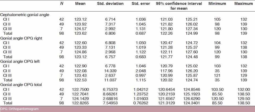
The mean value of the gonial angle in lateral cephalogram was 123.62° with a standard deviation of 6.80°. The mean value of the gonial angle in OPG was 122.82° with a standard deviation of 7.54°. In OPG, the mean value of the right gonial angle was 123.12° with a standard deviation of 6.7° and the mean value of the left gonial angle was 122.53° with a standard deviation of 11.0° [Table 1]. One-way ANOVA demonstrated no significant differences between the values of gonial angles determined by lateral cephalogram and OPG. Also, in OPG, there was no significant difference between the right and left gonial angles [Table 2]. As no statistically significant difference was seen by ANOVA, the post hoc test for multiple comparisons was not applied.
Table 1.
Mean, standard deviation and standard error of OPG and cephalometric gonial angle values in subjects distributed on the basis of malocclusion

Table 2.
ANOVA comparing cephalometric gonial angle, OPG gonial angle right, OPG gonial angle left and OPG gonial angle total

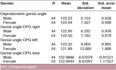
The subjects were then divided based on gender into males and females and compared using the T-test to check any overall gender difference for the value of gonial angle on OPG or cephalogram. In lateral cephalograms, the gonial angle in females was 123.94° and that in males was 123.23°. In panoramic radiography, the gonial angle in females was 122.69°and that in males was 122.98°. Table 3 shows that there was no statistically significant difference between the gonial angle values taken on lateral cephalograms and OPG.
Table 3.
Means, standard deviation and standard error of gonial angle based on gender

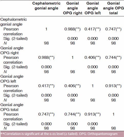
Pearson correlation was applied to check for correlation between cephalometric and OPG gonial angle value. Table 4 shows a high correlation between the values taken on both the radiographs.
Table 4.
Correlations of cephalometric gonial angle, gonial angle OPG right, gonial angle OPG left, gonial angle OPG total

DISCUSSION
The goal of this study was to enhance the panoramic radiograph's clinical use by determining its potential for evaluating craniofacial specifications. Even though there are a number of published articles on magnification and image distortion in panoramic radiographs, there are only a few studies involving the use of panoramic radiographs in evaluating dentoskeletal specifications and gonial angle measurements.
The results of the study demonstrate that there are no statistically significant differences in the values of gonial angle measured on cephalogram and OPG. Therefore, it is possible to use OPG for measuring the gonial angle with equal accuracy as cephalogram. Rather, it may be desirable to make the gonial angle measurements on the OPG as both right and left gonial angles can be viewed separately and clearly on the OPG. This fact was established in the study conducted by Mattila et al.[3] They took measurements of gonial angle on cephalograms, OPG and dried skulls and concluded that the measurements on OPG for right and left gonial angles conform to the angles measured on dry skulls. They further concluded that means of the measurements made on cephalograms and OPG show that the measurements made on OPG are more accurate. The present study shows the same results. But, still, the gonial angle measurements are routinely made on cephalogram rather than OPG. The results of the present study demonstrate that OPG can be used to make these measurements as often as lateral cephalograms, especially in cases where the outlines of two sides are not clearly visible and in asymmetry cases before PA cephalograms are taken. The present results are substantiated by Larheim and Svanaes (1986)[4] and Akcam et al.[5]
Alhaija[6] evaluated the potential of panoramic radiographs to measure mandibular inclination and steepness. A high correlation between the measurements taken from both radiographs was found. They concluded that panoramic radiographs are a useful tool for the measurement of gonial angle, which is an indicator of mandibular steepness and, subsequently, mandibular growth direction. The ability to determine growth direction from the OPG will be useful because majority of the dentists request an OPG for patients during routine dental examination. This will enable the dental professional to spot vertical growth problems using a readily available tool.
Fatahi and Babouei (2007)[7] evaluated the reliability of the cephalometric measurements when determined from an OPG. Comparison of actual measurements obtained from dry skulls and panoramic radiographic measurements revealed the highest correlation between panoramic and cephalometric radiographs in gonial angle, whereas the least correlation was seen in the length of the mandibular body. In different growth patterns, it was seen that gonial angle and ramus height showed the highest correlation between the two radiographs. They concluded that the ability to determine growth direction from the OPG should be useful because majority of the dentists request an OPG for patients during routine dental examination.
Kurt et al.[8] used OPGs to evaluate mandibular asymmetry in Class II subdivision malocclusion patients by measuring condylar, ramal, condylar-ramal asymmetry index values and gonial angle measurements. They concluded that acceptable results can be achieved with panoramic radiographs. The added advantages of panoramic radiographs is that they are non-invasive, have a favorable cost-benefit relationship and expose subjects to a relatively low dose of radiations.
Shahabi et al.[9] compared the external gonial angle determined from the lateral cephalograms and panoramic radiographs in Class I patients. Based on the obtained results, they concluded that panoramic radiography can be used to determine the gonial angle as accurately as a lateral cephalogram.
Jena et al.[10] concluded that OPGs can be used for vertical and angular measurements as well as evaluation of side to side mandibular asymmetry.
Ongkosuwito et al. (2009)[11] concluded that an OPG is as reliable as a lateral cephalogram for linear measurements of the mandible, i.e., condylion-gonion, gonion-menton and condylion-menton.
CONCLUSION
Panoramic radiography can be used to determine the gonial angle as accurately as a lateral cephalogram as there are no significant differences in the gonial angle values as measured on cephalogram and OPG. In addition, OPG forms an additional tool for easier and more accurate determination of both right and left gonial angles of a patient without interferences due to superimposed images of anatomical structures in a lateral cephalogram. For determination of the gonial angle, an OPG may be a better choice than a lateral cephalogram. Thus, the present study substantiates the possibility of enhancing the clinical versatility of the panoramic radiograph, which is an indispensable tool for dental diagnosis.
Footnotes
Source of Support: Nil.
Conflict of Interest: None declared.
REFERENCES
- 1.Sarver DM, Proffit WR. Orthodontics current principles and techniques. 4th ed. Amsterdam, Netherlands: Mosby Elsevier; 2009. Special considerations in Diagnosis and Treatment Planning. 2009. [Google Scholar]
- 2.Proffit WR, Sarver DM, Ackeram JL. Contemporary orthodontics. 4th ed. Amsterdam, Netherlands: Mosby Elsevier; 2009. Orthodontic Diagnosis: The development of a problem list. [Google Scholar]
- 3.Mattila K, Altonen M, Haavikko K. Determination of gonial angle from the orthopantomogram. Angle Orthod. 1977;47:107–10. doi: 10.1043/0003-3219(1977)047<0107:DOTGAF>2.0.CO;2. [DOI] [PubMed] [Google Scholar]
- 4.Larheim TA, Svanaes DB. Reproducibility of rotational panoramic radiography: Mandibular linear dimensions and angles. Am J Orthod Dentofacial Orthop. 1986;90:45–51. doi: 10.1016/0889-5406(86)90026-0. [DOI] [PubMed] [Google Scholar]
- 5.Akcam MO, Altiok T, Ozdiler E. Panoramic radiographs: A tool for investigating skeletal pattern. Am J Orthod Dentofacial Orthop. 2003;123:175–81. doi: 10.1067/mod.2003.3. [DOI] [PubMed] [Google Scholar]
- 6.Alhaija ES. Panoramic radiographs: Determination of mandibular steepness. J Clin Pediatr Dent. 2005;29:165–6. [PubMed] [Google Scholar]
- 7.Fatahi HR, Babouei EA. Evaluation of the precision of panoramic radiography in dimensional measurements and mandibular steepness in relation to lateral cephalomerty. J Mashhad Dent Sch Fall. 2007;31:223–30. [Google Scholar]
- 8.Kurt G, Uysal T, Sisman Y, Ramoglu SI. Mandibular Asymmetry in Class II Subdivision Malocclusion. Angle Orthod. 2008;78:32–7. doi: 10.2319/021507-73.1. [DOI] [PubMed] [Google Scholar]
- 9.Shahabi M, Ramazanzadeh BA, Mokhber N. Comparison between the external gonial angle in panoramic radiographs and lateral cephalograms of adult patients with Class I malocclusion. J Oral Sci. 2009;51:425–9. doi: 10.2334/josnusd.51.425. [DOI] [PubMed] [Google Scholar]
- 10.Jena AK, Singh SP, Utreja AK. Effects of sagittal maxillary growth hypoplasia severity on mandibular asymmetry in unilateral cleft lip and palate subjects. Angle Orthod. 2011;81:872–7. doi: 10.2319/110610-646.1. [DOI] [PMC free article] [PubMed] [Google Scholar]
- 11.Ongkosuwito EM, Dieleman MM, Kuijpers-Jagtman AM, Mulder PG, van Neck JW. Linear mandibular measurements: Comparison Between orthopantomograms and lateral cephalograms. Cleft Palate Craniofac J. 2009;46:147–53. doi: 10.1597/07-123.1. [DOI] [PubMed] [Google Scholar]


