Abstract
Background/Aims
Upregulation of aldo-keto reductase 1B10 (AKR1B10) through the mitogenic activator protein-1 signaling pathway might promote hepatocarcinogenesis and tumor progression. The goal of this study was to evaluate the prognostic significance of AKR1B10 protein expression in patients with hepatocellular carcinoma after surgery.
Methods
A tissue microarray was used to detect the expression level of AKR1B10 protein in tumors from 255 patients with hepatocellular carcinoma who underwent curative hepatectomy. The impact of AKR1B10 expression on the survival of patients was analyzed. The median follow-up period was 119.8 months.
Results
High AKR1B10 protein expression was observed in 125 of the 255 patients with hepatocellular carcinoma (49.0%). High AKR1B10 expression was significantly associated with a lack of invasion of the major portal vein (p=0.022), a lack of intrahepatic metastasis (p=0.010), lower the American Joint Committee on Cancer T stage (p=0.016), lower the Barcelona Clinic Liver Cancer stage (p=0.006), and lower α-fetoprotein levels (p=0.020). High AKR1B10 expression was also correlated with a lack of early recurrence (p=0.022). Multivariate analyses of survival revealed that intrahepatic metastases and lower albumin levels were independent predictors of both shorter recurrence-free survival and shorter disease-specific survival. High AKR1B10 expression was an independent predictor of both longer recurrence-free survival (p=0.024) and longer disease-specific survival (p=0.046).
Conclusions
High AKR1B10 protein expression might be useful as a marker of a favorable prognosis in patients with hepatocellular carcinoma after curative hepatectomy.
Keywords: AKR1B10, Carcinoma, hepatocellular, Survival
INTRODUCTION
Hepatic resection is a potentially curative treatment for hepatocellular carcinoma (HCC). However, the prognosis remains unsatisfactory as a result of frequent tumor recurrence and metastasis after hepatectomy.1,2 It is important to identify the biomarkers that predispose to tumor recurrence or disease-specific death. In recent years, molecular targeted therapy has offered new prospects and attracted a great deal of attention with regard to its use in the standardized treatment of HCC.3,4
The aldo-keto reductase 1B10 (AKR1B10) protein belongs to the AKR superfamily, a group of proteins implicated in xenobiotic detoxification, cell growth and proliferation, carcinogenesis, and cancer therapeutics.5,6 AKR1B10 mRNA was expressed most abundantly in normal small intestine and colon, and a low level of its mRNA was found in normal liver, thymus, prostate, testis, and skeletal muscle.7 AKR1B10 was overexpressed in human HCC, nonsmall cell lung carcinoma, and colorectal cancer.8–10 In human lung carcinoma cells and colon carcinoma cells, small-interfering RNA-induced AKR1B10 silencing resulted in carbonyl sensitivity and apoptotic cell death secondary to lipid depletion and mitochondrial dysfunction.7,11 Small-interfering RNA-mediated knockdown of AKR1B10 inhibited the proliferation of HCC cells and the growth of HCC xenografts transplanted into immunodeficient mice.12 Mitogenic epidermal growth factor stimulated AKR1B10 expression in HCC cells via activator protein-1 signaling, which might promote HCC development and progression.13 Recent study showed that positive immunoreactivity for AKR1B10 was found in 77 of the 116 HCC cases (66.4%) which was consistent with the results of the Western blot analysis of frozen tissues.13 Heringlake et al.14 reported that high AKR1B10 expression in HCCs was significantly associated with early tumor stage and a better overall survival compared with low AKR1B10 expression. However, the prognostic significance of AKR1B10 protein expression in HCC remains unclear.
In this study, we investigated AKR1B10 protein expression by immunohistochemistry in order to evaluate the prognostic significance of AKR1B10 in 255 HCC patients with long-term follow-up.
MATERIALS AND METHODS
1. Patients and histopathology
A total of 255 primary HCC tissues were collected from patients who underwent curative hepatectomy at the Samsung Medical Center, Seoul, Korea from July 2000 to May 2006. We defined curative resection as the complete resection of all tumor nodules with clear microscopic resection margins and no residual tumors as indicated by a computed tomography scan 1 month after surgery. This study was approved by the Institutional Review Board of the Samsung Medical Center. The patients’ ages ranged from 17 to 76 years with an average of 52.3 years. Chronic hepatitis B virus infection was detected in 195 patients (76.5%) and chronic hepatitis C virus infection in 26 patients (10.2%). No viral marker was recognized in 34 patients (13.3%). None of the patients had received preoperative chemotherapy. Tumor differentiation was defined according to the Edmondson grading system.15 Microvascular invasion was considered present when at least one or more endothelial cells or the tunica media of the vessel surrounded a neoplastic cell group. Intrahepatic metastasis and multicentric occurrence were defined according to the criteria of the Liver Cancer Study Group of Japan.16 Briefly, intrahepatic metastasis is defined as: 1) portal vein tumor thrombi or cancer lesions that have putatively proliferated from a tumor thrombus; 2) groups of cancer lesions that are most abundant adjacent to the largest main lesion and decrease in number with distance from the main lesion; or 3) small solitary cancer lesions located adjacent to the largest main lesion and of the same histological type that are definitely smaller than the main tumor and differentiated to the same degree or less differentiated than the main lesion. Multicentric occurrence is defined as: 1) adenomatous hyperplasia or early HCCs that preserve the existing liver architecture; 2) well differentiated HCCs found at the edge of moderately or poorly differentiated cancer tissues; or 3) multiple HCC lesions that cannot be classified as metastasis based on the above criteria. Tumor staging was based on both the American Joint Committee on Cancer (AJCC) staging system17 and Barcelona Clinic Liver Cancer (BCLC) staging classification.18 We defined lower AJCC T-stage as stage 1, lower BCLC stage as stage 0 and A, and lower α-fetoprotein level as ≤200 ng/mL. Tumor recurrences detected ≤2 years after tumor removal were regarded as early recurrences, whereas those detected after 2 years were regarded as late recurrences.19 Recurrent tumors arising from residual tumor cells disseminated in the remnant organ are usually observed within 2 years following surgery. On the other hand, de novo tumors independently of the completely resected primary tumors are observed clinically more than 2 years after surgery.20
Patients were followed up with assays of serum α-fetoprotein levels and three phase dynamic computed tomography scans every 3 months after surgery. When tumor recurrence was suspected, magnetic resonance imaging was performed to confirm the diagnosis. The follow-up period for recurrence was at least 34 months and the median follow-up period for overall survival was 119.8 months (range, 14 to 151.4 months). Recurrence-free survival (RFS) was defined as the interval between the date of surgery and the date of intrahepatic or extrahepatic recurrence. We chose disease-specific death (HCC-related death) as the clinical endpoint for survival analysis, defined as: 1) tumor occupying more than 80% of the liver; 2) portal venous tumor thrombus proximal to the second bifurcation; 3) obstructive jaundice due to the tumor; 4) distant metastases; or 5) variceal hemorrhage with portal venous tumor thrombus proximal to the first bifurcation.21 Disease-specific survival (DSS) was defined as the interval between the date of surgery and the date of HCC-related death. Tumor recurrence was detected in 183 patients (71.8%), early recurrence in 139 patients (54.5%), late recurrence in 44 patients (17.3%), and 100 patients (39.2%) died of HCC. Nineteen of the 119 deaths in this study were due to non-HCC causes. Twelve of the 19 deaths were due to hepatic failure, five due to nonhepatic causes, and two due to unknown causes.
2. Tissue microarrays and immunohistochemistry
All H&E-stained slides of the surgical specimens were reviewed and the representative tumor areas free from necrosis or hemorrhage were marked in paraffin-embedded blocks. Two tissue cores with a diameter of 2.0 mm were obtained from each block and re-embedded in microarray blocks. As controls, two cores of normal liver tissue from 12 patients with metastatic colonic carcinoma of the liver were included in each microarray block.
Immunohistochemical staining for AKR1B10 was performed as described previously.22 Antigen retrieval was carried out with 0.01 mol/L citrate buffer (pH 6.0) for 30 minutes in a pressure cooker. The sections were incubated with a primary antibody against AKR1B10 (mouse monoclonal antibody, clone 1A6, 1:800; Abnova Corp., Taipei, Taiwan) for 30 minutes at room temperature. Negative controls were performed by replacing the primary antibody with isotype-matched irrelevant antibody. The positive control (human normal colon) showed cytoplasmic AKR1B10 expression in the crypt epithelial cells. To validate the concordance between tissue microarrays and whole tumor sections, we used immunohistochemistry to detect the expression of AKR1B10 in 40 corresponding whole tumor sections randomly chosen from the 255 cases.
AKR1B10 expression was scored according to the proportion of stained tumor cells by two independent pathologists (C.K.P. and S.Y.H.) without any clinical or pathologic information (0, 0%; 1, 1%–25%; 2, 26%–50%; 3, 51%–75%; 4, >75%), and any discrepancies were resolved by consensus. A nearly homogeneous immunostaining was observed without any predominant expression pattern in whole tumor section of HCC, which was consistent with previous report.13 The immunoreactivity of tumor was graded as low expression (0%–50% stained tumor cells regardless of staining intensity) or high expression (>50% stained tumor cells). Duplicate tissue cores for each tumor showed high levels of homogeneity for proportion of stained cells. When there were differences between duplicate tissue cores, the higher score was taken.
3. Statistical analysis
For statistical analyses we used IBM SPSS version 18 software (IBM, Armonk, NY, USA). The chi-square test and Fisher exact test were used for comparison of variables. Univariate and multivariate analyses for prediction of tumor recurrence were performed using the logistic regression model. RFS and DSS were analyzed using the Kaplan-Meier method and the differences were compared by the log-rank test. Identification of relevant prognostic factors was performed with univariate and multivariate Cox regression analyses. Variables that were significantly associated with survival in univariate analysis were introduced into multivariate analysis. p-values less than 0.05 were considered statistically significant.
RESULTS
1. AKR1B10 protein expression in HCC
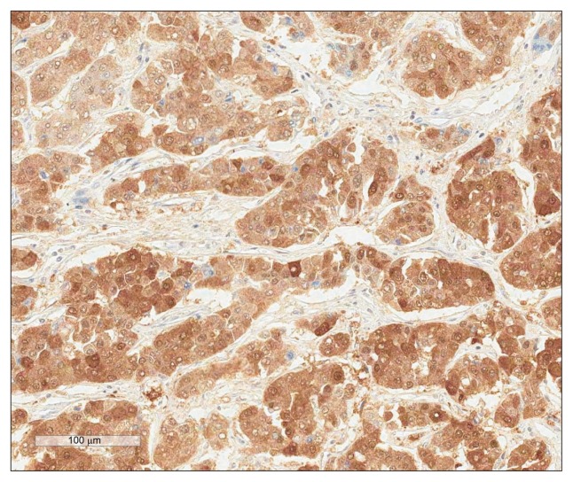
In normal livers, AKR1B10 protein was detected on the cytoplasm in 0% to 5% of hepatocytes with weak or moderate staining intensity. In HCC, immunoreactivity for AKR1B10 was observed in the cytoplasm of tumor cells with moderate staining intensity. In a few cases nuclear expression was also found. Thirty three HCCs (12.9%) lacked any AKR1B10 expression. High AKR1B10 protein expression was observed in 125 of the 255 HCCs (49.0%) (Fig. 1). High AKR1B10 expression was significantly associated with no major portal vein invasion (p=0.022), no intrahepatic metastasis (p=0.010), lower AJCC T-stage (p=0.016), lower BCLC stage (p=0.006), and lower α-fetoprotein level (p=0.020). High AKR1B10 expression was correlated with no early recurrence (p=0.022), but not with the late recurrence (p=0.255) (Table 1).
Fig. 1.
Immunostaining of Aldo-Keto Reductase 1B10 showing cytoplasmic immunoreactivity in hepatocellular carcinoma tissue (horse-radish peroxidase stain, ×200).
Table 1.
Correlation between Aldo-Keto Reductase 1B10 Expression and the Clinicopathologic Features of 255 Hepatocellular Carcinomas
| Variable | No. | High AKR1B10 expression | p-value |
|---|---|---|---|
| Age, yr | |||
| ≤55 | 150 | 81 (54.0) | 0.057 |
| >55 | 105 | 44 (41.9) | |
| Gender | |||
| Female | 47 | 20 (42.6) | 0.326 |
| Male | 208 | 105 (50.5) | |
| Tumor size, cm | |||
| ≤5.0 | 159 | 83 (52.2) | 0.191 |
| >5.0 | 96 | 42 (43.8) | |
| Edmondson grade | |||
| I | 24 | 7 (29.2) | 0.067 |
| II | 175 | 93 (53.1) | |
| III | 56 | 25 (44.6) | |
| Microvascular invasion | |||
| Negative | 112 | 62 (55.4) | 0.073 |
| Positive | 143 | 63 (44.1) | |
| Major portal vein invasion | |||
| Negative | 243 | 123 (50.6) | 0.022 |
| Positive | 12 | 2 (16.7) | |
| Intrahepatic metastasis | |||
| Negative | 192 | 103 (53.6) | 0.010 |
| Positive | 63 | 22 (34.9) | |
| Multicentric occurrence | |||
| Negative | 243 | 122 (50.2) | 0.088 |
| Positive | 12 | 3 (25.0) | |
| AJCC T-stage | |||
| 1 | 108 | 61 (56.5) | 0.049 |
| 2 | 97 | 47 (48.5) | |
| 3 | 44 | 14 (31.8) | |
| 4 | 6 | 3 (50.0) | |
| BCLC stage | |||
| 0–A | 138 | 77 (55.8) | 0.006 |
| B | 103 | 48 (44.7) | |
| C | 14 | 2 (14.3) | |
| Albumin level, g/dL | |||
| >3.5 | 228 | 111 (48.7) | 0.756 |
| ≤3.5 | 27 | 14 (51.9) | |
| AFP level, ng/mL* | |||
| ≤200 | 148 | 82 (55.4) | 0.020 |
| >200 | 97 | 39 (40.2) | |
| Etiology | |||
| Nonviral | 34 | 17 (50.0) | 0.144 |
| HBV | 195 | 100 (51.3) | |
| HCV | 26 | 8 (30.8) | |
| Liver cirrhosis | |||
| Negative | 129 | 61 (47.3) | 0.575 |
| Positive | 126 | 64 (50.8) | |
| Early recurrence (≤2 yr) | |||
| Negative | 116 | 66 (56.9) | 0.022 |
| Positive | 139 | 59 (42.4) | |
| Late recurrence (>2 yr) | |||
| Negative | 211 | 100 (47.4) | 0.255 |
| Positive | 44 | 25 (56.8) | |
Data are presented as number (%).
AKR1B10, Aldo-Keto Reductase 1B10; AJCC, American Joint Committee on Cancer; BCLC, Barcelona Clinic Liver Cancer; AFP, α-fetoprotein; HBV, hepatitis B virus; HCV, hepatitis C virus.
Information was available on 245 cases.
2. Prediction of early recurrence in HCC
In univariate analyses, early recurrence was significantly associated with larger tumor size (p=0.001), Edmondson grade III (p=0.005), microvascular invasion (p<0.001), major portal vein invasion (p=0.029), intrahepatic metastasis (p<0.001), higher AJCC T-stage (p<0.001), higher BCLC stage (p<0.001), viral etiology (p=0.018), and low AKR1B10 expression (p=0.022). As AJCC T-stage and BCLC stage were associated with vascular invasion, we did not make multiple analyses with these indices to avoid potential bias. In multivariate analyses, intrahepatic metastasis (p<0.001) was independent predictor of early recurrence (Table 2).
Table 2.
Univariate and Multivariate Logistic Regression Models for the Prediction of Early Tumor Recurrence in 255 Patients with Hepatocellular Carcinoma
| Variable | Univariate model | Multivariate model | ||||
|---|---|---|---|---|---|---|
|
|
|
|||||
| Coefficient | OR (95% CI) | p-value | Coefficient | OR (95% CI) | p-value | |
| Age, yr (≤55 vs >55) | 0.181 | 1.12 (0.73–1.98) | 0.480 | |||
| Gender (female vs male) | 0.065 | 1.07 (0.56–2.01) | 0.841 | |||
| Tumor size, cm (≤5.0 vs >5.0) | 0.879 | 2.41 (1.42–4.09) | 0.001 | 0.135 | 1.14 (0.58–2.25) | 0.696 |
| Edmondson grade (I+II vs III) | 0.926 | 2.53 (1.33–4.80) | 0.005 | 0.542 | 1.72 (0.81–3.64) | 0.157 |
| Microvascular invasion (no vs yes) | 1.327 | 3.77 (2.24–6.36) | <0.001 | 0.480 | 1.62 (0.85–3.08) | 0.143 |
| Major portal vein invasion (no vs yes) | 2.291 | 9.88 (1.26–77.74) | 0.029 | −0.276 | 0.76 (0.07–7.79) | 0.816 |
| Intrahepatic metastasis (no vs yes) | 2.766 | 15.90 (6.10–41.41) | <0.001 | 2.313 | 10.11 (3.23–31.61) | <0.001 |
| Multicentric occurrence (no vs yes) | 0.536 | 1.71 (0.50–5.83) | 0.391 | |||
| AJCC T-stage (1 vs 2+3+4) | 1.397 | 4.05 (2.39–6.85) | <0.001 | |||
| BCLC stage (0+A vs B+C) | 1.274 | 3.58 (2.12–6.04) | <0.001 | |||
| Albumin level, g/dL (>3.5 vs ≤3.5) | 0.962 | 2.62 (1.07–6.43) | 0.036 | |||
| AFP level, ng/mL* (≤200 vs >200) | 0.456 | 1.58 (0.94–2.66) | 0.086 | |||
| Etiology (nonviral vs viral) | 0.907 | 2.48 (1.17–5.26) | 0.018 | 0.857 | 2.36 (0.98–5.65) | 0.054 |
| Liver cirrhosis (no vs yes) | 0.401 | 1.49 (0.91–2.45) | 0.113 | |||
| AKR1B10 expression (low vs high) | −0.582 | 0.56 (0.34–0.92) | 0.022 | −0.418 | 0.66 (0.37–1.17) | 0.153 |
OR, odds ratio; CI; confidence interval; AJCC, American Joint Committee on Cancer; BCLC, Barcelona Clinic Liver Cancer; AFP, α-fetoprotein; AKR1B10, Aldo-Keto Reductase 1B10.
Information was available on 245 cases.
3. Survival analysis
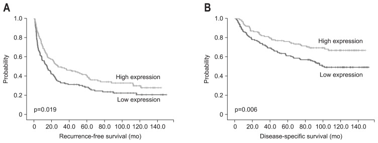
The RFS and DSS rates for 255 HCC patients were 39.9% and 76.1% at 3 years, 34.7% and 68.6% at 5 years, 28.8% and 63.8% at 7 years, and 27.7% and 57.8% at 9 years, respectively. On univariate analyses, larger tumor size, Edmondson grade III, microvascular invasion, major portal vein invasion, intra-hepatic metastasis, higher AJCC T-stage, higher BCLC stage, lower albumin level, and higher α-fetoprotein level showed unfavorable influences on both RFS and DSS. Viral etiology and liver cirrhosis showed unfavorable influences on RFS. High AKR1B10 expression showed a favorable influence on both RFS (p=0.020) and DSS (p=0.007) (Table 3). The 5-year RFS rate of the AKR1B10-high expression group was significantly higher than that of the AKR1B10-low expression group (40.0% vs 27.7%) (Fig. 2A). The mean RFS of AKR1B10-high expression group and AKR1B10-low expression group were 60.6 and 46.0 months, respectively. The 5-year DSS rate of the AKR1B10-high expression group was significantly higher than that of the AKR1B10-low expression group (76.2% vs 61.3%) (Fig. 2B). The mean DSS of AKR1B10-high expression group and AKR1B10-low expression group were 111.8 and 93.7 months, respectively.
Table 3.
Univariate Analyses of the Recurrence-Free Survival and the Disease-Specific Survival in 255 Patients with Hepatocellular Carcinoma
| Variable | Recurrence-free survival | Disease-specific survival | ||
|---|---|---|---|---|
|
|
|
|||
| HR (95% CI) | p-value | HR (95% CI) | p-value | |
| Age, yr (≤55 vs >55) | 0.97 (0.72–1.30) | 0.839 | 0.94 (0.63–1.40) | 0.747 |
| Gender (female vs male) | 0.98 (0.68–1.41) | 0.898 | 1.28 (0.76–2.16) | 0.357 |
| Tumor size, cm (≤5.0 vs >5.0) | 1.62 (1.21–2.18) | 0.001 | 2.51 (1.70–3.73) | <0.001 |
| Edmondson grade (I+II vs III) | 1.88 (1.35–2.62) | <0.001 | 2.12 (1.39–3.25) | 0.001 |
| Microvascular invasion (no vs yes) | 2.15 (1.59–2.91) | <0.001 | 3.03 (1.95–4.72) | <0.001 |
| Major portal vein invasion (no vs yes) | 3.82 (2.06–7.06) | <0.001 | 6.37 (3.27–12.42) | <0.001 |
| Intrahepatic metastasis (no vs yes) | 4.27 (3.08–5.91) | <0.001 | 5.79 (3.86–8.67) | <0.001 |
| Multicentric occurrence (no vs yes) | 1.57 (0.85–2.89) | 0.147 | 0.87 (0.32–2.37) | 0.785 |
| AJCC T-stage (1 vs 2+3+4) | 2.24 (1.65–3.04) | <0.001 | 3.18 (2.02–5.01) | <0.001 |
| BCLC stage (0+A vs B+C) | 1.99 (1.49–2.66) | <0.001 | 3.32 (2.20–5.02) | <0.001 |
| Albumin level, g/dL (>3.5 vs ≤3.5) | 1.87 (1.22–2.88) | 0.004 | 2.45 (1.45–4.15) | 0.001 |
| AFP level, ng/mL* (≤200 vs >200) | 1.62 (1.20–2.18) | 0.002 | 1.49 (1.00–2.22) | 0.052 |
| Etiology (nonviral vs viral) | 1.88 (1.14–3.09) | 0.014 | 1.53 (0.79–2.93) | 0.206 |
| Liver cirrhosis (no vs yes) | 1.37 (1.03–1.83) | 0.034 | 1.13 (0.76–1.67) | 0.553 |
| AKR1B10 expression (low vs high) | 0.71 (0.53–0.95) | 0.020 | 0.57 (0.38–0.86) | 0.007 |
HR, hazard ratio; CI, confidence interval; AJCC, American Joint Committee on Cancer; BCLC, Barcelona Clinic Liver Cancer; AFP, α-fetoprotein; AKR1B10, Aldo-Keto Reductase 1B10.
Information was available on 245 cases.
Fig. 2.
Kaplan-Meier survival curves showing recurrence-free survival (A) and disease-specific survival (B) according to Aldo-Keto Reductase 1B10 expression in 255 hepatocellular carcinomas.
On multivariate analyses, intrahepatic metastasis and lower albumin level were independent predictors of both shorter RFS and shorter DSS. Liver cirrhosis was an independent predictor of shorter RFS. High AKR1B10 expression was an independent predictor of both longer RFS (p=0.024) and longer DSS (p=0.046). AKR1B10-high expression patients were less likely to suffer from recurrence than AKR1B10-low expression patients (hazard ratio, 0.70) and were less likely to suffer from disease-specific death than AKR1B10-low expression patients (hazard ratio, 0.65) (Table 4).
Table 4.
Multivariate Analyses of the Recurrence-Free Survival and the Disease-Specific Survival in 255 Patients with Hepatocellular Carcinoma
| Variable | Recurrence-free survival | Disease-specific survival | ||
|---|---|---|---|---|
|
|
|
|||
| HR (95% CI) | p-value | HR (95% CI) | p-value | |
| Tumor size, cm (≤5.0 vs >5.0) | 1.11 (0.78–1.59) | 0.561 | 1.40 (0.85–2.29) | 0.183 |
| Edmondson grade (I+II vs III) | 1.15 (0.79–1.67) | 0.454 | 1.18 (0.74–1.89) | 0.487 |
| Microvascular invasion (no vs yes) | 1.36 (0.93–1.98) | 0.109 | 1.48 (0.84–2.59) | 0.175 |
| Major portal vein invasion (no vs yes) | 0.89 (0.43–1.83) | 0.754 | 1.50 (0.71–3.17) | 0.294 |
| Intrahepatic metastasis (no vs yes) | 3.22 (2.11–4.90) | <0.001 | 3.57 (2.12–6.00) | <0.001 |
| Albumin level, g/dL (>3.5 vs ≤3.5) | 1.71 (1.09–2.67) | 0.019 | 2.32 (1.35–4.00) | 0.002 |
| Etiology (nonviral vs viral) | 1.49 (0.88–2.52) | 0.135 | - | - |
| Liver cirrhosis (no vs yes) | 1.42 (1.03–1.96) | 0.034 | 1.53 (0.99–2.38) | 0.057 |
| AKR1B10 expression (low vs high) | 0.70 (0.52–0.95) | 0.024 | 0.65 (0.42–0.99) | 0.046 |
HR, hazard ratio; CI, confidence interval; AKR1B10, Aldo-Keto Reductase 1B10.
DISCUSSION
AKR1B10 upregulation was an independent risk factor for HCC development in a matched case-control study.23 AKR1B10 promotes cancer cell growth and survival by increasing lipid synthesis and eliminating cytotoxic carbonyls.24 Enhanced lipogenesis is important early event in cancer development, including HCC.25
In this study, most of the HCC tissues expressed higher levels of AKR1B10 protein than normal liver, with high AKR1B10 expression in 49.0% (125 of 255) of HCCs. High AKR1B10 expression was significantly associated with lower AJCC T-stage and lower BCLC stage, which was consistent with previous reports.14,26 High AKR1B10 expression was correlated with no early recurrence, but not with the late recurrence. The underlying mechanism on this inverse correlation with tumor stage remains speculative. One possible explanation might be the complicated expression regulation of AKR1B10 at the promoter level. In the AKR1B10 promoter, multiple putative oncogenic and tumor suppressor protein-binding sites were recognized, including c-Ets-1, C/EBP, and p53.27 Deregulation or nonspecific switching on/off of certain transcriptional mechanisms, which occurs in more malignant HCC cells, might downregulate the expression of AKR1B10.
To the best of our knowledge, this is the first report to show high AKR1B10 expression as an independent predictor of both longer RFS and longer DSS. In recent studies, high AKR1B10 expression was associated with a better overall survival compared to low expression14 and patients with AKR1B10-strongly positive HCCs showed a better DSS compared with patients with negative HCCs.26 These findings indicate that AKR1B10 is a new clinically useful marker for predicting the prognosis of HCC after curative hepatectomy and the results could help clinicians identify patients at low risk of recurrence after surgery. AKR1B10 could be used as an immunohistochemical biomarker to detect patients with a good prognosis.
In conclusion, this study demonstrated that AKR1B10 protein expression in HCC tissues might be a clinically useful predictive marker for good prognosis of HCC after curative hepatectomy in a large number of HCC patients with long-term follow-up. Further studies are warranted for the validation of AKR1B10 as a novel prognostic biomarker in HCCs.
Footnotes
CONFLICTS OF INTEREST
No potential conflict of interest relevant to this article was reported.
REFERENCES
- 1.Poon RT. Prevention of recurrence after resection of hepatocellular carcinoma: a daunting challenge. Hepatology. 2011;54:757–759. doi: 10.1002/hep.24569. [DOI] [PubMed] [Google Scholar]
- 2.Llovet JM, Schwartz M, Mazzaferro V. Resection and liver transplantation for hepatocellular carcinoma. Semin Liver Dis. 2005;25:181–200. doi: 10.1055/s-2005-871198. [DOI] [PubMed] [Google Scholar]
- 3.Inagaki Y, Tang W, Zhang L, Du G, Xu W, Kokudo N. Novel aminopeptidase N (APN/CD13) inhibitor 24F can suppress invasion of hepatocellular carcinoma cells as well as angiogenesis. Biosci Trends. 2010;4:56–60. [PubMed] [Google Scholar]
- 4.Song P, Tang W, Tamura S, et al. The management of hepatocellular carcinoma in Asia: a guideline combining quantitative and qualitative evaluation. Biosci Trends. 2010;4:283–287. [PubMed] [Google Scholar]
- 5.Lee KW, Ko BC, Jiang Z, Cao D, Chung SS. Overexpression of al-dose reductase in liver cancers may contribute to drug resistance. Anticancer Drugs. 2001;12:129–132. doi: 10.1097/00001813-200102000-00005. [DOI] [PubMed] [Google Scholar]
- 6.Barski OA, Tipparaju SM, Bhatnagar A. The aldo-keto reductase superfamily and its role in drug metabolism and detoxification. Drug Metab Rev. 2008;40:553–624. doi: 10.1080/03602530802431439. [DOI] [PMC free article] [PubMed] [Google Scholar]
- 7.Cao D, Fan ST, Chung SS. Identification and characterization of a novel human aldose reductase-like gene. J Biol Chem. 1998;273:11429–11435. doi: 10.1074/jbc.273.19.11429. [DOI] [PubMed] [Google Scholar]
- 8.Scuric Z, Stain SC, Anderson WF, Hwang JJ. New member of al-dose reductase family proteins overexpressed in human hepatocellular carcinoma. Hepatology. 1998;27:943–950. doi: 10.1002/hep.510270408. [DOI] [PubMed] [Google Scholar]
- 9.Fukumoto S, Yamauchi N, Moriguchi H, et al. Overexpression of the aldo-keto reductase family protein AKR1B10 is highly correlated with smokers’ non-small cell lung carcinomas. Clin Cancer Res. 2005;11:1776–1785. doi: 10.1158/1078-0432.CCR-04-1238. [DOI] [PubMed] [Google Scholar]
- 10.Loeffler-Ragg J, Mueller D, Gamerith G, et al. Proteomic identification of aldo-keto reductase AKR1B10 induction after treatment of colorectal cancer cells with the proteasome inhibitor bortezomib. Mol Cancer Ther. 2009;8:1995–2006. doi: 10.1158/1535-7163.MCT-08-0987. [DOI] [PubMed] [Google Scholar]
- 11.Yan R, Zu X, Ma J, Liu Z, Adeyanju M, Cao D. Aldo-keto reductase family 1 B10 gene silencing results in growth inhibition of colorectal cancer cells: implication for cancer intervention. Int J Cancer. 2007;121:2301–2306. doi: 10.1002/ijc.22933. [DOI] [PubMed] [Google Scholar]
- 12.Satow R, Shitashige M, Kanai Y, et al. Combined functional genome survey of therapeutic targets for hepatocellular carcinoma. Clin Cancer Res. 2010;16:2518–2528. doi: 10.1158/1078-0432.CCR-09-2214. [DOI] [PubMed] [Google Scholar]
- 13.Liu Z, Yan R, Al-Salman A, et al. Epidermal growth factor induces tumour marker AKR1B10 expression through activator protein-1 signalling in hepatocellular carcinoma cells. Biochem J. 2012;442:273–282. doi: 10.1042/BJ20111322. [DOI] [PubMed] [Google Scholar]
- 14.Heringlake S, Hofdmann M, Fiebeler A, Manns MP, Schmiegel W, Tannapfel A. Identification and expression analysis of the aldo-ketoreductase1-B10 gene in primary malignant liver tumours. J Hepatol. 2010;52:220–227. doi: 10.1016/j.jhep.2009.11.005. [DOI] [PubMed] [Google Scholar]
- 15.Edmondson HA, Steiner PE. Primary carcinoma of the liver: a study of 100 cases among 48,900 necropsies. Cancer. 1954;7:462–503. doi: 10.1002/1097-0142(195405)7:3<462::AID-CNCR2820070308>3.0.CO;2-E. [DOI] [PubMed] [Google Scholar]
- 16.Liver Cancer Study Group of Japan. General rules for the clinical and pathological study of primary liver cancer. 2nd ed. Tokyo: Kanehara; 2003. [Google Scholar]
- 17.Edge SB American Joint Committee on Cancer; American Cancer Society; American College of Surgeons. AJCC cancer staging manual. 7th ed. New York: Springer; 2010. [Google Scholar]
- 18.Llovet JM, Brú C, Bruix J. Prognosis of hepatocellular carcinoma: the BCLC staging classification. Semin Liver Dis. 1999;19:329–338. doi: 10.1055/s-2007-1007122. [DOI] [PubMed] [Google Scholar]
- 19.Imamura H, Matsuyama Y, Tanaka E, et al. Risk factors contributing to early and late phase intrahepatic recurrence of hepatocellular carcinoma after hepatectomy. J Hepatol. 2003;38:200–207. doi: 10.1016/S0168-8278(02)00360-4. [DOI] [PubMed] [Google Scholar]
- 20.Shimada M, Hamatsu T, Yamashita Y, et al. Characteristics of multicentric hepatocellular carcinomas: comparison with intrahepatic metastasis. World J Surg. 2001;25:991–995. doi: 10.1007/s00268-001-0068-6. [DOI] [PubMed] [Google Scholar]
- 21.Hoshida Y, Villanueva A, Kobayashi M, et al. Gene expression in fixed tissues and outcome in hepatocellular carcinoma. N Engl J Med. 2008;359:1995–2004. doi: 10.1056/NEJMoa0804525. [DOI] [PMC free article] [PubMed] [Google Scholar]
- 22.Ahn S, Hyeon J, Park CK. Metadherin is a prognostic predictor of hepatocellular carcinoma after curative hepatectomy. Gut Liver. 2013;7:206–212. doi: 10.5009/gnl.2013.7.2.206. [DOI] [PMC free article] [PubMed] [Google Scholar]
- 23.Sato S, Genda T, Hirano K, et al. Up-regulated aldo-keto reductase family 1 member B10 in chronic hepatitis C: association with serum alpha-fetoprotein and hepatocellular carcinoma. Liver Int. 2012;32:1382–1390. doi: 10.1111/j.1478-3231.2012.02827.x. [DOI] [PMC free article] [PubMed] [Google Scholar]
- 24.Wang C, Yan R, Luo D, Watabe K, Liao DF, Cao D. Aldo-keto reductase family 1 member B10 promotes cell survival by regulating lipid synthesis and eliminating carbonyls. J Biol Chem. 2009;284:26742–26748. doi: 10.1074/jbc.M109.022897. [DOI] [PMC free article] [PubMed] [Google Scholar]
- 25.Yahagi N, Shimano H, Hasegawa K, et al. Co-ordinate activation of lipogenic enzymes in hepatocellular carcinoma. Eur J Cancer. 2005;41:1316–1322. doi: 10.1016/j.ejca.2004.12.037. [DOI] [PubMed] [Google Scholar]
- 26.Schmitz KJ, Sotiropoulos GC, Baba HA, et al. AKR1B10 expression is associated with less aggressive hepatocellular carcinoma: a clinicopathological study of 168 cases. Liver Int. 2011;31:810–816. doi: 10.1111/j.1478-3231.2011.02511.x. [DOI] [PubMed] [Google Scholar]
- 27.Liu Z, Zhong L, Krishack PA, et al. Structure and promoter characterization of aldo-keto reductase family 1 B10 gene. Gene. 2009;437:39–44. doi: 10.1016/j.gene.2009.02.007. [DOI] [PMC free article] [PubMed] [Google Scholar]


