



ABSTRACT
Mitochondrial dynamics, including fusion, fission and translocation, are crucial to cellular homeostasis, with roles in cellular polarity, stress response and apoptosis. Mitochondrial fission has received particular attention, owing to links with several neurodegenerative diseases. A central player in fission is the cytoplasmic dynamin-related GTPase Drp1, which oligomerizes at the fission site and hydrolyzes GTP to drive membrane ingression. Drp1 recruitment to the outer mitochondrial membrane (OMM) is a key regulatory event, which appears to require a pre-constriction step in which the endoplasmic reticulum (ER) and mitochondrion interact extensively, a process termed ERMD (ER-associated mitochondrial division). It is unclear how ER–mitochondrial contact generates the force required for pre-constriction or why pre-constriction leads to Drp1 recruitment. Recent results, however, show that ERMD might be an actin-based process in mammals that requires the ER-associated formin INF2 upstream of Drp1, and that myosin II and other actin-binding proteins might be involved. In this Commentary, we present a mechanistic model for mitochondrial fission in which actin and myosin contribute in two ways; firstly, by supplying the force for pre-constriction and secondly, by serving as a coincidence detector for Drp1 binding. In addition, we discuss the possibility that multiple fission mechanisms exist in mammals.
KEY WORDS: Actin, Mitochondrial fission, Myosin
Introduction
First described as bioblasts in 1890, mitochondria were renamed eight years later as a combination of the Greek terms ‘mitos’, meaning thread, and ‘chondros’ for granule (Benda, 1898). Originating from an endosymbiotic relationship, the mitochondrial genome and cardiolipin-rich inner mitochondrial membrane (IMM) are remnants of the bacterial endosymbiont (Mileykovskaya and Dowhan, 2009), whereas the outer mitochondrial membrane (OMM) likely came from the original host. A major function of the mitochondrion is ATP production, and large amounts of NAD+ and FADH undergo redox cycles in the mitochondrial matrix. In this large flux of electrons, some can become lost, creating free radicals. This process, coupled with limited DNA repair mechanisms, increases the mutational susceptibility of the mitochondrial genome (Youle and van der Bliek, 2012), raising the question of how mitochondrial DNA damage is mitigated.
In 1914, Lewis and Lewis noted that mitochondria were incredibly dynamic – constantly moving, fusing and undergoing fission (Lewis and Lewis, 1914). However, no immediate significance was attributed to these dynamics. Within the past 20 years, there has been a renewed interest, owing to the clear role of mitochondrial dynamics in protection from oxidative damage. The picture emerging is that there are several possible responses to mitochondrial oxidative damage: (1) fusion with a healthy mitochondrion to spread the damage; (2) segregation of damaged components followed by fission of the damaged segment from the healthy segment, with subsequent mitophagy to remove the damaged fission product; or (3) apoptosis, in which the fission machinery also appears to play a role. In addition, mitochondrial dynamics are required for proper mitochondrial distribution during cell division and cell polarization. Owing to the highly polarized state of neurons, mitochondrial dynamics are intimately associated with the pathogenesis of many neurodegenerative diseases, including Alzheimer's, Huntington's, Parkinson's, ALS and Charcot-Marie-Tooth disease. A number of excellent recent reviews cover these subjects in detail (Archer, 2013; Chan, 2012; Friedman and Nunnari, 2014; Hoppins and Nunnari, 2012; Nunnari and Suomalainen, 2012; Vafai and Mootha, 2012; Youle and van der Bliek, 2012).
This Commentary focuses on the participation of the actin cytoskeleton in mitochondrial fission, and we construct a mechanistic model for how actin might produce constrictive force. One goal of our model is to highlight the relevant sizes of the key players, because the dimensions of the ER, mitochondrion and fission apparatus put constraints on the possible mechanisms. For instance, the narrow widths of mammalian mitochondria from many cell types, with diameters frequently in the range of only 150–300 nm, restrict the possible ways in which force can be applied by actin and myosin (Goldstein et al., 1984; Hu et al., 2013; Jans et al., 2013; Kim et al., 2012; Noske et al., 2008; Perkins and Ellisman, 2011; Vafai and Mootha, 2012). In addition, we present the possibility that there might be multiple mechanisms by which actin participates in fission.
Mitochondrial fission – factors and outstanding questions
Before discussing the role of actin, we describe a central protein in mitochondrial fission, Drp1. Drp1 is a dynamin-related GTPase conserved throughout eukaryotes (also referred to as Dnm1, Dlp1, Dvlp1, Dnm1l and Dymple), with work in yeast and Caenorhabditis elegans originally identifying it as a mitochondrial fission factor (Bleazard et al., 1999; Boldogh et al., 2001; Labrousse et al., 1999; Otsuga et al., 1998). The crystal structure of human Drp1 shows that it is elongated, with the GTPase domain at one end (Fröhlich et al., 2013). Purified Drp1 is an X-shaped dimer that assembles into higher-order oligomers under a number of conditions, and it has the ability to tubulate anionic lipid membranes (Fröhlich et al., 2013; Ingerman et al., 2005; Koirala et al., 2013; Macdonald et al., 2014). GTP hydrolysis causes constriction of the tubulated membrane (Koirala et al., 2013; Mears et al., 2011). Drp1 accumulates at mitochondrial fission sites, and its constriction activity appears to be a driving force in fission (Fig. 1A).
Fig. 1.
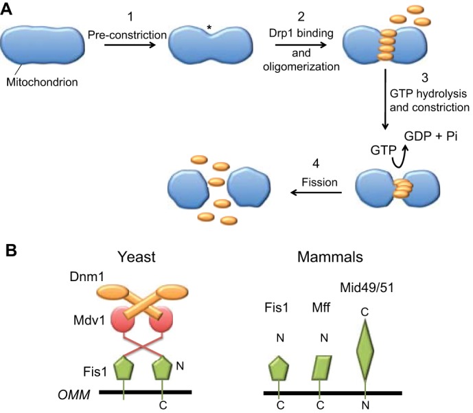
Role of Drp1 in mitochondrial fission. (A) Schematic illustration of the general steps involved in mitochondrial fission. Step 1: a fission site is marked by an unknown mechanism, and this site undergoes ‘pre-constriction’ (asterisk) prior to the arrival of Drp1 (orange). Step 2: Drp1 binds to the pre-constriction site and oligomerizes. Step 3: GTP hydrolysis by the oligomerized Drp1 causes constriction of the fission site. Step 4: fission occurs by an unknown mechanism. (B) Dnm1/Drp1 receptors in budding yeast (left) and in mammals (right). In budding yeast, the OMM protein Fis1 binds to the dimeric adaptor protein Mdv1, which in turn binds to the Dnm1 dimer (its GTPase domain is represented by the oval). A second adaptor protein, Caf4, can act in place of Mdv1. In mammals, four possible OMM proteins have been postulated to act as Drp1 receptors – Fis1, Mff, MiD49 and MiD51. No adaptor proteins that are homologous to Mdv1 or Caf4 have been identified in mammals.
Major questions in the field are: how is fission initiated, what defines a fission site and how is Drp1 recruited? It has been shown that some constriction still occurs at specific points along the mitochondrion in cells that are compromised for Drp1 activity, leading to the hypothesis that a Drp1-independent ‘pre-constriction’ event is necessary for Drp1 recruitment (Fig. 1A) (Friedman et al., 2011; Koch et al., 2004; Labrousse et al., 1999; Legesse-Miller et al., 2003). However, this model then raises other questions. How does pre-constriction occur? How does pre-constriction trigger Drp1 recruitment?
Furthermore, what are the Drp1 ‘receptors’ that bind to Drp1 on the OMM? In yeast, Dnm1 recruitment is relatively well defined: the single-pass OMM protein Fis1 binds to one of two adaptor proteins, Mdv1 or Caf4 (Fig. 1B) (Griffin et al., 2005; Guo et al., 2012; Tieu et al., 2002), either of which then recruits Dnm1. In mammals, the picture is less clear. Although metazoans possess Fis1, they lack obvious adaptor homologs, and the effects of Fis1 on fission have been variable between studies (Gandre-Babbe and van der Bliek, 2008; Koirala et al., 2013; Losón et al., 2013; Otera et al., 2010; Palmer et al., 2013). However, exciting recent work has demonstrated a role for Fis1 during mitophagy (Shen et al., 2014; Yamano et al., 2014). Three other single-pass OMM proteins have been identified as potential Drp1 receptors – Mff, MiD49 and MiD51 (Gandre-Babbe and van der Bliek, 2008; Otera et al., 2010; Palmer et al., 2011) (Fig. 1B). Below, we discuss the possibility that these multiple receptors represent mechanistic variations of mitochondrial fission.
The question of what activates Drp1 recruitment has been addressed in an elegant study that showed fission often occurs at mitochondrial–ER contact sites – areas where the ER is wrapped around mitochondria (Friedman et al., 2011) – in a process called ERMD (ER-associated mitochondrial division). Interestingly, in Drp1-deficient cells, the ER still wraps around mitochondria and these sites still constrict. At least one Drp1 receptor, Mff, has been shown to be enriched at contact points (Friedman et al., 2011), but the mechanism leading to its enrichment is unknown. Overall, there are many questions regarding how the ER mediates pre-constriction, including questions about the mechanism providing the force for pre-constriction.
Evidence for actin, INF2 and myosin in mitochondrial fission
The first suggestion that actin might play a role in mitochondrial fission came from the observation that actin-depolymerizing drugs inhibit Drp1 recruitment and the reduction in mitochondrial length that is mediated by several mitochondrial poisons in CV1-4A monkey kidney cells (De Vos et al., 2005). Subsequently, it was shown that changes in the levels or distribution of actin filaments cause alterations in mitochondrial length and in Drp1 recruitment in Drosophila neurons and Cos-1 cells (DuBoff et al., 2012). However, it was not clear how actin could influence mitochondrial fission or what would assemble these actin filaments.
A more recent study linked actin to ERMD through the formin INF2 (Korobova et al., 2013). Formins are actin-assembly factors that can accelerate both the nucleation and elongation of actin filaments through their binding to the fast-growing ‘barbed’ end of the filament (Fig. 2A) (Higgs, 2005). INF2 has additional effects on actin filaments (see Box 1). INF2 is expressed as two isoforms that differ at their C-termini. The INF2-ER variant (also called INF2-CAAX) contains an 18-amino-acid C-terminus that is post-translationally prenylated, whereas the INF2-Cyto variant (also called INF2-nonCAAX) contains a non-prenylated nine-amino-acid C-terminus. This difference essentially creates two different proteins, with INF2-ER being ER-bound and INF2-Cyto being cytosolic (Chhabra et al., 2009; Ramabhadran et al., 2011). Whereas suppression of INF2-Cyto causes Golgi fragmentation (Ramabhadran et al., 2011), that of INF2-ER results in mitochondrial elongation (Korobova et al., 2013). Actin filaments accumulate at ER–mitochondrial contact sites that undergo constriction (Korobova et al., 2013; M. Karbowski, personal communication). Furthermore, two lines of evidence suggest that INF2 acts upstream of Drp1; INF2 depletion decreases mitochondrially-associated Drp1, and Drp1 inhibition reduces mitochondrial fragmentation by constitutively active INF2 (Korobova et al., 2013).
Fig. 2.

Mechanisms of actin-based membrane constriction. (A) The effects of formins on actin. Upper panel, a formin dimer (blue) can enhance the nucleation of actin monomers (red). Subsequently, it remains at the barbed end of the elongating filament, regulating elongation rate by controlling monomer addition. For more detail regarding formin biochemistry, see Higgs, 2005 and Chesarone et al., 2010. Lower panel, INF2 is additionally able to sever filaments and enhance their depolymerization through its ability to bind to the sides of filaments by encircling them. Upon ATP hydrolysis and phosphate release from actin subunits in the filament, INF2 severs the filament and subsequently enhances depolymerization. For further details regarding INF2 biochemistry, see Gurel et al., 2014. (B) Non-muscle myosin II. Upper panel, the fundamental unit of myosin II is a multi-protein complex of two heavy chains, two essential light chains (ELC) and two regulatory light chains (RLC). The heavy chains are tightly dimerized in parallel by their coiled-coil tails of ∼160 nm length. We refer to this fundamental unit as the ‘dimer’ because the dimerized heavy chains dominate the structure. Lower panel, non-muscle myosin II can oligomerize further to create a bipolar filament with the motor domain heads at each end and a ‘bare zone’ in the center. In the presence of ATP, myosin II assumes a ‘10S’ compact structure, presumably by folding the tail into three segments. RLC phosphorylation allows for bipolar filament assembly from the compact structure in the presence of ATP. The latter is a biochemical observation and has not been documented in non-muscle cells. A ‘typical’ mitochondrial diameter is also shown for size comparison. (C) Actin-polymerization-based membrane deformation by monomer addition to membrane-abutting filament barbed ends. Left, the classic example of Arp2/3-complex-based dendritic nucleation is shown. Right, an example of a formin tethered to the membrane directing monomer addition to the barbed end. The Arp2/3 complex and formin are shown in blue. (D) Myosin-II-based membrane deformation. Motor activity of the bipolar myosin II filament on anti-parallel membrane-attached actin filaments causes the deformation and thus constriction of the membrane. In the example shown here, the barbed ends of the filament are tethered to the membrane by a formin.
Box 1. The biochemical properties of INF2.
INF2 is a formin protein. Formins are actin-binding proteins (15 in mammals) that generally act as actin filament assembly factors. As with most formins, INF2 accelerates the nucleation of new actin filaments in vitro, then remains bound to the elongating barbed end, moving processively with this end as new monomers are added. Through this mechanism, INF2 can regulate filament elongation rate and prevent elongation termination by capping proteins (Chesarone et al., 2010; Higgs, 2005). In cells, there is a question as to whether formins nucleate actin filaments or are elongation factors working downstream of other nucleation factors (Block et al., 2012; Quinlan, 2013; Quinlan et al., 2007). The answer to this question might depend on both the formin and the cellular context.
In addition to accelerating actin polymerization, INF2 also accelerates depolymerization, which is unique amongst formins (Chhabra and Higgs, 2006). How can INF2 do both? The switch between polymerization and depolymerization is triggered by the change in the nucleotide state of actin upon polymerization. After adding to the filament barbed end, the actin monomer hydrolyzes its bound ATP and releases the phosphate product. Both reactions (hydrolysis and phosphate release) are slower than polymerization, so that the ‘newer’ end of the filament is ATP bound whereas the ‘older’ end is ADP bound. INF2 severs ADP-bound filament regions, by encircling the filament and disrupting filament structure (Gurel et al., 2014). INF2 can sever at any location along the filament as long as phosphate has been released, but the requirement for phosphate release means that ‘older’ filament segments are favored for severing. Subsequent to severing, INF2 increases the depolymerization rate of the shorter filament segments by a mechanism that we do not fully understand. Recently, we have found that INF2 dramatically increases the ATP turnover rate of actin even when actin is not polymerized (P.S.G. and H.N.H., unpublished observations), a property that might contribute to depolymerization.
This unusual depolymerization activity might allow INF2 to mediate both filament assembly and disassembly at the mitochondrial fission site, resulting in highly transient filaments that exist only for the time required to mediate pre-constriction and Drp1 recruitment. In addition, the severing activity of INF2 might allow actin filament release from the ER and subsequent direct binding to the mitochondrion, as stated in step 4 of the mitokinesis model.
Finally, INF2 binds to microtubules with high affinity (Gaillard et al., 2011). The physiological relevance of this interaction is unclear, but the possibilities are intriguing, because both ER and mitochondria translocate along microtubules (Goyal and Blackstone, 2013).
Mounting evidence suggests that myosin II also acts in mitochondrial fission. There are three mammalian non-muscle myosin II paralogs, IIA, IIB and IIC, with IIA and IIB being the most widely expressed. All mammalian non-muscle myosin II paralogs form anti-parallel filaments that contain 14–30 motor-containing heads at each end (Billington et al., 2013) (Fig. 2B). Suppression of myosin regulatory light chain (MRLC) in Cos7 cells (DuBoff et al., 2012) or of myosin IIA or IIB heavy chains in U2OS cells (Korobova et al., 2014) causes mitochondrial elongation and decreases the amount of mitochondrially bound Drp1. Two myosin-directed small molecule inhibitors also cause mitochondrial elongation – blebbistatin (Korobova et al., 2014) and ML-7 (DuBoff et al., 2012). Myosin II inhibition also reverses the effect of constitutively active INF2 (short mitochondria) (Korobova et al., 2014). Active MRLC localizes to constriction sites in an INF2-dependent manner (Korobova et al., 2014), suggesting that myosin II recruitment requires INF2-mediated polymerization of actin filaments. Results from Drosophila show that the amount of actin associated with mitochondria is reduced in myosin mutant flies (DuBoff et al., 2012), suggesting a reciprocal relationship between the accumulation of actin and myosin.
Finally, there is increasing evidence that Drp1 interacts with actin filaments. In Drosophila extracts, Drp1 co-precipitates with actin filaments (DuBoff et al., 2012). Myosin II appears to enhance this interaction, because myosin mutants reduce the co-precipitation of Drp1 and actin (DuBoff et al., 2012). Using purified proteins, we have found evidence that mammalian Drp1 directly binds to actin filaments with sub-micromolar affinity, increasing the GTP hydrolysis rate of Drp1 (A.L.H. and H.N.H., unpublished observations). The ability to bind to actin filaments might be common to the dynamin family, as both dynamin 1 and 2 are also able to bind to actin filaments (Gu et al., 2010).
Mechanistic model for actomyosin-mediated mitochondrial fission
The results discussed above imply that INF2, actin and myosin II work together to facilitate the accumulation of Drp1 at the fission site, perhaps by driving pre-constriction. How might this force be generated? Actin-based force generation on membranes has been the subject of intense research for many years, with two general mechanisms being proposed: (1) that actin polymerization ‘pushes’ the membrane in front of it, or (2) that myosin motors ‘pull’ the actin filament and attached membrane, thereby producing a constrictive force in the case of bipolar myosin II filaments (Fig. 2C,D). Examples of actin-polymerization-induced force are endocytosis and leading edge protrusion during cell motility, whereas myosin-II-based forces power stress fiber contraction, cell cortex contraction and the retrograde flow of actin networks (Blanchoin et al., 2014).
Cytokinesis is another myosin-dependent event, resulting in membrane constriction and division. The best-studied model for cytokinesis is fission yeast, for which a clear set of molecular factors and sequence of events has been defined (Lee et al., 2012; Pollard, 2010). Myosin II accumulates at the prospective cleavage furrow in precursor structures, called ‘nodes’. Formin-mediated actin polymerization from these nodes results in their myosin-II-mediated focusing into the more compact cytokinetic ring. Constriction of this ring results in plasma membrane ingression (Fig. 3). Although myosin motor activity is required early in the process, constriction of the full ring does not appear to be driven by myosin, but by other forces, such as septation (Proctor et al., 2012).
Fig. 3.
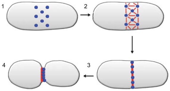
Model for fission yeast cytokinesis. Step 1: at the presumptive cleavage site on the plasma membrane, ∼130 ‘nodes’ (blue) assemble the cytokinetic components; each node contains two formin dimers and eight myosin II dimers. Step 2: the formins nucleate actin filaments (red), which remain attached to the formin at their barbed ends. The pointed end of the actin filament interacts with myosin II on a neighboring node by a ‘search and capture’ mechanism. Step 3: myosin II activity along the actin filaments causes the nodes to condense into a compact ring structure. Step 4: further myosin II activity causes the ring to further condense, thereby causing constriction of the plasma membrane. Additional force-generating mechanisms might exist that become more important at subsequent stages of constriction and fission (Proctor et al., 2012).
Based on the fact that some of the proteins involved in cytokinesis and mitochondrial fission are similar (actin, a formin, myosin II), we propose a mechanistic model that we call ‘mitokinesis’ (Fig. 4A). As discussed below, we think that there might be more than one mechanism by which actin participates in fission, including mechanisms involving actin-polymerization-driven force production. The model presented here, however, provides a starting point for elucidating the roles of INF2 and myosin II in what is perhaps one type of fission.
Fig. 4.

The mitokinesis model of mitochondrial fission. (A) Step 1: The ER (green) and mitochondrion (blue) interact at the future site of mitochondrial fission. Step 2: actin filaments (red) are nucleated and elongate with INF2 (gray donut) at their barbed ends, thus staying tethered to the ER through INF2. Step 3: Myosin II is recruited to the fission site. The recruited myosin might come from a cytosolic pool of free dimers or from pre-assembled bipolar filaments. Step 4 (pre-constriction): myosin II activity on anti-parallel actin filaments causes deformation of the network, resulting in constriction of both the surrounding ER and the underlying mitochondrion. Step 5 (coincidence detection): Drp1 (brown circles) binds and oligomerizes at the pre-constriction site, owing to coincidence detection of two signals – an OMM-bound Drp1 receptor (purple) and actin filaments. Step 6: GTPase activity of Drp1 causes increased mitochondrial constriction. Step 7: The actual membrane fission process occurs, and components of the fission complex (actin, myosin, Drp1 oligomer) disassemble. (B) Two possible models for myosin II arrangement during mitokinesis. Side views of the enlarged fission site at Step 3 and 4 of the model depicted in A are shown. In both models, ER is not required to completely encircle the mitochondrion, but encircles sufficiently to allow a continuous actomyosin ring around the mitochondrion. (Bi) Bipolar arrangement. Myosin II is assembled in the form of bipolar filaments around the mitochondrion and acts on actin filaments that are bound by INF2 at the ER membrane to constrict both ER and the mitochondrion. (Bii) Nodal arrangement. Several nodes assemble on the ER membrane and each contains INF2 and myosin II. Actin filaments that are assembled at one node bind to myosin II at neighboring nodes. Myosin II activity then pulls the nodes together, in turn constricting both the ER and mitochondrial membranes. In this model, similar to models of fission yeast cytokinesis (see Fig. 3), the myosin II is organized as individual dimers that are attached to the nodes by their tails. Possibly, the tails are also folded so that the myosin II dimer is shorter than its extended length of 175 nm.
Step 1 – Initial ER–mitochondrion interaction
The elegant work from Friedman et al. clearly shows that the ER makes extensive contact with mitochondria at many mitochondrial constriction sites (Friedman et al., 2011). The mechanisms that target and deliver the ER to mitochondria are unknown in mammals; however, we suspect that the cytoskeleton is likely to be involved and we predict this to be an exciting area of future research. We postulate that a major consequence of this contact is to activate INF2-mediated production of actin filaments. The molecules that mediate ER–mitochondrial contacts in mammals are unclear (discussed further below).
How would ER–mitochondrial contact activate INF2? INF2 is regulated by autoinhibition, similar to other formins, but the autoinhibitory mechanism for INF2 is not straightforward. In the case of formins such as mDia1, the N-terminal diaphanous inhibitory domain (DID) binds tightly to the C-terminal diaphanous autoregulatory domain (DAD) and potently inhibits actin polymerization activity (Li and Higgs, 2003; Li and Higgs, 2005). Although INF2 possesses both DID and DAD, these domains interact with much lower affinity as compared with those of mDia1 (Ramabhadran et al., 2013). Furthermore, the DAD of INF2 binds to actin monomers, and this interaction competes with the DID–DAD interaction (Ramabhadran et al., 2013). Nevertheless, INF2 is clearly in an ‘off’ state on the bulk ER, and inhibition requires the DID–DAD interaction (Ramabhadran et al., 2013). We therefore propose that additional inhibitory factors exist, either in the cytoplasm or at the ER surface, and that a factor localized at the OMM acts as an INF2 activator. This molecule might even be one of the Drp1 receptors. In this regard, it is interesting that MID51 could be involved, as its overexpression causes actin filament accumulation around mitochondria (Palmer et al., 2011).
Step 2 – Actin filament production at the fission site
ER–mitochondrial contact results in actin filament nucleation through INF2 or other proteins. Whatever the nucleator, the result is that the filament barbed end remains tethered to the ER membrane through INF2. Although INF2 can also bind to the side of filament (Gurel et al., 2014) (Fig. 2A), binding to the barbed end is the initial interaction. Possible roles for side binding in filament disassembly are discussed in Step 7.
Step 3 – Myosin recruitment to the fission site
Following actin recruitment, myosin II assembles into the pre-constriction complex. How is myosin II recruited? Typically, non-muscle myosin II is activated by phosphorylation of the MRLC, which stimulates both bipolar filament assembly and motor activity (Billington et al., 2013; Vicente-Manzanares et al., 2009). In other myosin-II-requiring processes, MRLC phosphorylation is mediated by myosin regulatory light chain kinase (MLCK, also known as MRLK or MYLK), Rho kinase (ROCK) or possibly by myotonic dystrophy kinase-related Cdc42-binding kinase (MRCK) (Unbekandt and Olson, 2014; Vicente-Manzanares et al., 2009). It has been shown that inhibition of MLCK induces long mitochondria (DuBoff et al., 2012) and that phosphorylated MRLC is enriched at constriction sites (Korobova et al., 2014); therefore, MLCK activity appears to be necessary for the recruitment of active myosin II to the fission site, although the mechanisms that activate MLCK at this site are unclear.
Step 4 – Pre-constriction through acto-myosin based contraction
Myosin II is a barbed-end-directed motor and is typically associated with constrictive forces, such as in muscle or stress fibers. However, the relative size of the myosin II molecule and the mitochondrion provide significant constraints for the arrangement of myosin II during fission. We envisage two potential arrangements – a bipolar and a nodal arrangement (Fig. 4B). In both cases, constriction occurs through complete encirclement of the mitochondrion by the actomyosin network, but ER need only encircle the mitochondrion to an extent that allows assembly of the continuous actomyosin network.
Bipolar arrangement
The canonical structure of active myosin II is as a bipolar filament, with multiple motor heads at each end. Motor activity on antiparallel actin filaments acts like a tightening belt (Fig. 2D), constricting both the ER (to which the filaments are bound) and the underlying mitochondrion. In the model shown in Fig. 4Bi, actin filaments would only be attached to the ER, as attachment to both ER and mitochondrion would produce an outward force on the OMM. Alternatively, the actin filaments could be attached only to the OMM, but in this case there would have to be a mechanism to release their barbed ends from INF2 at the ER. Release could occur through INF2-mediated filament severing.
One challenge with such a bipolar arrangement is that the relative size of the non-muscle myosin II bipolar filament (∼300 nm) is of the same order of magnitude as the diameter of many mammalian mitochondria, which is generally <300 nm (Fig. 2B). These dimensions create a significant limit on the spatial arrangement of the myosin II bipolar filament at the fission site (Fig. 4Bi). Because pre-constriction represents only partial membrane ingression, the multiple motor heads of the thick myosin filament should, in principle, be able to accommodate this degree of constriction.
Nodal arrangement
This model uses aspects of the node-based model that has been described for fission yeast cytokinesis (Lee et al., 2012; Pollard, 2010), in which a loose cluster of nodes containing formin and myosin II develops at the cleavage site. In the case of mitochondrial fission, these nodes would assemble on the ER. Myosin II activity on actin filaments emanating from neighboring nodes would condense the ER and, thus, the underlying mitochondrion (Fig. 4Bii). Alternatively, the nodes could assemble on the OMM, but again this would require release of the filament barbed end from ER, as well as subsequent filament binding to a factor located at the OMM. The severing activity of INF2 could contribute to filament release from ER.
One feature of the nodal model is that myosin II might not be organized as a bipolar filament, but as individual myosin II dimers attached to the node through their tails. There is evidence for such an arrangement in fission yeast cytokinesis, through use of single-molecule high-resolution colocalization (SHREC) (Laporte et al., 2011). In this study, a distance of 70 nm between the myosin head and its tail at the membrane has been measured, suggesting that the coiled-coil tail of myosin might be folded back on itself, perhaps similar to an inactive compact conformation of many myosin II molecules identified in vitro (Billington et al., 2013; Craig et al., 1983) and in smooth muscle cells (Milton et al., 2011). Recently, activated myosin II dimers have been identified in mammalian culture cells (Shutova et al., 2014).
One difference between fission yeast cytokinesis and mammalian mitochondrial fission is that the narrow ER tubule (50 nm in diameter) would not be able to accommodate the wide band of nodes that are observed at the onset of fission yeast cytokinesis (see Fig. 3). Nevertheless, it is conceivable that a narrower band could produce the same effect. Also, ER can transition between tubules and sheets, which are wider (Voeltz and Prinz, 2007). Some aspect of the tubule–sheet transition could occur here.
Step 5 – Drp1 recruitment to the pre-constriction site
Our recent evidence that Drp1 can bind to actin filaments directly (A.L.H. and H.N.H., unpublished observations) suggests that actin might bind to Drp1 at the fission site. We envisage that Drp1 recruitment occurs by coincidence detection of two interactions, one with a Drp1 receptor (Mff, MiD49/51 or Fis1) and the other with actin filaments. Coincidence detection is a common theme in protein activation; one example is the activation of WASP/N-WASP by Cdc42, polyphosphoinositides and SH3 domain-containing proteins (Prehoda and Lim, 2002; Prehoda et al., 2000). Similar interactions could also orient Drp1 in order to enhance productive oligomerization. Recent results suggest that there are possible subtypes of Drp1 oligomers with differential properties – a cytoplasmic pool and a membrane-bound pool (Macdonald et al., 2014). For dynamins in general, oligomerization promotes GTP hydrolysis by allowing the GTPase domains to interact (Bui and Shaw, 2013). A function of actin filaments might therefore be to organize Drp1 oligomers into a GTPase-competent oligomerization state. Indeed, we observe that actin filaments increase the GTP hydrolysis rate of Drp1 approximately threefold (A.L.H. and H.N.H., unpublished observations).
An additional interaction might occur with phospholipids at the OMM. Unlike dynamin, Drp1 does not have a clear lipid-binding domain, but studies using purified Drp1 have shown that anionic lipids increase its GTP hydrolysis rate, suggesting that it has some affinity for anionic lipids (Fröhlich et al., 2013; Lackner et al., 2009; Macdonald et al., 2014). Cardiolipin and phosphatidic acid appear to be particularly effective in this respect (Macdonald et al., 2014). It remains to be seen whether all three interactions (actin filaments, receptor protein and anionic lipids) can occur on Drp1 simultaneously or whether perhaps actin and lipids act as alternative coincidence detectors, in combination with specific Drp1 receptors on the OMM. Also of interest is whether all eight currently identified Drp1 splice variants (Strack et al., 2013) interact with actin or cardiolipin similarly.
Step 6 – Drp1-mediated constriction
The generally accepted model for dynamin action is that GTP hydrolysis causes a change in the orientation of a domain adjacent to its GTPase domain, akin to a power stroke for a myosin motor, and that this causes constriction of the oligomeric ring (Bui and Shaw, 2013). One question of relevance to mitochondrial fission is how small a constricted Drp1 ring can become? In the case of yeast Dnm1, GTP hydrolysis results in an approximate twofold decrease in ring diameter, to 68 nm (Mears et al., 2011). Mammalian Drp1 forms rings as small as 15 nm in the presence of the cytoplasmic domain of MiD49 (Koirala et al., 2013). The diameter of the Drp1-constricted ring is interesting to consider, as it adds another size constraint, potentially limiting the number of membranes within the ring.
Step 7 – Membrane fission and fission complex disassembly
At present, the mechanism underlying the final phase of mitochondrial fission, the recombination of both the OMM and the IMM, remains unclear. It has been implied that the actual fission event might occur ‘passively’, simply owing to close membrane apposition, and that the OMM and IMM break and recombine in the correct manner. Although possible, this scenario appears unlikely given the potential consequences of OMM or IMM leakage (such as cytochrome C leakage, leading to apoptosis, or proton gradient dissipation, leading to reduced mitochondrial efficiency). Furthermore, GTP-induced constriction of the yeast Dnm1 ring is transient (Mears et al., 2011), suggesting that Drp1/Dnm1-mediated constriction might not be sufficient for fission.
Although plants have a system for regulating IMM recombination, mediated by the GTPase FtsZ [(Osteryoung and Pyke, 2014; Yoshida et al., 2012); see Box 2], no system in metazoans or yeast was identified until recently. There is now evidence that the GTPase Opa1, originally thought to mediate IMM fusion only, might play a role in fission when it is proteolytically processed in a specific manner (Anand et al., 2014). Furthermore, IMM fission and recombination might occur prior to OMM constriction, as there is evidence for extensive vesiculation of the matrix compartment during apoptosis (Sun et al., 2007), and this might also occur in a more controlled manner in other fission events. Nevertheless, the question remains of how the OMM undergoes controlled fission and/or recombination. Again, a comparison with cytokinesis might be instructive, and we now know that this process is tightly regulated by the ESCRT complex (Chen et al., 2012). Although we raise this possibility, there is no current evidence for this mechanism.
Box 2. Interspecies diversity of mitochondrial fission mechanisms.
Although Drp1 is a key component of mitochondrial fission in all eukaryotes studied, there are several clear differences in other components between species.
Plants and algae
Much has been learned about chloroplast and mitochondrial fission in plants and certain red algae (Osteryoung and Pyke, 2014; Yoshida et al., 2012). Two major structural features arise during fission. First, a proteinaceous ring assembles within the IMM, a major component of which is the GTPase FtsZ. Second, an additional proteinaceous ring, called the PD or MD, assembles outside the OMM. Drp1 is recruited to this PD/MD ring, first as individual punctate entities then as a ring-like structure. Importantly, plant and algal plastids are significantly larger (1–10 µm diameter) than mammalian mitochondria.
Budding yeast
A major difference between yeast and the plant/alga systems is that yeast (and mammals) possess no FtsZ homolog, although recent evidence suggests that Opa1 might participate in IMM fission (Anand et al., 2014). The OMM constricts at sites of ER contact, with the ER wrapping around the mitochondrion. Varying degrees of wrapping occur (the greatest observed being 91%), and the degree of constriction is roughly proportional to the degree of wrapping (Friedman et al., 2011). Unconstricted yeast mitochondria are 200–300 nm in diameter, reducing to 146 nm upon ER association. Dnm1 enriches at this site, but it is not clear whether initial constriction is Dnm1-dependent. Fis1 is the receptor for Dnm1 on the OMM, but also requires a cytosolic adaptor (Griffin et al., 2005; Guo et al., 2012; Tieu et al., 2002). The ER–mitochondrion association is mediated by ERMES, a four-protein complex including two integral OMM proteins and one integral ER protein. ERMES acts in mitochondrial DNA (nucleoid) distribution during fission (Murley et al., 2013). Interestingly, ERMES is required for interaction between actin filaments and mitochondria (Boldogh et al., 1998). Mitochondrial motility is actin-dependent in budding yeast, and ERMES mutant strains lose this actin-dependent mitochondrial motility (Boldogh et al., 2003; Burgess et al., 1994; Sogo and Yaffe, 1994). ERMES is present at a subset of, but not at all, fission sites (Murley et al., 2013), which could suggest additional fission mechanisms in yeast.
Finally, we mention the slime mold Physarum. Although little is known about mitochondrial fission here, the observation that fission is inhibited by cytochalasin B (Kuroiwa and Kuroiwa, 1980) suggests that actin might be involved.
It is also unclear how the Drp1 ring disassembles. After GTP-induced constriction, the yeast Dnm1 ring relaxes but does not disassemble in its purified form (Mears et al., 2011). Presumably, the Dnm1 subunits of the ring are in the GDP- or nucleotide-free state at this point, but this appears to be insufficient for disassembly. It will be interesting to examine the combination of membrane-bound Drp1 receptors and actin filaments on Drp1 ring dynamics.
How do the actin filaments disassemble? One possibility is INF2 itself might mediate disassembly, through its ability to accelerate both actin assembly and disassembly (Chhabra and Higgs, 2006; Gurel et al., 2014). The switch between assembly and disassembly is controlled by the nucleotide state of the actin filament (see Box 1), which could serve as a timer for disassembly. If so, the filaments might be extremely transient. Another possible function of INF2-mediated depolymerization, not mutually exclusive with the first, is in the actual constriction process, to reduce the build-up of filaments as constriction progresses. A similar function of cofilin might be important in cytokinesis (Chen and Pollard, 2011). Regardless of the exact mechanistic details that the above model presents, there are a number of other issues that are raised by the discovery of an actin-based component to mitochondrial fission. We will discuss some of these in the next section.
Other proteins involved in actin-dependent mitochondrial fission
It is highly likely that other proteins are involved in this mechanism of mitochondrial fission, as it is difficult to imagine that only INF2, myosin II, Drp1 and a Drp1 receptor can fulfill all required functions. Additional proteins have been implicated in mediating ER–mitochondrion interactions, allowing the ER to wrap around the mitochondrion. In budding yeast, the ER–mitochondrion encounter structure (ERMES) serves this function (Murley et al., 2013), but there are no clear mammalian ERMES homologs. Mammalian proteins known or suspected to mediate ER–mitochondrion interactions include PACS proteins (Simmen et al., 2005) and Mfn2 [a mitochondrial fusion GTPase (de Brito and Scorrano, 2008)], but it is unclear whether these act in fission. Depending on the mechanism of INF2 activation at the ER–mitochondrion interface, additional proteins might not be necessary if INF2 itself provides transient ER–mitochondrial tethering. Additionally, Drp1 itself could serve as the tether through its ability to bind to both actin filaments and an OMM receptor protein.
Are any additional actin-binding proteins involved in the process? At a minimum, one likely protein is profilin, an abundant cytosolic actin monomer binding protein. Profilin binds to almost all formins, including INF2. The consequence of profilin binding is an increase in the actin filament elongation rate from formin-bound barbed ends (Paul and Pollard, 2009). Other classes of actin-binding proteins that might be utilized for mitochondrial fission are filament-stabilizing proteins, filament bundlers and filament-depolymerizing factors (Blanchoin et al., 2014). In fission yeast cytokinesis, the list of essential actin-binding proteins includes one of each class – tropomyosin, IQGAP and cofilin (Pollard, 2010). Because mammals contain homologs of each of these in the form of multiple genes and/or splice variants, it is possible that these factors are involved in mitochondrial fission. Indeed, there is some evidence that cofilin participates in mitochondrial dynamics in neurons (Beck et al., 2012).
Another possibly relevant actin-binding protein is Spire, an actin-nucleation factor known to interact with formins of the FMN/Cappucino family (Quinlan, 2013; Quinlan et al., 2007). A particular splice variant of Spire is enriched on mitochondria, and suppression of Spire function causes fission defects (Uri Manor and Jennifer Lippincott-Schwartz, personal communication). Spire and INF2 can interact directly, and Spire overexpression causes increased association of mitochondria with INF2-containing ER. How Spire and INF2 functionally interact in mitochondrial fission is unclear, but one possibility is that Spire is actually the actin nucleation factor, with INF2 serving subsequently as an ER-tethered elongation factor that secures barbed ends to ER, as well as participating in depolymerization.
Are microtubules involved in mitochondrial fission? Both INF2 and Drp1 can interact with microtubules. INF2 binds to microtubules directly and in a manner that does not compete with its actin polymerization or depolymerization activities (Gaillard et al., 2011). Cellular localization studies show that Drp1 can distribute along microtubules (Yoon et al., 1998), and a recent study has shown that two of the eight Drp1 splice variants bind to microtubules in cells (Strack et al., 2013). These results provide food for thought as to whether there is a possibility for Drp1-mediated crosstalk between microtubules and actin filaments. However, the association of Drp1 with microtubules or mitochondria appears to be mutually exclusive, as the expression of microtubule-binding isoforms of Drp1 causes elongated mitochondria. As shown in this study, CDK-mediated phosphorylation of Drp1 reduces its binding to microtubules, thereby allowing it to associate with mitochondria (Strack et al., 2013). Taken together, the available evidence suggests that microtubule binding would be inhibitory to mitochondrial fission for some but not all Drp1 isoforms. It is also unclear whether Drp1 binds to microtubules directly or through a microtubule-associated protein, but the former possibility appears to be more likely. A possibly confounding issue is that metazoan mitochondria undergo extensive microtubule-based transport, particularly in neurons (Hollenbeck and Saxton, 2005), and it has been suggested that microtubule-based transport might influence fission without directly being involved in the fission process (Liu et al., 2009).
The possibility of multiple mammalian mitochondrial fission mechanisms
Although all known mitochondrial fission requires Drp1, eukaryotes vary greatly in the mechanisms used to recruit Drp1 (Box 2). Mammals are distinct from these eukaryotes in several respects: they do not have FtsZ (found in plants), the PD/MD ring (plants), ERMES subunits (yeast) or adaptors for the Fis1–Drp1 interaction (yeast), and the majority of known mitochondrial translocation is microtubule-based (Hollenbeck and Saxton, 2005) (as opposed to that of budding yeast). Thus, there are aspects of mitochondrial fission and dynamics that are not universal between species.
We suspect that multiple mitochondrial fission mechanisms exist in mammals, for the following reasons; (1) mitochondria vary functionally and morphologically, (2) mutations in mitochondrial fission or fusion proteins often affect only a limited number of tissues, (3) mitochondria undergo fission for a variety of purposes, and (4) there is extensive heterogeneity in key fission proteins. Many of these issues have been discussed in detail recently (Friedman and Nunnari, 2014; Hoppins and Nunnari, 2012; Vafai and Mootha, 2012; Youle and van der Bliek, 2012), and we especially recommend Vafai and Mootha (2012) for its discussion of mitochondrial complexity and heterogeneity. Here, we emphasize key points that are not covered as extensively in these reviews.
Mitochondrial heterogeneity
Mammalian mitochondria clearly vary in morphology and function, both between cells and within a single cell. Having said that, some details of mitochondrial heterogeneity are poorly understood. One morphological aspect of direct relevance to fission is mitochondrial diameter. Diameters range between 150 and 300 nm in a variety of cells (Goldstein et al., 1984; Hu et al., 2013; Jans et al., 2013; Kim et al., 2012; Noske et al., 2008; Perkins and Ellisman, 2011; Vafai and Mootha, 2012), with a narrow range of variation within a single cell type. These diameters are far narrower than the 500–1000 nm estimates given in some textbooks and reviews. There are suggestions that mitochondria in certain tissues might have diameters of >500 nm (see Fig. 3A in Vafai and Mootha, 2012), but we have been unable to find systematic studies confirming these sizes. Even a variability of 150 nm between cell types might necessitate mechanistic differences in fission, both in the pre-constriction mechanism (given the size of myosin II) and in Drp1-mediated constriction (given the mechanics of force generation by the Drp1 oligomer).
Mitochondrial length can also be highly variable and is clearly dependent on mitochondrial ‘health’, which influences the fission/fusion balance (Youle and van der Bliek, 2012; Hoppins and Nunnari, 2012; Friedman and Nunnari, 2014; Nunnari and Suomalainen, 2012; Vafai and Mootha, 2012). Having said that, the mechanisms by which changes in mitochondrial homeostasis signal changes in fission and fusion might be quite heterogeneous. Tangential to this discussion, a little-examined phenomenon is mitochondrial ‘branching’. Mitochondria can often exist in branched networks but the mechanisms involved are poorly understood. What makes a mitochondrion become branched instead of linear? What specifies the branch points?
Mitochondria are also functionally heterogeneous. For example, there is clear metabolic diversity between tissues (e.g. slow-twitch muscle is efficient at β-oxidation whereas neuronal cells are not). This heterogeneity is exemplified in mitochondrial proteomes, where ∼25% of the ∼1400 proteins vary between tissues (Vafai and Mootha, 2012). Even within an individual cell, mitochondria can vary (Chang and Reynolds, 2006; Cogswell et al., 1993; Collins and Bootman, 2003). The varying metabolic functions and requirements of individual mitochondria might necessitate differential morphology and, hence, mechanistic differences in fission.
Effects of mutations of mitochondrial fission or fusion proteins
One remarkable aspect of human mitochondria is that mutations in ubiquitous proteins can lead to tissue-specific diseases (Vafai and Mootha, 2012). An example related to mitochondrial dynamics is that mutations in two GTPases involved in fusion lead to distinct neuronal abnormalities, with mutations in Mfn2 leading to Charcot-Marie-Tooth disease (CMTD) and mutations in Opa1 leading to optic nerve atrophy (Chen and Chan, 2009). For mitochondrial fission, mutations in INF2 can lead to either focal segmental glomerulosclerosis (FSGS), a kidney disease (Brown et al., 2010), or to a combination of FSGS and CMTD (Boyer et al., 2011). One possibility is that these tissue-specific effects might reflect the relevance of an INF2-dependent fission mechanism in these tissues.
Heterogeneity of fission stimuli
The mitochondrial fission machinery is used for at least two purposes – to maintain the cellular distribution of mitochondria and to promote cellular homeostasis. The latter purpose fulfills two roles – fission that leads to mitophagy and fission components that act in apoptosis. Because there has been much discussion of these pathways (Youle and van der Bliek, 2012; Hoppins and Nunnari, 2012; Friedman and Nunnari, 2014; Nunnari and Suomalainen, 2012; Archer, 2013), we will not address them in depth. We mention, however, that Mff and Fis1 appear to act in distinct steps of mitophagy, with Mff being required for an initial Drp1-dependent fission step, whereas Fis1 is required for a second fission step (Shen et al., 2014; Yamano et al., 2014). Both steps appear to occur at ER–mitochondrion contact sites and require Drp1 (Shen et al., 2014). There has also been evidence for actin involvement at mitochondria in both apoptosis and mitochondrial distribution. For apoptosis, both cofilin and CAP1 (actin-binding proteins) translocate to mitochondria in response to certain apoptotic stimuli (Wang et al., 2008; Roh et al., 2013; Li et al., 2013). In addition, a pro-apoptotic factor, KIP2, has clear effects on the actin cytoskeleton that are necessary for mitochondrial apoptotic signaling (Kavanagh et al., 2012). For cellular distribution, recent results show actin filament accumulation on the OMM during mitochondrial fission at the G2/S transition (M. Karbowski, personal communication).
Heterogeneity of key fission proteins
Two key components of mitochondrial fission are highly heterogeneous – Drp1 receptors and Drp1 itself. This heterogeneity occurs at several levels – multiple proteins serving the same apparent function (Drp1 receptors), multiple splice variants (of both Drp1 and Drp1 receptors) and multiple post-translational modifications (Drp1). These variations could simply suggest regulatory heterogeneity for a common fission process, but might also suggest mechanistic heterogeneity in the fission process itself.
At least four proteins can act as Drp1 receptors on the OMM (Fig. 1B) – Fis1, Mff, MiD49 and MiD51. We also mention a fifth OMM protein, GDAP1, that is enriched in neurons and appears to play a role in fission, but its role is as yet undefined (Niemann et al., 2005; Niemann et al., 2009). Interestingly, GDAP1 mutations can lead to CMTD. Evidence for differential function between the known Drp1 receptors is beginning to accumulate. For example, both Mff and MiD49/51 have been shown to mediate mitochondrial fission independently, but only Mff (not MiD49/51) has a role in peroxisome fission (Losón et al., 2013; Palmer et al., 2013). By contrast, MiD51, but not Mff, Fis1 or MiD49, appears to be essential for fission that is stimulated by the electron transport chain inhibitor antimycin A (Losón et al., 2014).
Variation in Drp1-dependent fission pathways might also occur as a consequence of the eight Drp1 splice variants known to exist in mammalian cells. The expression levels of these isoforms vary significantly between tissues, thereby providing the possibility of altered mechanistic preferences between tissues (Strack et al., 2013). It is unclear whether microtubule-binding splice variants of Drp1 might engage in crosstalk with actin or might compete with actin. We point out that several Drp1 receptors also exist as multiple splice variants (Gandre-Babbe and van der Bliek, 2008).
In addition, we have entirely ignored the role of post-translational modifications of Drp1 in regulating fission, although it is well known that Drp1 is extensively modified by phosphorylation, sumoylation, ubiquitylation, and S-nitrosylation (Braschi et al., 2009; Chang and Blackstone, 2010). Drp1 phosphorylation affects its function in several ways (Losón et al., 2013; Shen et al., 2014; Strack et al., 2013). Interestingly, ROCK phosphorylates and activates Drp1, and triggers mitochondrial fission in response to hyperglycemia in kidney podocytes and endothelial cells (Wang et al., 2012). Because ROCK can also activate myosin II, it could activate two components of the fission process in this case.
Predicting possible routes to mitochondrial fission
We suspect there are two levels of variation in mitochondrial fission; (1) Drp1-dependent versus Drp1-independent pathways, and (2) variations within a Drp1-dependent pathway. There is no clear evidence for Drp1-independent pathways, apart from the observation that Drp1-null mouse embryonic fibroblasts (MEFs) are viable (Losón et al., 2013), suggesting that some fission takes place in the absence of Drp1 to maintain partitioning during division.
Regarding the second level of variation, one could envisage multiple mechanisms to recruit Drp1 at fission sites, specified both by the particular OMM-bound Drp1 receptor and by the specific coincidence detector, such as actin filaments or cardiolipin. Both actin filaments and cardiolipin stimulate the GTPase activity of Drp1 [(Macdonald et al., 2014); our unpublished results] and could accumulate at the fission site through distinct signals (ER contact for actin, IMM–OMM communication for cardiolipin). It is unclear which Drp1 receptor(s) would operate with which coincidence detector, although it is interesting that overexpression of MiD51 causes actin filament accumulation around mitochondria (Palmer et al., 2011). Of note, MiD49 and MiD51 recruit Drp1 to mitochondria in its inactive phosphorylated state (Losón et al., 2013), suggesting that MiD49 or MiD51 might serve to prime certain sites for fission, which is then triggered upon a second signal (dephosphorylation of Drp1).
Finally, more than one actin-dependent fission pathway might exist, including those that do not use INF2 and/or myosin II. Three results support this statement. First, myosin V mutants cause mitochondrial elongation in Drosophila neurons (Pathak et al., 2010), raising the possibility of myosin-V-dependent fission. Second, budding yeast do not express INF2 yet clearly undergo ERMD, perhaps using actin-binding subunits of the ERMES complex (Murley et al., 2013). Third, recent evidence suggests that cortactin plays a role in mammalian fission under certain conditions (M. Karbowski, personal communication). Cortactin is a protein generally linked to polymerization-based force generation through the Arp2/3 complex (Blanchoin et al., 2014), so this might represent an alternative pathway to the INF2–myosin II pathway. Alternatively, the Arp2/3 complex might be the actin nucleator in the INF2–myosin II pathway, although one study has shown that Arp2/3 complex inhibition has no apparent effect on mitochondrial size (Korobova et al., 2014).
We should also mention myosin 19, which binds to mitochondria and mediates their translocation (Quintero et al., 2009). Although there is no current evidence for a role of myosin 19 in mitochondrial fission, some of the studies in the literature (including ours) (Korobova et al., 2014; DuBoff et al., 2012) could have inadvertently inhibited myosin 19 instead of (or in addition to) myosin II, because myosin 19 has the same regulatory light chain (RLC) as myosin II (Lu et al., 2014). However, the effects observed upon suppression of the myosin II heavy chain suggest that the fission effects are indeed specific for myosin II (Korobova et al., 2014). A general take-home message is that there are many myosins (over 20 classes), and in many cases their functions and relevant light chains are unknown (Berg et al., 2001). Of note here is the presumed myosin-II-specific inhibitor blebbistatin, which has only been tested against four myosin classes (Limouze et al., 2004).
Final thoughts
In summary, we hypothesize that multiple pathways for mammalian mitochondrial fission might exist. Most mechanisms are likely to depend on Drp1 and could use coincidence detection of two signals, an OMM Drp1 receptor and a second signal (e.g. actin filaments or OMM-exposed cardiolipin). Finally, signals such as actin filaments might work similarly to the PD ring found for plant plastids (see Box 2) to template productive Drp1 oligomerization at the fission site.
One issue to bear in mind when examining a role for actin filaments in any membrane-based process is that they might be short and extremely transient. For this reason, their presence might be easily overlooked amongst other more abundant actin-based structures. Frustrations over identifying the presence or morphology of actin filaments have been common historically, examples being leading edge actin filament morphology, nuclear actin filaments and actin in Plasmodium (Belin and Mullins, 2013; Kudryashev et al., 2010; Ydenberg et al., 2011). Many imaging techniques, especially electron microscopy, present challenges for imaging short low-abundance filament populations (Kudryashev et al., 2010; Lehrer, 1981; Maupin and Pollard, 1983). For this reason, and because both actin-dependent and actin-independent mechanisms could exist, we predict some controversy over the role of actin in mitochondrial fission in the immediate future.
Acknowledgments
We would like to thank many people for discussion, advice and factual in-put for this work, including: Dan Billadeau (Mayo Clinic, Rochester, MN), David Drubin (University of California, Berkley, CA), Tsuneyoshi Kuroiwa Rikkyo University, Tokyo, Japan), Matt Lord (University of Vermont, Burlington, VT), Katherine Osteryoung (Michigan State University, East Lansing, MI), Guy Perkins (University of California, San Diego, CA), Tom Pollard (Yale University, New Haven, CT), Janet Shaw (University of Utah, Salt Lake City, UT), Stefan Strack (University of Iowa, Iowa City, IA) and Mark Terasaki (University of Connecticut, Storrs, CT). We thank Vernel Solo (local resident of Hanover, NH) for teaching us new things.
Footnotes
Competing interests
The authors declare no competing interests.
Funding
H.N.H. is funded by the National Institutes of Health [grant numbers GM069818, DK088826 and GM160000]; and A.L.H. is supported by a National Science Foundation Pre-doctoral Fellowship. Deposited in PMC for release after 12 months.
References
- Anand R., Wai T., Baker M. J., Kladt N., Schauss A. C., Rugarli E., Langer T. (2014). The i-AAA protease YME1L and OMA1 cleave OPA1 to balance mitochondrial fusion and fission. J. Cell Biol. 204, 919–929. 10.1083/jcb.201308006 [DOI] [PMC free article] [PubMed] [Google Scholar]
- Archer S. L. (2013). Mitochondrial dynamics – mitochondrial fission and fusion in human diseases. N. Engl. J. Med. 369, 2236–2251. 10.1056/NEJMra1215233 [DOI] [PubMed] [Google Scholar]
- Beck H., Flynn K., Lindenberg K. S., Schwarz H., Bradke F., Di Giovanni S., Knöll B. (2012). Serum Response Factor (SRF)-cofilin-actin signaling axis modulates mitochondrial dynamics. Proc. Natl. Acad. Sci. USA 109, E2523–E2532. 10.1073/pnas.1208141109 [DOI] [PMC free article] [PubMed] [Google Scholar]
- Belin B. J., Mullins R. D. (2013). What we talk about when we talk about nuclear actin. Nucleus 4, 291–297. 10.4161/nucl.25960 [DOI] [PMC free article] [PubMed] [Google Scholar]
- Benda C. (1898). Arch. Anat. Physiol. 73, 393–398 [Google Scholar]
- Berg J. S., Powell B. C., Cheney R. E. (2001). A millennial myosin census. Mol. Biol. Cell 12, 780–794. 10.1091/mbc.12.4.780 [DOI] [PMC free article] [PubMed] [Google Scholar]
- Billington N., Wang A., Mao J., Adelstein R. S., Sellers J. R. (2013). Characterization of three full-length human nonmuscle myosin II paralogs. J. Biol. Chem. 288, 33398–33410. 10.1074/jbc.M113.499848 [DOI] [PMC free article] [PubMed] [Google Scholar]
- Blanchoin L., Boujemaa-Paterski R., Sykes C., Plastino J. (2014). Actin dynamics, architecture, and mechanics in cell motility. Physiol. Rev. 94, 235–263. 10.1152/physrev.00018.2013 [DOI] [PubMed] [Google Scholar]
- Bleazard W., McCaffery J. M., King E. J., Bale S., Mozdy A., Tieu Q., Nunnari J., Shaw J. M. (1999). The dynamin-related GTPase Dnm1 regulates mitochondrial fission in yeast. Nat. Cell Biol. 1, 298–304. 10.1038/13014 [DOI] [PMC free article] [PubMed] [Google Scholar]
- Block J., Breitsprecher D., Kühn S., Winterhoff M., Kage F., Geffers R., Duwe P., Rohn J. L., Baum B., Brakebusch C. et al. (2012). FMNL2 drives actin-based protrusion and migration downstream of Cdc42. Curr. Biol. 22, 1005–1012. 10.1016/j.cub.2012.03.064 [DOI] [PMC free article] [PubMed] [Google Scholar]
- Boldogh I., Vojtov N., Karmon S., Pon L. A. (1998). Interaction between mitochondria and the actin cytoskeleton in budding yeast requires two integral mitochondrial outer membrane proteins, Mmm1p and Mdm10p. J. Cell Biol. 141, 1371–1381. 10.1083/jcb.141.6.1371 [DOI] [PMC free article] [PubMed] [Google Scholar]
- Boldogh I. R., Yang H. C., Nowakowski W. D., Karmon S. L., Hays L. G., Yates J. R., 3rd, Pon L. A. (2001). Arp2/3 complex and actin dynamics are required for actin-based mitochondrial motility in yeast. Proc. Natl. Acad. Sci. USA 98, 3162–3167. 10.1073/pnas.051494698 [DOI] [PMC free article] [PubMed] [Google Scholar]
- Boldogh I. R., Nowakowski D. W., Yang H. C., Chung H., Karmon S., Royes P., Pon L. A. (2003). A protein complex containing Mdm10p, Mdm12p, and Mmm1p links mitochondrial membranes and DNA to the cytoskeleton-based segregation machinery. Mol. Biol. Cell 14, 4618–4627. 10.1091/mbc.E03-04-0225 [DOI] [PMC free article] [PubMed] [Google Scholar]
- Boyer O., Nevo F., Plaisier E., Funalot B., Gribouval O., Benoit G., Cong E. H., Arrondel C., Tête M. J., Montjean R. et al. (2011). INF2 mutations in Charcot-Marie-Tooth disease with glomerulopathy. N. Engl. J. Med. 365, 2377–2388. 10.1056/NEJMoa1109122 [DOI] [PubMed] [Google Scholar]
- Braschi E., Zunino R., McBride H. M. (2009). MAPL is a new mitochondrial SUMO E3 ligase that regulates mitochondrial fission. EMBO Rep. 10, 748–754. 10.1038/embor.2009.86 [DOI] [PMC free article] [PubMed] [Google Scholar]
- Brown E. J., Schlöndorff J. S., Becker D. J., Tsukaguchi H., Tonna S. J., Uscinski A. L., Higgs H. N., Henderson J. M., Pollak M. R. (2010). Mutations in the formin gene INF2 cause focal segmental glomerulosclerosis. Nat. Genet. 42, 72–76. 10.1038/ng.505 [DOI] [PMC free article] [PubMed] [Google Scholar]
- Bui H. T., Shaw J. M. (2013). Dynamin assembly strategies and adaptor proteins in mitochondrial fission. Curr. Biol. 23, R891–R899. 10.1016/j.cub.2013.08.040 [DOI] [PMC free article] [PubMed] [Google Scholar]
- Burgess S. M., Delannoy M., Jensen R. E. (1994). MMM1 encodes a mitochondrial outer membrane protein essential for establishing and maintaining the structure of yeast mitochondria. J. Cell Biol. 126, 1375–1391. 10.1083/jcb.126.6.1375 [DOI] [PMC free article] [PubMed] [Google Scholar]
- Chan D. C. (2012). Fusion and fission: interlinked processes critical for mitochondrial health. Annu. Rev. Genet. 46, 265–287. 10.1146/annurev-genet-110410-132529 [DOI] [PubMed] [Google Scholar]
- Chang C. R., Blackstone C. (2010). Dynamic regulation of mitochondrial fission through modification of the dynamin-related protein Drp1. Ann. N. Y. Acad. Sci. 1201, 34–39. 10.1111/j.1749-6632.2010.05629.x [DOI] [PMC free article] [PubMed] [Google Scholar]
- Chang D. T., Reynolds I. J. (2006). Differences in mitochondrial movement and morphology in young and mature primary cortical neurons in culture. Neuroscience 141, 727–736. 10.1016/j.neuroscience.2006.01.034 [DOI] [PubMed] [Google Scholar]
- Chen H., Chan D. C. (2009). Mitochondrial dynamics—fusion, fission, movement, and mitophagy—in neurodegenerative diseases. Hum. Mol. Genet. 18 R2, R169–R176. 10.1093/hmg/ddp326 [DOI] [PMC free article] [PubMed] [Google Scholar]
- Chen Q., Pollard T. D. (2011). Actin filament severing by cofilin is more important for assembly than constriction of the cytokinetic contractile ring. J. Cell Biol. 195, 485–498. 10.1083/jcb.201103067 [DOI] [PMC free article] [PubMed] [Google Scholar]
- Chen C. T., Hehnly H., Doxsey S. J. (2012). Orchestrating vesicle transport, ESCRTs and kinase surveillance during abscission. Nat. Rev. Mol. Cell Biol. 13, 483–488. 10.1038/nrm3395 [DOI] [PMC free article] [PubMed] [Google Scholar]
- Chesarone M. A., DuPage A. G., Goode B. L. (2010). Unleashing formins to remodel the actin and microtubule cytoskeletons. Nat. Rev. Mol. Cell Biol. 11, 62–74. 10.1038/nrm2816 [DOI] [PubMed] [Google Scholar]
- Chhabra E. S., Higgs H. N. (2006). INF2 Is a WASP homology 2 motif-containing formin that severs actin filaments and accelerates both polymerization and depolymerization. J. Biol. Chem. 281, 26754–26767. 10.1074/jbc.M604666200 [DOI] [PubMed] [Google Scholar]
- Chhabra E. S., Ramabhadran V., Gerber S. A., Higgs H. N. (2009). INF2 is an endoplasmic reticulum-associated formin protein. J. Cell Sci. 122, 1430–1440. 10.1242/jcs.040691 [DOI] [PMC free article] [PubMed] [Google Scholar]
- Cogswell A. M., Stevens R. J., Hood D. A. (1993). Properties of skeletal muscle mitochondria isolated from subsarcolemmal and intermyofibrillar regions. Am. J. Physiol. 264, C383–C389 [DOI] [PubMed] [Google Scholar]
- Collins T. J., Bootman M. D. (2003). Mitochondria are morphologically heterogeneous within cells. J. Exp. Biol. 206, 1993–2000. 10.1242/jeb.00244 [DOI] [PubMed] [Google Scholar]
- Craig R., Smith R., Kendrick-Jones J. (1983). Light-chain phosphorylation controls the conformation of vertebrate non-muscle and smooth muscle myosin molecules. Nature 302, 436–439. 10.1038/302436a0 [DOI] [PubMed] [Google Scholar]
- de Brito O. M., Scorrano L. (2008). Mitofusin 2 tethers endoplasmic reticulum to mitochondria. Nature 456, 605–610. 10.1038/nature07534 [DOI] [PubMed] [Google Scholar]
- De Vos K. J., Allan V. J., Grierson A. J., Sheetz M. P. (2005). Mitochondrial function and actin regulate dynamin-related protein 1-dependent mitochondrial fission. Curr. Biol. 15, 678–683. 10.1016/j.cub.2005.02.064 [DOI] [PubMed] [Google Scholar]
- DuBoff B., Götz J., Feany M. B. (2012). Tau promotes neurodegeneration via DRP1 mislocalization in vivo. Neuron 75, 618–632. 10.1016/j.neuron.2012.06.026 [DOI] [PMC free article] [PubMed] [Google Scholar]
- Friedman J. R., Nunnari J. (2014). Mitochondrial form and function. Nature 505, 335–343. 10.1038/nature12985 [DOI] [PMC free article] [PubMed] [Google Scholar]
- Friedman J. R., Lackner L. L., West M., DiBenedetto J. R., Nunnari J., Voeltz G. K. (2011). ER tubules mark sites of mitochondrial division. Science 334, 358–362. 10.1126/science.1207385 [DOI] [PMC free article] [PubMed] [Google Scholar]
- Fröhlich C., Grabiger S., Schwefel D., Faelber K., Rosenbaum E., Mears J., Rocks O., Daumke O. (2013). Structural insights into oligomerization and mitochondrial remodelling of dynamin 1-like protein. EMBO J. 32, 1280–1292. 10.1038/emboj.2013.74 [DOI] [PMC free article] [PubMed] [Google Scholar]
- Gaillard J., Ramabhadran V., Neumanne E., Gurel P., Blanchoin L., Vantard M., Higgs H. N. (2011). Differential interactions of the formins INF2, mDia1, and mDia2 with microtubules. Mol. Biol. Cell 22, 4575–4587. 10.1091/mbc.E11-07-0616 [DOI] [PMC free article] [PubMed] [Google Scholar]
- Gandre-Babbe S., van der Bliek A. M. (2008). The novel tail-anchored membrane protein Mff controls mitochondrial and peroxisomal fission in mammalian cells. Mol. Biol. Cell 19, 2402–2412. 10.1091/mbc.E07-12-1287 [DOI] [PMC free article] [PubMed] [Google Scholar]
- Goldstein S., Moerman E. J., Porter K. (1984). High-voltage electron microscopy of human diploid fibroblasts during ageing in vitro. Morphometric analysis of mitochondria. Exp. Cell Res. 154, 101–111. 10.1016/0014-4827(84)90671-2 [DOI] [PubMed] [Google Scholar]
- Goyal U., Blackstone C. (2013). Untangling the web: mechanisms underlying ER network formation. Biochim. Biophys. Acta 1833, 2492–2498. 10.1016/j.bbamcr.2013.04.009 [DOI] [PMC free article] [PubMed] [Google Scholar]
- Griffin E. E., Graumann J., Chan D. C. (2005). The WD40 protein Caf4p is a component of the mitochondrial fission machinery and recruits Dnm1p to mitochondria. J. Cell Biol. 170, 237–248. 10.1083/jcb.200503148 [DOI] [PMC free article] [PubMed] [Google Scholar]
- Gu C., Yaddanapudi S., Weins A., Osborn T., Reiser J., Pollak M., Hartwig J., Sever S. (2010). Direct dynamin-actin interactions regulate the actin cytoskeleton. EMBO J. 29, 3593–3606. 10.1038/emboj.2010.249 [DOI] [PMC free article] [PubMed] [Google Scholar]
- Guo Q., Koirala S., Perkins E. M., McCaffery J. M., Shaw J. M. (2012). The mitochondrial fission adaptors Caf4 and Mdv1 are not functionally equivalent. PLoS ONE 7, e53523. 10.1371/journal.pone.0053523 [DOI] [PMC free article] [PubMed] [Google Scholar]
- Gurel P. S., Ge P., Grintsevich E. E., Shu R., Blanchoin L., Zhou Z. H., Reisler E., Higgs H. N. (2014). INF2-mediated severing through actin filament encirclement and disruption. Curr. Biol. 24, 156–164. 10.1016/j.cub.2013.12.018 [DOI] [PMC free article] [PubMed] [Google Scholar]
- Higgs H. N. (2005). Formin proteins: a domain-based approach. Trends Biochem. Sci. 30, 342–353. 10.1016/j.tibs.2005.04.014 [DOI] [PubMed] [Google Scholar]
- Hollenbeck P. J., Saxton W. M. (2005). The axonal transport of mitochondria. J. Cell Sci. 118, 5411–5419. 10.1242/jcs.02745 [DOI] [PMC free article] [PubMed] [Google Scholar]
- Hoppins S., Nunnari J. (2012). Cell Biology. Mitochondrial dynamics and apoptosis—the ER connection. Science 337, 1052–1054. 10.1126/science.1224709 [DOI] [PubMed] [Google Scholar]
- Hu M., Crawford S. A., Henstridge D. C., Ng I. H., Boey E. J., Xu Y., Febbraio M. A., Jans D. A., Bogoyevitch M. A. (2013). p32 protein levels are integral to mitochondrial and endoplasmic reticulum morphology, cell metabolism and survival. Biochem. J. 453, 381–391. 10.1042/BJ20121829 [DOI] [PMC free article] [PubMed] [Google Scholar]
- Ingerman E., Perkins E. M., Marino M., Mears J. A., McCaffery J. M., Hinshaw J. E., Nunnari J. (2005). Dnm1 forms spirals that are structurally tailored to fit mitochondria. J. Cell Biol. 170, 1021–1027. 10.1083/jcb.200506078 [DOI] [PMC free article] [PubMed] [Google Scholar]
- Jans D. C., Wurm C. A., Riedel D., Wenzel D., Stagge F., Deckers M., Rehling P., Jakobs S. (2013). STED super-resolution microscopy reveals an array of MINOS clusters along human mitochondria. Proc. Natl. Acad. Sci. USA 110, 8936–8941. 10.1073/pnas.1301820110 [DOI] [PMC free article] [PubMed] [Google Scholar]
- Kavanagh E., Vlachos P., Emourgeon V., Rodhe J., Joseph B. (2012). P57 (KIP2) control of actin cytoskeleton dynamics is responsible for its mitochondrial pro-apoptotic effect. Cell Death Dis. 3, e311. 10.1038/cddis.2012.51 [DOI] [PMC free article] [PubMed] [Google Scholar]
- Kim H. W., Oh S. H., Kim J. W., Cho B., Park I. S., Sun W., Rhyu I. J. (2012). Efficient and accurate analysis of mitochondrial morphology in a whole cell with a high-voltage electron microscopy. J. Electron Microsc. (Tokyo) 61, 127–131. 10.1093/jmicro/dfs001 [DOI] [PubMed] [Google Scholar]
- Koch A., Schneider G., Lüers G. H., Schrader M. (2004). Peroxisome elongation and constriction but not fission can occur independently of dynamin-like protein 1. J. Cell Sci. 117, 3995–4006. 10.1242/jcs.01268 [DOI] [PubMed] [Google Scholar]
- Koirala S., Guo Q., Kalia R., Bui H. T., Eckert D. M., Frost A., Shaw J. M. (2013). Interchangeable adaptors regulate mitochondrial dynamin assembly for membrane scission. Proc. Natl. Acad. Sci. USA 110, E1342–E1351. 10.1073/pnas.1300855110 [DOI] [PMC free article] [PubMed] [Google Scholar]
- Korobova F., Ramabhadran V., Higgs H. N. (2013). An actin-dependent step in mitochondrial fission mediated by the ER-associated formin INF2. Science 339, 464–467. 10.1126/science.1228360 [DOI] [PMC free article] [PubMed] [Google Scholar]
- Korobova F., Gauvin T. J., Higgs H. N. (2014). A role for myosin II in mammalian mitochondrial fission. Curr. Biol. 24, 409–414. 10.1016/j.cub.2013.12.032 [DOI] [PMC free article] [PubMed] [Google Scholar]
- Kudryashev M., Lepper S., Baumeister W., Cyrklaff M., Frischknecht F. (2010). Geometric constrains for detecting short actin filaments by cryogenic electron tomography. PMC Biophys 3, 6. [DOI] [PMC free article] [PubMed] [Google Scholar]
- Kuroiwa T., Kuroiwa H. (1980). Inhibition of Physarum mitochondrial division by cytochalasin B. Experientia 36, 193–194. 10.1007/BF01953726 [DOI] [PubMed] [Google Scholar]
- Labrousse A. M., Zappaterra M. D., Rube D. A., van der Bliek A. M. (1999). C. elegans dynamin-related protein DRP-1 controls severing of the mitochondrial outer membrane. Mol. Cell 4, 815–826. 10.1016/S1097-2765(00)80391-3 [DOI] [PubMed] [Google Scholar]
- Lackner L. L., Horner J. S., Nunnari J. (2009). Mechanistic analysis of a dynamin effector. Science 325, 874–877. 10.1126/science.1176921 [DOI] [PMC free article] [PubMed] [Google Scholar]
- Laporte D., Coffman V. C., Lee I. J., Wu J. Q. (2011). Assembly and architecture of precursor nodes during fission yeast cytokinesis. J. Cell Biol. 192, 1005–1021. 10.1083/jcb.201008171 [DOI] [PMC free article] [PubMed] [Google Scholar]
- Lee I. J., Coffman V. C., Wu J. Q. (2012). Contractile-ring assembly in fission yeast cytokinesis: Recent advances and new perspectives. Cytoskeleton 69, 751–763. 10.1002/cm.21052 [DOI] [PMC free article] [PubMed] [Google Scholar]
- Legesse-Miller A., Massol R. H., Kirchhausen T. (2003). Constriction and Dnm1p recruitment are distinct processes in mitochondrial fission. Mol. Biol. Cell 14, 1953–1963. 10.1091/mbc.E02-10-0657 [DOI] [PMC free article] [PubMed] [Google Scholar]
- Lehrer S. S. (1981). Damage to actin filaments by glutaraldehyde: protection by tropomyosin. J. Cell Biol. 90, 459–466. 10.1083/jcb.90.2.459 [DOI] [PMC free article] [PubMed] [Google Scholar]
- Lewis M. R., Lewis W. H. (1914). Mitochondria in Tissue Culture. Science 39, 330–333. 10.1126/science.39.1000.330 [DOI] [PubMed] [Google Scholar]
- Li F., Higgs H. N. (2003). The mouse Formin mDia1 is a potent actin nucleation factor regulated by autoinhibition. Curr. Biol. 13, 1335–1340. 10.1016/S0960-9822(03)00540-2 [DOI] [PubMed] [Google Scholar]
- Li F., Higgs H. N. (2005). Dissecting requirements for auto-inhibition of actin nucleation by the formin, mDia1. J. Biol. Chem. 280, 6986–6992. 10.1074/jbc.M411605200 [DOI] [PubMed] [Google Scholar]
- Li G. B., Cheng Q., Liu L., Zhou T., Shan C. Y., Hu X. Y., Zhou J., Liu E. H., Li P., Gao N. (2013). Mitochondrial translocation of cofilin is required for allyl isothiocyanate-mediated cell death bia ROCK1/PTEN/PI3K signaling pathway. Cell Commun. Signal. 11, 50. 10.1186/1478-811X-11-50 [DOI] [PMC free article] [PubMed] [Google Scholar]
- Limouze J., Straight A. F., Mitchison T., Sellers J. R. (2004). Specificity of blebbistatin, an inhibitor of myosin II. J. Muscle Res. Cell Motil. 25, 337–341. 10.1007/s10974-004-6060-7 [DOI] [PubMed] [Google Scholar]
- Liu X., Weaver D., Shirihai O., Hajnóczky G. (2009). Mitochondrial ‘kiss-and-run’: interplay between mitochondrial motility and fusion-fission dynamics. EMBO J. 28, 3074–3089. 10.1038/emboj.2009.255 [DOI] [PMC free article] [PubMed] [Google Scholar]
- Losón O. C., Song Z., Chen H., Chan D. C. (2013). Fis1, Mff, MiD49, and MiD51 mediate Drp1 recruitment in mitochondrial fission. Mol. Biol. Cell 24, 659–667. 10.1091/mbc.E12-10-0721 [DOI] [PMC free article] [PubMed] [Google Scholar]
- Losón O. C., Liu R., Rome M. E., Meng S., Kaiser J. T., Shan S. O., Chan D. C. (2014). The mitochondrial fission receptor MiD51 requires ADP as a cofactor. Structure 22, 367–377. 10.1016/j.str.2014.01.001 [DOI] [PMC free article] [PubMed] [Google Scholar]
- Lu Z., Ma X. N., Zhang H. M., Ji H. H., Ding H., Zhang J., Luo D., Sun Y., Li X. D. (2014). Mouse Myosin-19 Is a Plus-end-directed, High-duty Ratio Molecular Motor. J. Biol. Chem. 289, 18535–18548. 10.1074/jbc.M114.569087 [DOI] [PMC free article] [PubMed] [Google Scholar]
- Macdonald P. J., Stepanyants N., Mehrotra N., Mears J. A., Qi X., Sesaki H., Ramachandran R. (2014). A dimeric equilibrium intermediate nucleates Drp1 reassembly on mitochondrial membranes for fission. Mol. Biol. Cell 25, 1905–1915. 10.1091/mbc.E14-02-0728 [DOI] [PMC free article] [PubMed] [Google Scholar]
- Maupin P., Pollard T. D. (1983). Improved preservation and staining of HeLa cell actin filaments, clathrin-coated membranes, and other cytoplasmic structures by tannic acid-glutaraldehyde-saponin fixation. J. Cell Biol. 96, 51–62. 10.1083/jcb.96.1.51 [DOI] [PMC free article] [PubMed] [Google Scholar]
- Mears J. A., Lackner L. L., Fang S., Ingerman E., Nunnari J., Hinshaw J. E. (2011). Conformational changes in Dnm1 support a contractile mechanism for mitochondrial fission. Nat. Struct. Mol. Biol. 18, 20–26. 10.1038/nsmb.1949 [DOI] [PMC free article] [PubMed] [Google Scholar]
- Mileykovskaya E., Dowhan W. (2009). Cardiolipin membrane domains in prokaryotes and eukaryotes. Biochim. Biophys. Acta 1788, 2084–2091. 10.1016/j.bbamem.2009.04.003 [DOI] [PMC free article] [PubMed] [Google Scholar]
- Milton D. L., Schneck A. N., Ziech D. A., Ba M., Facemyer K. C., Halayko A. J., Baker J. E., Gerthoffer W. T., Cremo C. R. (2011). Direct evidence for functional smooth muscle myosin II in the 10S self-inhibited monomeric conformation in airway smooth muscle cells. Proc. Natl. Acad. Sci. USA 108, 1421–1426. 10.1073/pnas.1011784108 [DOI] [PMC free article] [PubMed] [Google Scholar]
- Murley A., Lackner L. L., Osman C., West M., Voeltz G. K., Walter P., Nunnari J. (2013). ER-associated mitochondrial division links the distribution of mitochondria and mitochondrial DNA in yeast. eLife 2, e00422. 10.7554/eLife.00422 [DOI] [PMC free article] [PubMed] [Google Scholar]
- Niemann A., Ruegg M., La Padula V., Schenone A., Suter U. (2005). Ganglioside-induced differentiation associated protein 1 is a regulator of the mitochondrial network: new implications for Charcot-Marie-Tooth disease. J. Cell Biol. 170, 1067–1078. 10.1083/jcb.200507087 [DOI] [PMC free article] [PubMed] [Google Scholar]
- Niemann A., Wagner K. M., Ruegg M., Suter U. (2009). GDAP1 mutations differ in their effects on mitochondrial dynamics and apoptosis depending on the mode of inheritance. Neurobiol. Dis. 36, 509–520. 10.1016/j.nbd.2009.09.011 [DOI] [PubMed] [Google Scholar]
- Noske A. B., Costin A. J., Morgan G. P., Marsh B. J. (2008). Expedited approaches to whole cell electron tomography and organelle mark-up in situ in high-pressure frozen pancreatic islets. J. Struct. Biol. 161, 298–313. 10.1016/j.jsb.2007.09.015 [DOI] [PMC free article] [PubMed] [Google Scholar]
- Nunnari J., Suomalainen A. (2012). Mitochondria: in sickness and in health. Cell 148, 1145–1159. 10.1016/j.cell.2012.02.035 [DOI] [PMC free article] [PubMed] [Google Scholar]
- Osteryoung K. W., Pyke K. A. (2014). Division and dynamic morphology of plastids. Annu. Rev. Plant Biol. 65, 443–472. 10.1146/annurev-arplant-050213-035748 [DOI] [PubMed] [Google Scholar]
- Otera H., Wang C., Cleland M. M., Setoguchi K., Yokota S., Youle R. J., Mihara K. (2010). Mff is an essential factor for mitochondrial recruitment of Drp1 during mitochondrial fission in mammalian cells. J. Cell Biol. 191, 1141–1158. 10.1083/jcb.201007152 [DOI] [PMC free article] [PubMed] [Google Scholar]
- Otsuga D., Keegan B. R., Brisch E., Thatcher J. W., Hermann G. J., Bleazard W., Shaw J. M. (1998). The dynamin-related GTPase, Dnm1p, controls mitochondrial morphology in yeast. J. Cell Biol. 143, 333–349. 10.1083/jcb.143.2.333 [DOI] [PMC free article] [PubMed] [Google Scholar]
- Palmer C. S., Osellame L. D., Laine D., Koutsopoulos O. S., Frazier A. E., Ryan M. T. (2011). MiD49 and MiD51, new components of the mitochondrial fission machinery. EMBO Rep. 12, 565–573. 10.1038/embor.2011.54 [DOI] [PMC free article] [PubMed] [Google Scholar]
- Palmer C. S., Elgass K. D., Parton R. G., Osellame L. D., Stojanovski D., Ryan M. T. (2013). Adaptor proteins MiD49 and MiD51 can act independently of Mff and Fis1 in Drp1 recruitment and are specific for mitochondrial fission. J. Biol. Chem. 288, 27584–27593. 10.1074/jbc.M113.479873 [DOI] [PMC free article] [PubMed] [Google Scholar]
- Pathak D., Sepp K. J., Hollenbeck P. J. (2010). Evidence that myosin activity opposes microtubule-based axonal transport of mitochondria. J. Neurosci. 30, 8984–8992. 10.1523/JNEUROSCI.1621-10.2010 [DOI] [PMC free article] [PubMed] [Google Scholar]
- Paul A. S., Pollard T. D. (2009). Review of the mechanism of processive actin filament elongation by formins. Cell Motil. Cytoskeleton 66, 606–617. 10.1002/cm.20379 [DOI] [PMC free article] [PubMed] [Google Scholar]
- Perkins G. A., Ellisman M. H. (2011). Mitochondrial configurations in peripheral nerve suggest differential ATP production. J. Struct. Biol. 173, 117–127. 10.1016/j.jsb.2010.06.017 [DOI] [PMC free article] [PubMed] [Google Scholar]
- Pollard T. D. (2010). Mechanics of cytokinesis in eukaryotes. Curr. Opin. Cell Biol. 22, 50–56. 10.1016/j.ceb.2009.11.010 [DOI] [PMC free article] [PubMed] [Google Scholar]
- Prehoda K. E., Lim W. A. (2002). How signaling proteins integrate multiple inputs: a comparison of N-WASP and Cdk2. Curr. Opin. Cell Biol. 14, 149–154. 10.1016/S0955-0674(02)00307-1 [DOI] [PubMed] [Google Scholar]
- Prehoda K. E., Scott J. A., Mullins R. D., Lim W. A. (2000). Integration of multiple signals through cooperative regulation of the N-WASP-Arp2/3 complex. Science 290, 801–806. 10.1126/science.290.5492.801 [DOI] [PubMed] [Google Scholar]
- Proctor S. A., Minc N., Boudaoud A., Chang F. (2012). Contributions of turgor pressure, the contractile ring, and septum assembly to forces in cytokinesis in fission yeast. Curr. Biol. 22, 1601–1608. 10.1016/j.cub.2012.06.042 [DOI] [PMC free article] [PubMed] [Google Scholar]
- Quinlan M. E. (2013). Direct interaction between two actin nucleators is required in Drosophila oogenesis. Development 140, 4417–4425. 10.1242/dev.097337 [DOI] [PMC free article] [PubMed] [Google Scholar]
- Quinlan M. E., Hilgert S., Bedrossian A., Mullins R. D., Kerkhoff E. (2007). Regulatory interactions between two actin nucleators, Spire and Cappuccino. J. Cell Biol. 179, 117–128. 10.1083/jcb.200706196 [DOI] [PMC free article] [PubMed] [Google Scholar]
- Quintero O. A., DiVito M. M., Adikes R. C., Kortan M. B., Case L. B., Lier A. J., Panaretos N. S., Slater S. Q., Rengarajan M., Feliu M. et al. (2009). Human Myo19 is a novel myosin that associates with mitochondria. Curr. Biol. 19, 2008–2013. 10.1016/j.cub.2009.10.026 [DOI] [PMC free article] [PubMed] [Google Scholar]
- Ramabhadran V., Korobova F., Rahme G. J., Higgs H. N. (2011). Splice variant-specific cellular function of the formin INF2 in maintenance of Golgi architecture. Mol. Biol. Cell 22, 4822–4833. 10.1091/mbc.E11-05-0457 [DOI] [PMC free article] [PubMed] [Google Scholar]
- Ramabhadran V., Hatch A. L., Higgs H. N. (2013). Actin monomers activate inverted formin 2 by competing with its autoinhibitory interaction. J. Biol. Chem. 288, 26847–26855. 10.1074/jbc.M113.472415 [DOI] [PMC free article] [PubMed] [Google Scholar]
- Roh S. E., Woo J. A., Lakshmana M. K., Uhlar C., Ankala V., Boggess T., Liu T., Hong Y. H., Mook-Jung I., Kim S. J., Kang D. E. (2013). Mitochondrial dysfunction and calcium deregulation by the RanBP9-cofilin pathway. FASEB J. 27, 4776–4789. 10.1096/fj.13-234765 [DOI] [PMC free article] [PubMed] [Google Scholar]
- Shen Q., Yamano K., Head B. P., Kawajiri S., Cheung J. T., Wang C., Cho J. H., Hattori N., Youle R. J., van der Bliek A. M. (2014). Mutations in Fis1 disrupt orderly disposal of defective mitochondria. Mol. Biol. Cell 25, 145–159. 10.1091/mbc.E13-09-0525 [DOI] [PMC free article] [PubMed] [Google Scholar]
- Shutova M. S., Spessott W. A., Giraudo C. G., Svitkina T. (2014). Endogenous species of mammalian nonmuscle myosin IIA and IIB include activated monomers and heteropolymers. Curr. Biol. Epub ahead of print. 10.1016/j.cub.2014.07.070 [DOI] [PMC free article] [PubMed] [Google Scholar]
- Simmen T., Aslan J. E., Blagoveshchenskaya A. D., Thomas L., Wan L., Xiang Y., Feliciangeli S. F., Hung C. H., Crump C. M., Thomas G. (2005). PACS-2 controls endoplasmic reticulum-mitochondria communication and Bid-mediated apoptosis. EMBO J. 24, 717–729. 10.1038/sj.emboj.7600559 [DOI] [PMC free article] [PubMed] [Google Scholar]
- Sogo L. F., Yaffe M. P. (1994). Regulation of mitochondrial morphology and inheritance by Mdm10p, a protein of the mitochondrial outer membrane. J. Cell Biol. 126, 1361–1373. 10.1083/jcb.126.6.1361 [DOI] [PMC free article] [PubMed] [Google Scholar]
- Strack S., Wilson T. J., Cribbs J. T. (2013). Cyclin-dependent kinases regulate splice-specific targeting of dynamin-related protein 1 to microtubules. J. Cell Biol. 201, 1037–1051. 10.1083/jcb.201210045 [DOI] [PMC free article] [PubMed] [Google Scholar]
- Sun M. G., Williams J., Munoz-Pinedo C., Perkins G. A., Brown J. M., Ellisman M. H., Green D. R., Frey T. G. (2007). Correlated three-dimensional light and electron microscopy reveals transformation of mitochondria during apoptosis. Nat. Cell Biol. 9, 1057–1065. 10.1038/ncb1630 [DOI] [PubMed] [Google Scholar]
- Tieu Q., Okreglak V., Naylor K., Nunnari J. (2002). The WD repeat protein, Mdv1p, functions as a molecular adaptor by interacting with Dnm1p and Fis1p during mitochondrial fission. J. Cell Biol. 158, 445–452. 10.1083/jcb.200205031 [DOI] [PMC free article] [PubMed] [Google Scholar]
- Unbekandt M., Olson M. F. (2014). The actin-myosin regulatory MRCK kinases: regulation, biological functions and associations with human cancer. J. Mol. Med. 92, 217–225. 10.1007/s00109-014-1133-6 [DOI] [PMC free article] [PubMed] [Google Scholar]
- Vafai S. B., Mootha V. K. (2012). Mitochondrial disorders as windows into an ancient organelle. Nature 491, 374–383. 10.1038/nature11707 [DOI] [PubMed] [Google Scholar]
- Vicente-Manzanares M., Ma X., Adelstein R. S., Horwitz A. R. (2009). Non-muscle myosin II takes centre stage in cell adhesion and migration. Nat. Rev. Mol. Cell Biol. 10, 778–790. 10.1038/nrm2786 [DOI] [PMC free article] [PubMed] [Google Scholar]
- Voeltz G. K., Prinz W. A. (2007). Sheets, ribbons and tubules - how organelles get their shape. Nat. Rev. Mol. Cell Biol. 8, 258–264. 10.1038/nrm2119 [DOI] [PubMed] [Google Scholar]
- Wang C., Zhou G. L., Vedantam S., Field J. (2008). Mitochondrial shuttling of CAP1 promotes actin-and cofilin-dependent apoptosis. J. Cell. Sci. 121, 2913–2920. 10.1242/jcs.023911 [DOI] [PMC free article] [PubMed] [Google Scholar]
- Wang W., Wang Y., Long J., Wang J., Haudek S. B., Overbeek P., Chang B. H., Schumacker P. T., Danesh F. R. (2012). Mitochondrial fission triggered by hyperglycemia is mediated by ROCK1 activation in podocytes and endothelial cells. Cell Metab. 15, 186–200. 10.1016/j.cmet.2012.01.009 [DOI] [PMC free article] [PubMed] [Google Scholar]
- Yamano K., Fogel A. I., Wang C., van der Bliek A. M., Youle R. J. (2014). Mitochondrial Rab GAPs govern autophagosome biogenesis during mitophagy. eLife 3, e01612. 10.7554/eLife.01612 [DOI] [PMC free article] [PubMed] [Google Scholar]
- Ydenberg C. A., Smith B. A., Breitsprecher D., Gelles J., Goode B. L. (2011). Cease-fire at the leading edge: new perspectives on actin filament branching, debranching, and cross-linking. Cytoskeleton 68, 596–602. 10.1002/cm.20543 [DOI] [PMC free article] [PubMed] [Google Scholar]
- Yoon Y., Pitts K. R., Dahan S., McNiven M. A. (1998). A novel dynamin-like protein associates with cytoplasmic vesicles and tubules of the endoplasmic reticulum in mammalian cells. J. Cell Biol. 140, 779–793. 10.1083/jcb.140.4.779 [DOI] [PMC free article] [PubMed] [Google Scholar]
- Yoshida Y., Miyagishima S. Y., Kuroiwa H., Kuroiwa T. (2012). The plastid-dividing machinery: formation, constriction and fission. Curr. Opin. Plant Biol. 15, 714–721. 10.1016/j.pbi.2012.07.002 [DOI] [PubMed] [Google Scholar]
- Youle R. J., van der Bliek A. M. (2012). Mitochondrial fission, fusion, and stress. Science 337, 1062–1065. 10.1126/science.1219855 [DOI] [PMC free article] [PubMed] [Google Scholar]
