Abstract
Stylosanthes sp. is the most important forage legume in tropical areas worldwide. Stylosanthes anthracnose, which is mainly caused by Colletotrichum gloeosporioides, is a globally severe disease in stylo production. Little progress has been made in anthracnose molecular pathogenesis research. In this study, Agrobacterium tumefaciens-mediated transformation was used to transform Stylosanthes colletotrichum strain CH008. The major factors of the genetic transformation system of S. colletotrichum were optimized as follows: A. tumefaciens’ AGL-1 concentration (OD600), 0.8; concentration of Colletotrichum conidium, 1×10⁶ conidia/mL; acetosyringone concentration, 100 mmol/L; induction time, 6 h; co-culture temperature, 25°C; and co-culture time, 3 d. Thus, the transformation efficiency was increased to 300–400 transformants per 106 conidia. Based on the optimized system, a mutant library containing 4616 mutants was constructed, from which some mutants were randomly selected for analysis. Results show that the mutants were single copies that could be stably inherited. The growth rate, spore amount, spore germination rate, and appressorium formation rate in some mutants were significantly different from those in the wild-type strain. We then selected the most appropriate method for the preliminary screening and re-screening of each mutant’s pathogenic defects. We selected 1230 transformants, and obtained 23 strains with pathogenic defects, namely, 18 strains with reduced pathogenicity and five strains with lost pathogenicity. Thermal asymmetric interlaced PCR was used to identify the transfer DNA (T-DNA) integration site in the mutant that was coded 2430, and a sequence of 476 bp was obtained. The flanking sequence of T-DNA was compared with the Colletotrichum genome by BLAST, and a sequence of 401 bp was found in Contig464 of the Colletotrichum genome. By predicting the function of the flanking sequence, we discovered that T-DNA insertion in the promoter region of the putative gene had 79% homology with the aspartate aminotransferase gene in Magnaporthe oryzae (XP_003719674.1).
Introduction
Stylosanthes guianensis, a diverse tropical and subtropical forage legume, is native to South America, Central America, and Africa. It is used for grazing cattle and raising livestock. Species of Stylosanthes are used for soil improvement through nitrogen fixation, reclaiming degraded wastelands, and water and soil conservation [1]–[3].
Introduction of Stylosanthes sp. to China from Australia, Africa, and South America began in the late 1960s and has continued to the present. Stylosanthes sp. is principally grown in Hainan and Guangdong Provinces as an annual crop for cut-and-carry forage, leaf meal, and hay [4].
Anthracnose of Stylosanthes, mainly caused by Colletotrichum gloeosporioides (Penz.) Penz. & Sacc., has been the most significant biotic factor limiting the production, persistence, and utilization of Stylosanthes in several countries [5]. The fungus initially infects leaves via an appressorium that develops from the germinating spore on the plant surface, followed by turgor-driven penetration of the cuticle. Fungal colonization on the leaf tissue follows and is associated with host cell necrosis, leading to a blight-like symptom and the formation of spore masses as acervuli [6], [7]. Research in Australia, Colombia, Brazil, and China has identified two biotypes of C. gloeosporioides infecting Stylosanthes sp. [8]–[13]. Similarly, biotypes A and B and putative biotype C from Africa have been described [14]. The diversity among strains pathogenic on Stylosanthes and their relationship with other strains were analyzed at the molecular level using various markers, such as dsRNA [15], RFLP [16], [17], RAPD [14], [18]–[20], and ITS [21]. The diversity among the pathogen population from Brazil, Colombia, China, and India is extensive [19]. Agrobacterium tumefaciens-mediated transformation (ATMT) has been used to identify mutants of C. gloeosporioides impaired in pathogenicity to gain more insight into the molecular mechanisms of C. gloeosporioides pathogenesis [22].
ATMT is a suitable and efficient technique for insertion mutagenesis, genetic mapping, and related research in filamentous fungi [23]. ATMT has been used to transform over 50 different fungal species since it was first reported [24]. The advantages of ATMT are as follows: first, A. tumefaciens directly transforms fungal spores, hyphae, or tissues without protoplast preparation; second, the integration of transfer DNA (T-DNA) into the chromosome is random and generally involves a single copy, which can easily isolate and identify the insertion locus; third, ATMT is competent for the transformation of high-molecular-weight exogenous DNA [25], [26]. Agrobacterium-mediated T-DNA tagging has been developed as a powerful tool for both random and targeted gene disruption; it is increasingly being regarded as the system of choice for many fungi [24]. Agrobacterium-mediated T-DNA tagging is a high-throughput system for identifying and analyzing novel genes [27]–[29], and the key for its success is the discovery of T-DNA-inserted mutants with altered phenotypes.
Traditional control measures for Stylosanthes anthracnose mainly involve chemical prevention and agricultural measure control. The use of pesticide produces the most direct effect, but it may easily cause a series of problems, such as pollution of the ecological environment. Theoretically, the most cost-effective and efficient control method is the cultivation of disease-resistant S. guianensis varieties. However, this method requires intensive manpower, material resources, and time. A single disease-resistant variety of S. guianensis cannot overcome the diversity and variability of Stylosanthes Colletotrichum. The resistance of cultivated disease-resistant varieties can only keep about 5a because Colletotrichum suffers from variation easily. Therefore, exploration on pathogenesis and causes for variation in Stylosanthes colletotrichum at the levels of molecular biology and functional genomics is of scientific significance and application value to effectively cultivate new varieties of S. guianensis with long-term disease resistance, and formulate permanent strategies for the reasonable control of Stylosanthes anthracnose.
Based on the constructed anthracnose genetic transformation system of S. guianensis, this study aimed to clone genes related to pathopoiesia of pathogenic bacteria. In this study, we utilized and inserted A. tumefaciens-mediated T-DNA, which contains an anti-chlorimuronethyl gene, into genes of Stylosanthes colletotrichum gloeosporiodes Penz strain CH008 with strong pathogenicity to generate insertion mutations, construct a library of mutants from Stylosanthes colletotrichum gloeosporiodes Penz strain CH008, and provide many mutation materials for future studies on functional genes analysis. Based on the selection of mutants related to pathogenicity in the mutant library, PCR and Southern blot were used for molecular verification. Flanking sequences of T-DNA insertion in virulence genes were obtained via thermal asymmetric interlaced PCR (TAIL-PCR). However, sequencing work on the whole genome sequence of S. colletotrichum remains unfinished. To predict information such as related gene functions of pathopoiesia, we compared the flanking sequences with the Colletotrichum gene libraries of known sequences, and used BLAST to analyze the homologous sequences. The results of this study could provide a basis for further investigations on functions of disrupted genes.
Materials and Methods
No specific permission was required for the sampling locations of this study. Moreover, ethical approval for this study was not required because we did not handle or collect animals involved in any animal welfare regulations, and no endangered or protected species were used as samples in the experiments. The study was conducted in Institute of Tropical Bioscience and Biotechnology, Chinese Academy of Tropical Agricultural Sciences (CATAS), Haikou, Hainan Province.
Materials
Bacterial strains and plasmid for tests
The bacterial strain used was A. tumefaciens AGL-1. The bacterium included a pSULF.gfp plasmid, which uses pCAMBIAl300 as a framework and contains the ILV1 gene of chlorimuronethyl resistance marker and binary carriers of reporter gene GFP. The strain was constructed by the Sainsbury Laboratory of John Innes Center in Norwich, Britain [30], and donated by Professor He Zhaozu of Hainan University. The recipient bacterium was collected from main flower and grass planting areas in China. Colletotrichum strain CH008 with strong pathogenicity was obtained via purification and single spore isolation [20].
Culture medium
PDA and LB were prepared by conventional methods. Details about the preparation of minimal medium (MM), induction medium (IM), and selective medium can be found in the literature [31], [32].
S. guianensis for tests
Highly sensitive S. guianensis variant AFT3309 was collected from the Forage Germplasm Garden of CATAS. The collected leaves were seven-day-old ternate compound leaves. Each compound leaf contained three small leaves. One small leaf was connected to a wild-type strain with strong pathogenicity, whereas the other two were connected to a transformant strain.
Methods
Construction of T-DNA insertion mutant library of S. colletotrichum
ATMT for S. colletotrichum was performed according to the methods of Hu XW [33] and Lin CH [34].
Determination of optimum working concentration of antibiotics (chlorimuronethyl). DCM plates with various concentrations of chlorimuronethyl (0, 5, 10, 15, 20, 30, 40, 50, and 60 µg/mL) were prepared. Approximately 1 µL of activated spore suspension of S. colletotrichum strain CH008 (concentration, 106 spores/mL) was dropped into the center of DCM containing various concentrations of chlorimuronethyl (each concentration gradient was investigated in triplicate), and cultivated at 28°C for 5 d to observe the growth of C. gloeosporiodes Penz and determine the working concentration.
Effects of acceptor materials on transformation. We used six different levels (0.2, 0.4, 0.6, 0.8, 1.0, and 1.2) for the OD600 value of A. tumefaciens, and four different levels (104, 105, 106, and 107 spores/mL) of S. colletotrichum gloeosporiodes spore fluid concentration. The effects of 24 different combinations on the transformation efficiency were examined. Experiments were performed in triplicate.
Effects of Agrobacterium induction time on transformation. The effects of seven different induction times of Agrobacterium (3, 5, 6, 7, 8, 10, and 12 h) on the transformation efficiency were determined. Experiments were performed in triplicate.
Effects of acetosyringone (AS) concentrations on transformation. The effects of four different levels of AS concentration (0, 100, 150, and 200 µmol/L) on the conversion efficiency were determined. Experiments were performed in triplicate.
Effects of co-culture time on transformation. The effects of seven different co-culture times (1, 2, 3, 4, 5, 6, and 7 d) on the conversion efficiency were determined. Experiments were performed in triplicate. Co-culture was conducted following the methods of Merer [35].
Effects of co-culture temperature on transformation during co-culture. The effects of five different temperatures (20°C, 22°C, 25°C, 26°C, and 28°C) on the conversion efficiency were determined. Experiments were performed in triplicate. Co-culture was performed according to the methods of Merer [35].
Verification of transformants.
A. PCR
Total DNA of S. colletotrichum gloeosporiodes Penz transformants was extracted according to the conventional hexadecyltrimethylammonium bromide (CTAB) method. According to the GFP gene sequence in the plasmid pSuLF·GFP, primer pairs were designed. The primers were as follows:
GFP-F: 5′-TACTGCAGATGGTGAGCAAGGGCGAG-3′
GFP-R: 5′-CGGGATCCCTTGTACAGCTCGTCCATG-3′
For PCR reaction, a 20 µL system was used. The following reaction conditions were used: pre-degeneration at 94°C for 3 min, degeneration at 94°C for 45 s, annealing at 58°C for 45 s, extension at 72°C for 1 min, and final extension at 72°C for 10 min after 30 cycles. Samples were stored at 10°C.
B. Fluorescence microscopy test
Thirty transformants from the mutant library were randomly selected and cultivated on PDA with illumination at 28°C. Sterile water (1 mL) was dropped on the bacterial colony, and a pipette was used to carefully blow and mix the colony. Subsequently, 3 µL of the bacterial colony was placed on a clean glass slide. The slide was covered with a cover slip, and examined by confocal fluorescence microscopy. An excitation wavelength of 400–500 nm was used.
Analysis of T-DNA insertion mutant library of S. colletotrichum
Analysis of the copy number of T-DNA insertion mutants (Southern hybridization)
Mycelia of Colletotrichum mutants were inoculated to PDA/CM fluid culture medium containing 200 µg/mL cephalosporin, 50 µg/mL tetracycline, and 10 µg/mL chlorimuronethyl. The medium was shaken at 28°C and 150 rpm for 7 d. Filter paper was used for filtration, and mycelial pellets were collected. Total DNA of S. colletotrichum gloeosporiodes transformants was extracted according to the conventional CTAB method. A detail protocol of Southern blot was performed following the specifications of DIG-High Prime DNA Labeling and Detection Starter Kit I.
Analysis of the growth rate of mutants
Purified untransformed CH008 was cultivated, and 30 transformants were randomly selected and cultivated on PDA plates for 5 d. The concentration of spore fluid was adjusted to 104 spores/mL. A pipette was used to extract 2 µL of spore fluid, which was inoculated onto a PDA plate. The plate was incubated at 28°C. After 4 d of cultivation, the colony diameter was measured once a day. The difference in colony diameter of two adjacent days was determined to represent the colony growth rate, and each strain was analyzed three times. The growth of colonies was observed for 9 d, and photos of their morphology were taken.
Sporulation quantity and spore morphology of mutants
Thirty transformants and untransformed CH008 were cultivated according to the methods specified above. After 6 d, sterile water was used to dilute spore fluid. A blood counting chamber was used for counting. Spore morphology was observed, and differences in spore morphology were recorded.
Conidial germination and appressorium formation of mutants
Fresh conidia suspension liquid was obtained using a water washing method. Its concentration was adjusted to 1.0×10⁵ and 1.0×10⁶ conidia/mL. Suspension liquid was dropped to a clean glass slide, blotted with a piece of bibulous filter paper, and incubated at 28°C. A total of 100 conidia were statistically analyzed, and the appressorium formation rate of germinated spores was recorded. Analyses were performed in triplicate. Samples were observed at 4, 6, 8, 10, and 12 h.
Selection of pathogenicity-defective transformants of S. colletotrichum mutant library and analysis of flanking sequences at T-DNA insertion sites
Preliminary screening and re-screening of pathogenic defects of mutants
For preliminary screening, two inoculation methods, namely, spore fluid and mycelium cake, were used. In the spore fluid method, sterile water was used to wash Colletotrichum conidium cultivated on PDA for 5 d to prepare 5×10⁵ CFU/mL suspension liquid. Conidium liquid (1 µL) was dropped on S. guianensis leaves with punctured parts and normal ones. Mycelium cakes at the edge of Colletotrichum bacterial colony were cultivated on PDA for 5 d (diameter, 5 mm). The surface of a mycelium cake, which contained hyphae, was attached to S. guianensis leaves with punctured parts and normal ones to observe morbidity. This experiment was performed in triplicate. The re-screening method was similar to preliminary screening, except that it was performed using potted and complete plants. According to the comparison of different inoculation methods, the most appropriate one was selected and applied for preliminary screening and re-screening of the pathogenic defects of each mutant.
Cloning of flanking sequences at T-DNA insertion sites of mutant strains with pathogenic defects
TAIL-PCR was used for amplification of the flanking sequences at T-DNA insertion sites. Random primers, composition of nested primers at left and right boundaries, and PCR procedures were based on the methods of Mullins [36]. After the PCR products accepted a 1.0% AGE test, cloning and sequencing were conducted. BLAST comparison was implemented for the obtained sequence.
Results
Construction of T-DNA insertion mutant library of S. colletotrichum
Optimum concentration of chlorimuronethyl
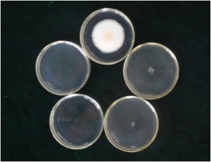
In accordance with Figure 1, S. colletotrichum CH008 was inhibited at 5 µg/mL chlorimuronethyl and this strain nearly showed no growth at 10 µg/mL chlorimuronethyl. Thus, the final concentration selected by the culture medium was 10 (preliminary screening) and 20 µg/mL (re-screening).
Figure 1. Tolerance test of a wild-type strain on medium containing concentration gradients of chlorimuronethyl.
Note (from the top, clockwise): DCM plates (concentrations of 0, 5, 10, 20, and 30 µg/mL); cultivation was carried out at 28°C for 4 d.
Effects of acceptor materials on transformation
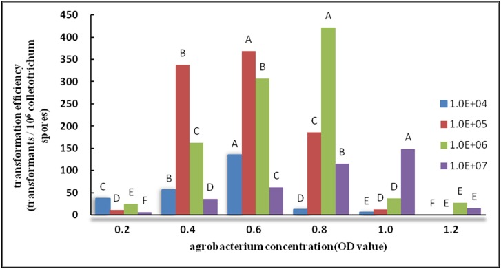
Results of the differences in concentrations of A. tumefaciens and Colletotrichum spore liquid were analyzed by ANOVA (similar to Duncan’s new multiple range method below). According to the related results, the difference in experimental results was significant at p<0.01. Based on Figure 2, each OD value had a corresponding and appropriate spore liquid concentration within an appropriate range of OD600 (0.4–0.8). A high concentration of spore liquid resulted in a high transformation efficiency. In particular, when Agrobacterium OD600 was equal to 0.8 and the number of Colletotrichum CH008 spores was 106 spores/mL, the results were significantly higher than the other combinations, and the transformation efficiency peaked. This result may be related to the growth cycle of Agrobacterium and features of Colletotrichum CH008 spores. When the OD600 values of Agrobacterium were 1.0 and 1.2, the transformation efficiency showed a reducing trend with false-positive results.
Figure 2. Effect of Agrobacterium concentration on transformation efficiency.
Note: In the same series (four series), the same capital letters indicate no significant difference (p>0.01), whereas different capital letters indicate significant differences (p<0.01).
Effects of induction time on transformation
By inducing the activation and expression of genes at the Vir region of Agrobacterium, AS promoted T-DNA processing and transfer so that Agrobacterium T-DNA could enter the target genome and integrate with it more easily. An appropriate induction time was directly related to the ability of AS to activate the Vir region sufficiently, and affected the efficiency of final recombination. The effect was most significant in this experiment, and the transformation efficiency peaked at 6 h of induction and transformation (Figure 3). However, extending the induction time did not improve the transformation efficiency. This finding may be due to the fact that the culture time of Agrobacterium was too long, so Agrobacterium gradually entered a decline phase and affected the transformation efficiency.
Figure 3. Effect of induction time on transformation efficiency.
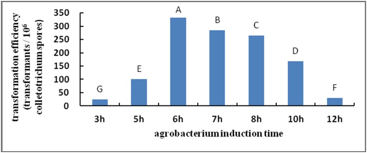
Note: Different capital letters indicate significant differences (p<0.01).
Effects of AS on transformation
When the AS concentration was 100 µmol/L (Figure 4), the results were significantly higher than those at other AS levels. However, the transformation efficiency did not improve as the concentration increased.
Figure 4. Effect of AS concentrations on transformation efficiency.
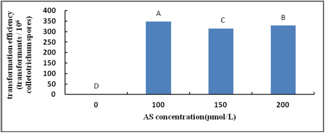
Note: Different capital letters indicate significant differences (p<0.01).
Effects of co-culture time on transformation
According to Figure 5, the transformation efficiency peaked when S. colletotrichum and Agrobacterium were co-cultured for 3–4 d, and the results were highly significant.
Figure 5. Effect of co-culture time on transformation efficiency.
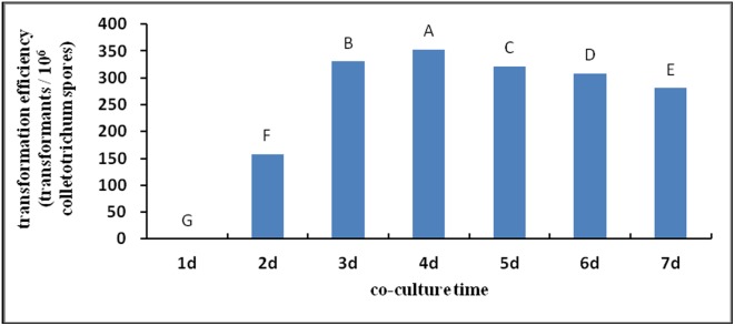
Note: Different capital letters indicate significant differences (p<0.01).
Effects of temperature on transformation during co-culture
In this experiment (Figure 6), the optimum transformation efficiency was obtained at a co-culture temperature of 25°C. Its effect was most significant at this temperature.
Figure 6. Effect of co-culture temperature on transformation efficiency.
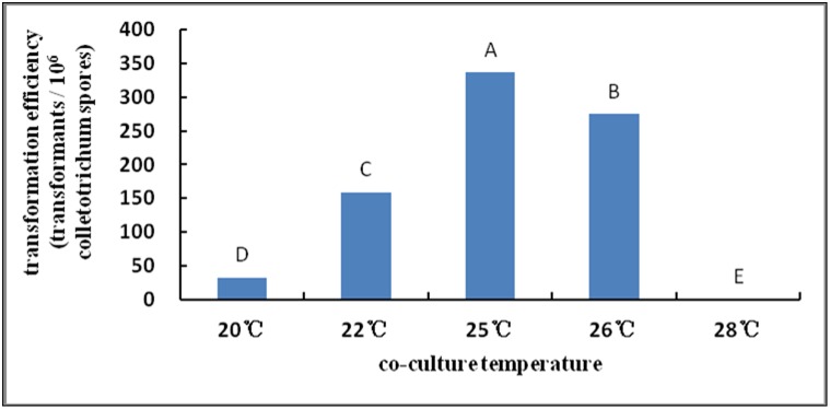
Note: Different capital letters indicate significant differences (p<0.01).
PCR
Among 20 selected converter strains, the positive control and all transformants demonstrated bright and clear strips at 750 bp. Thus, the T-DNA insertion rate was 100% (Figure 7).
Figure 7. Verification of PCR results.

Note: Marker v is on the left, followed by the positive control of plasmid and transformants, and untransformed CH 008 is on the right.
Verification of fluorescence of transformant’s conidia
Thirty transformants were randomly selected. After sporulation, conidia were examined under confocal fluorescence microscopy at an excitation wavelength of 400–500 nm (Figure 8). Spores of 13 transformants were bright under fluorescence, which indicates that GFP genes were carried to and integrated with S. colletotrichum gloeosporiodes Penz genome, and exhibited good expression.
Figure 8. GFP fluorescence of S. colletotrichum T-DNA transformants.

This study optimized the genetic transformation system conditions of S. colletotrichum. The optimized conditions were as follows: AGL-1 concentration of A. tumefaciens (OD600), 0.8; concentration of Colletotrichum conidium, 1×10⁶ conidia/mL; AS concentration, 100 mmol/L; induction time, 6 h; co-culture temperature, 25°C; and co-culture time, 3 d. The T-DNA insertion mutant library of S. colletotrichum was constructed successfully using the genetic transformation system. The transformation efficiency was determined to be 300–400 transformants/106 Colletotrichum spores.
Analysis of S. colletotrichum T-DNA insertion mutant libraries
The genetic transformation system was used to obtain 4616 S. colletotrichum genetic transformants. Some of these transformants were then selected for analysis.
Analysis of the copy number of T-DNA insertion mutants
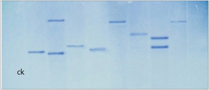
The results of Southern blot are shown in Figure 9. Among eight randomly selected transformants, six demonstrated a single strip and the remaining two had two strips. Untransformed CH 008 and one transformant had no strip. These results indicate that most T-DNA insertions had a single locus, which could aid in amplification for sequences of insertion sites via Tail-PCR.
Figure 9. Southern blot analysis of genomic DNA from untransformed CH008 (CK) and transformed isolates.
Genetic stability
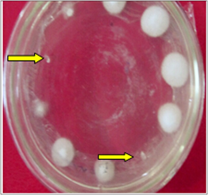
After cultivating untransformed and mutant strains in PDA plates without chlorimuronethyl for 10 generations, they were inoculated to screening plates with chlorimuronethyl. The transformants grew normally (Figure 9), whereas the untransformed strains did not grow (indicated by the arrow in Figure 10). These findings show that resistance could be stably inherited in transformants.
Figure 10. Determination of transformants’ genetic stability (arrows indicate wild strains).
Growth rate and colony morphology of transformants
Untransformed CH008 was cultivated, and 30 transformants were randomly selected. Thus, we observed an obvious difference in the growth rate of transformants. Specifically, mutant 1561 had the lowest growth rate, and its growth rate was much lower than that of untransformed CH008. Transformant 328 exhibited the highest growth rate (Table 1). Some strains demonstrated morphological variation, such as white hyphae that did not generate spores, greyish green hyphae, and yellow hyphae. However, the proportion of such strains was small.
Table 1. Comparison of the growth rate of untransformed strains and transformants.
| Approaching growth rate of untransformed strains | High growth rate of untransformed strains | Low growth rate of untransformed strains | |||
| Ch008 | 0.80 cm/d | 888 | 1.06 cm/d | 1561 | 0.37 cm/d |
| 1130 | 0.76 cm/d | 328 | 1.40 cm/d | 3532 | 0.53 cm/d |
| 993 | 0.83 cm/d | 2715 | 1.13 cm/d | 678 | 0.40cm/d |
| 1477 | 0.80 cm/d | 3590 | 1.20cm/d | 3425 | 0.60 cm/d |
| 3200 | 0.87 cm/d | 3416 | 1.23 cm/d | 1801 | 0.56 cm/d |
| 2181 | 0.73 cm/d | 1761 | 1.16 cm/d | 3393 | 0.46 cm/d |
| 3616 | 0.86 cm/d | 3605 | 1.00 cm/d | 604 | 0.43 cm/d |
Note: Data in the table are average values of three measurements.
Sporulation quantity of mutants
The sporulation quantities of wild strains and mutants were determined, and the results are shown in Table 2. The sporulation quantity of transformants 2181, 2881, 2561, 844, and 888 was much higher than that of untransformed CH008, whereas the sporulation quantity of transformants 1477, 1561, and 2097 was lower than that of untransformed CH008. Transformants 3443 and 993 showed nearly no sporulation quantity.
Table 2. Comparison of transformants’ ability to produce spores.
| Strain number | Ch008 | 2097 | 1447 | 1477 | 2181 | 961 | 2881 | 888 | 993 |
| Spore concentration | 1.05×10⁶ | 4.00×10⁴ | 2.15×10⁵ | 1.00×10⁶ | 2.15×10⁷ | 6.03×10⁴ | 1.67×10⁷ | 1.50×10⁵ | 0 |
| Strain number | 3590 | 3661 | 3605 | 2561 | 3443 | 844 | 1561 | 1761 | 604 |
| Spore concentration | 2.33×10⁶ | 2.34×10⁵ | 2.50×10⁵ | 3.45×10⁷ | 0 | 1.75×10⁷ | 2.35×10⁵ | 5.40×10⁷ | 1.70×10⁵ |
Conidial germination and appressorium formation in mutants
The results show that wild-type conidia and 10 randomly selected transformant spores could germinate after some time. The time that most of the transformants took to germinate was shorter than that of the wild-type strains, and the average length of germ tube growth in some transformants was longer than that of the wild-type strains. Three mutant strains demonstrated both low germination and appressorium formation rates, namely, t-960, t-604, and t-2327. By contrast, seven mutant spores (t-2393, t-2515, t-906, t-888, t-1130, t-2416, and t-3616) showed no significant difference from wild-type strains. In addition, wild-type strains and most transformants grew one to two germinal tubes from both ends of spores, and 80% of them demonstrated appressorium formation. Spore germination of mutant t-906 was abnormal; this mutant had three to four germinal tubes and its germination rate was high, but it germinated few appressoria (Table 3 and Figure 11).
Table 3. Comparison of conidial germination and germination rate.
| Strain name | Initial germination time | Germination rate | Length and number of germinal tubes |
| Ch008 | 8 h | 90–100 | 55–110 µm, 1 |
| t-2515 | 6 h | 80–90 | 110–300 µm, 2 |
| t-2393 | 8 h | 70–80 | 200–300 µm, 2 |
| t-2327 | 6 h | 40–50 | 300–400 µm, 1 |
| t-888 | 6 h | 70–80 | 200–250 µm, 2 |
| t-906 | 4 h | 95–100 | 300–400 µm, 3–4 |
| t-960 | 8 h | 60–70 | 30–45 µm, 1 |
| t-1130 | 8 h | 70–80 | 100–255 µm, 2 |
| t-2416 | 6 h | 90–100 | 110–350 µm, 2 |
| t-3616 | 6 h | 70–80 | 20–100 µm, 1 |
| t-604 | 4 h | 40–60 | 110–300 µm, 2 |
Figure 11. Conidial germination and appressorium formation after 8 h.

Selection of transformants of the S. colletotrichum mutant library with pathogenic defects and analysis of flanking sequences at T-DNA insertion sites
Preliminary screening of mutants with pathogenic defects
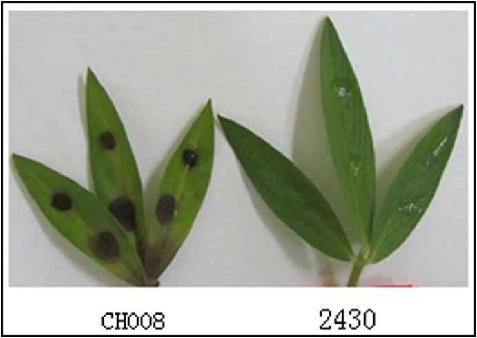
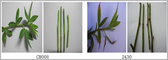
Comparison of inoculation methods showed that the most appropriate preliminary screening method was the use of the wild-type strain CH008 and transformant spore liquid to infect punctured parts of leaves of S. guianensis, and select mutants with lost pathogenicity. Using this method, we selected 1230 transformants and obtained 23 strains with pathogenic defects (18 strains with reduced pathogenicity and five strains with lost pathogenicity). Figure 12 shows that brown scabs formed at the infected part of wild strain CH008, whereas transformant 2430 lost its infection ability.
Figure 12. Determination of virulence during preliminary screening.
Re-screening of pathogenicity determination
For re-screening, we selected defective mutant strains whose pathogenicity was lost when preliminary screening was performed. The potted S. guianensis was inoculated again. Comparison of different re-screening inoculation methods showed that the most appropriate re-screening method was spraying the wild strain CH008 and transformant spore liquid to punctured parts of leaves of S. guianensis for verification and selection of mutants with lost pathogenicity. This method was used to re-screen five mutants with completely lost pathogenicity. The results were similar to those of preliminary screening. For example, mutant 2430 was used to infect S. guianensis plants. Two weeks later, pathogenic symptoms appeared. Scabs were observed on some leaves and stems of plants, and slowly expanded to most of the stem leaves (Figure 13).
Figure 13. Incidence of different parts of plants after re-screening and inoculation.
Cloning of T-DNA insertion flanking sequences of mutant strains with pathogenic defects
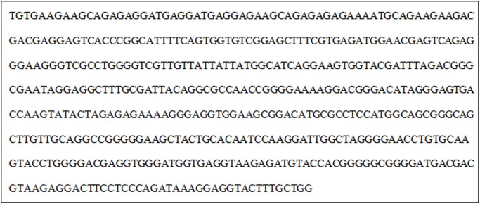
TAIL-PCR amplification was used in 15 mutants with reduced pathogenicity and defects, and three transformants with peculiar strips were obtained, i.e., Anthrax transformants 2430, 913, and 3521. Only transformant 2430 was successful in transformation and sequencing; and the flanking sequences of 476 bp were obtained (Figure 14).
Figure 14. Flanking sequences of T-DNA at the insertion sites of mutant 2430.
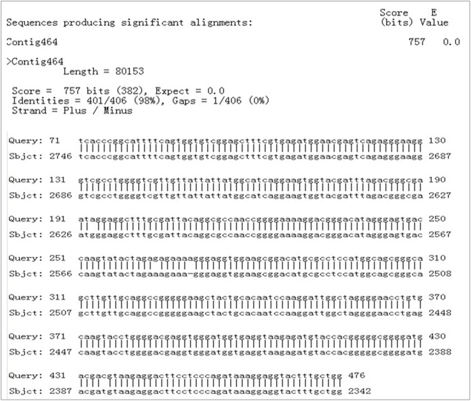
BLAST was used to compare the sequence with the sequenced Colletotrichum whole genome database (unpublished), and the website ‘The FGENESH Program’ (Softberry Inc., Mount Kisco, NY, USA; http://linux1.Softberry.com/berry.phtml) was used to predict its functions. Therefore, a hypothetical gene in the regional code of the nucleotide sequence was noted. BLAST was subsequently adopted to compare the sequences in NCBI; 401 nt was completely consistent with the partial sequence of Contig464 of the database (Figure 15). T-DNA was a promoter subregion of the predicted gene. The full length of the predicted gene was 1251 bp. The code of the predicted gene was 416 aa, and the amino acid homology between the predicted gene and Magnaporthe oryzae gene (XP_003719674.1) was 79%. This type of gene codes aspartate transaminase. This code may play an important role in the infection process of pathogeny. Insertion of exogenous sections destroyed the gene’s functions, so the mutants exhibited lost pathogenicity.
Figure 15. BLAST results of T-DNA flanking sequences in NCBI.
Discussion
In 2011, genetic transformation of over 60 fungi was realized successfully by ATMT [38]. When transformation is carried out, each fungus has optimum transformation conditions. The optimum transformation and screening conditions should be explored to construct a high-quality genetic mutant library with a large quantity in a short time period. Moreover, when high-quality and efficient transformation and optimization systems are used to construct necessary mutant libraries, manpower and material resources are not only saved but the library-establishment cycle is also shortened. Such systems can also reduce difficulty in screening and cloning related pathogenic genes [39].
The cultivation, growth status, and purity of Agrobacterium have significant effects on transformation, and they are important for preparing Agrobacterium infection liquid with high purity, vigorous growth, and powerful infection capability [40]. Agrobacterium in the middle and late logarithmic phases is considered to be the optimum infectious bacteria. However, the time at which different species of Agrobacterium reach the logarithmic phase, as well as their concentrations, can differ. In the present study, after AGL-1 was cultured in MM for 36 h and cultivated in IM for 6 h, its OD600 value ranged from 0.6 to 1.0. By selecting Agrobacterium within this range and adjusting to an appropriate concentration, the obtained transformation efficiency was high.
AS is currently the most common Vir gene inducer. Numerous studies showed an optimum inductive effect at a concentration of 50–200 µmol/L. When the pH of the culture medium containing AS is 5.0–5.6, the induction of genes at the Vir region of A. tumefaciens peaks [41]. IM in the present study showed a final selected concentration of 100 µmol/L and pH of 5.2. This finding verifies the conclusion drawn by Holford et al., who reported optimum transformation effects at pH 5.2 [42]. When the AS concentration was 100 µmol/L, the results were significantly higher than those at other levels. However, the transformation efficiency did not improve with increasing AS concentration. Excess AS concentrations may have toxic effects on explants, and influence further improvement in the transformation efficiency [41].
The co-culture time is one of the most important factors affecting the success of the transformation of A. tumefaciens. The process of transformation mediated by A. tumefaciens takes some time, and transformants are unable to form at a very short co-culture time [35], [43]–[45]. In the present study, the number of transformants increased with increasing co-culture time. The number of transformants peaked when S. colletotrichum and Agrobacterium were co-cultured for 3–4 d, and the results were highly significant. Continuous co-culture can result in generation of false-positive clones [46], resulting in very large bacterial colonies that cannot be selected easily [47]. Campoy et al. revealed that the number of transformants peaks when the co-culture temperature is consistent with the optimum growth temperature of acceptors; reducing or increasing the co-culture temperature reduces the number of transformants [48]. The results of this study were consistent with those observed by Campoy et al.
Furthermore, selection of transformation acceptors is critical. Many forms, such as protoplast, mycelium, and conidium, can be used as acceptor materials [26]. Acceptor strains can be transformed by exogenous genes only when they stay in the phase of cell division. High requirements must be met in the preparation of protoplast, and this process is quite complicated. Materials such as mycelium and conidium can also achieve ideal transformation efficiency. Very high cell concentration of acceptor fungi can lead to excess fungal growth, so transformants cannot be selected [24]; very high Agrobacterium concentrations can result in serious Agrobacterium pollution [37].
The results of Southern blot show that the T-DNA insertion rate in transformants was 90%. The transformants, whose T-DNA insertion was at a single site, accounted for about 67%. The rate of a single copy was much higher than that of Colletotrichum graminicola (16%), slightly higher than that of other anthracnose species (65%) [49], [50], but lower than that obtained by Jia Peisong [51] and Wang Haiyan [52] (100%). Single-site insertion in genome is usually expected because the derived phenotype is related to changes in single sites in the genome [24], which can help in finding marker genes from the fungal genome. Some studies implied that the insertion rate of T-DNA single copy numbers has an inverse relation with the co-culture time to some extent [53]. However, the co-culture time must not be shortened too much in single-copy insertion so that the transformation efficiency does not decrease. Moreover, Examination of a large number of mutants using PCR and Southern blot is time-consuming and laborious, and generally provides only a subjective measure of infectivity. Therefore it would be worthwhile to develop the high-throughput technologies for molecular verification of Stylosanthes anthracnose mutants, such as Luminex or transposon sequencing, Lin Tao et al. examined 434 signature-tagged mutagenesis mutants using Luminex-based multiplex PCR, which is an efficient and timesaving method [54]–[56].
This research showed that the growth rates and sporulation quantity of some transformants changed. T-DNA insertion causes inactivation for a certain gene of transformants, and this variation results in changes in other characters. However, most transformants did not demonstrate a significant difference from untransformed strains. Conidial generation is a premise for infection and pathopoiesia of many pathogenic bacteria; the quantity and germination rate of conidia, as well as appressorium formation, affect their pathogenicity to some extent [57], [58]. In the future, relations among the pathogenicity of transformants, growth rate of mutants, sporulation quantity, conidial germination, appressorium formation, and pathopoiesia will be further examined.
The flanking sequences of T-DNA insertion sites were cloned. TAIL-PCR is commonly used to amplify unknown flanking sequences of the known T-DNA sequence. This technique can design reliable random degenerate primers that are appropriate for the background of genomes that need to be detected [59]. The present study identified the optimum random primers (i.e., AD4 and AD8), and four right flanking sequences and one left flanking sequence were obtained. The reason for these results might be the high probability at which the left border of T-DNA is cut off, which was consistent with several fungal results that have been reported [36], [49], [60]. The left border of T-DNA may be unnecessary for T-DNA transfer, whereas the right border is essential for T-DNA transfer; thus, transfer starts at the right border and continues toward the left [61].
TAIL-PCR was used in the present study to amplify the flanking sequences of five mutant genomes, and five flanking sequences were obtained. For one sequence, BLAST comparison showed that its pathogenicity was lost and the flank section amplified by t-2430 was approximately 0.5 kb. By analyzing and comparing the obtained sequences, these genes and genes of aspartate transaminase coded by the pathogenic bacterium M. oryzae demonstrated high homology. This enzyme mainly exists in the mitochondria of cells, and exerts important catalytic actions during nitrogen metabolism. The insertion of exogenous sections may alter the functions of genes, affect the coding of transferase, disturb some important metabolisms of the mutant, and result in the loss of pathogenicity in pathogenic infection processes.
Acknowledgments
We thank the National Natural Science Foundation of China (NSFC, No. 31072076), Special Fund for Agro-scientific Research in the Public Interest (201303057), and The Major Technology Project of Hainan (ZDZX2013023-1) for funding this research.
Data Availability
The authors confirm that all data underlying the findings are fully available without restriction. All relevant data are within the paper.
Funding Statement
We thank the National Natural Science Foundation of China (NSFC, No. 31072076), Special Fund for Agro-scientific Research in the Public Interest (201303057) and The Major Technology Project of Hainan (ZDZX2013023-1) for funding this research. The funders had no role in study design, data collection and analysis, decision to publish, or preparation of the manuscript.
References
- 1.Williams RJ, Reid R, Schhultze-Kraft R, Sousa Costa NM, Thomas BD (1984) Natural distribution of Stylosanthes. In: Stace HM, Edye LA, editors. The Biology and Agronomy of Stylosanthes. pp. 73–101.
- 2. Jiang CS, Jia HS, Ma XR, Zou DM, Zhang YZ (2004) AFLP analysis of genetic variability among Stylosanthes guianensis accessions resistant and susceptible to the Stylo Anthracnose. Acta Botanica Sinica 46: 480–488. [Google Scholar]
- 3. Segenet K, Jiang CS, H GX, Gustavo S (2005) Genetic transformation of the tropical forage legume Stylosanthes guianensis with a confers resistance to Rhizoctonia foliar blight disease rice-chitinase gene. Afr J Biotechnol 4: 1025–1033. [Google Scholar]
- 4. Liu GD, Phaikaew C, Stur WW (1997) Status of Stylosanthes development in other countries: II Stylosanthes development and utilization in China and south-east Asia. Trop Grassl 31: 460–467. [Google Scholar]
- 5.Lenné JM, Calderon MA (1984) Disease and pest problems of Stylosanthes. In: Stace HM,Edye LA, editors. The Biology and Agronomy of Stylosanthes. 279–293.
- 6.Manners JM, Masel AM, Braithwaite KS, Irwin JAG (1992) Molecular analysis of Colletotrichum gloeosporioides pathogenic on the tropical pasture legumes Stylosanthes spp. In: Bailey JA, Jeger MJ, editors. Colletotrichum Biology, Pathology, and Control. pp. 250–268.
- 7.Manners JM, Stephenson SA, He C, Maclean DJ (2000) Gene transfer and expression in Colletotrichum gloeosporioides causing anthracnose on Stylosanthes. In: Prusky D, Freeman S, Dickman M, editors. Host Specificity, Pathology and Host–Pathogen Interactions of Colletotrichum. pp. 180–194.
- 8. Chakraborty S, Fernandes CD, Charchar MJDA, Thomas MR (2002) Pathogenic variation in Colletotrichum gloeosporioides infecting Stylosanthes spp. in a center of diversity in Brazil. Phytopathology 92: 553–562. [DOI] [PubMed] [Google Scholar]
- 9. Chakraborty S, Perott R, Charchar MJDA, Fernandes CD, Kelemu S (1997) Biodiversity epidemiology and virulence of Colletotrichum gloeosporioides. II Genetic and pathogenic diversity in Colletotrichum gloeosporioides isolates from eight species of Stylosanthes. . Trop Grassl 31: 387–393. [Google Scholar]
- 10. Chakraborty S, Perott R, Ellis N, Thomas MR (1999) New aggressive Colletotrichum gloeosporioides strains on Stylosanthes scabra detected by virulence and analysis. Plant Dis 83: 333–339. [DOI] [PubMed] [Google Scholar]
- 11. Iriwin JAG, Cameron DF (1978) Two diseases of Stylosanthes spp. caused by Colletotrichum gloeosporioides in Australia, and pathogenic specialization within one of the causal organisms. Aust J Agric Res 29: 305–317. [Google Scholar]
- 12. Kelemu S, Badel JL, Moreno CX, Miles JW (1996) Virulence spectrum of South American isolates of Colletotrichum gloeosporioides on selected Stylosanthes guianensis genotypes. Plant Dis. 80: 1355–1358. [Google Scholar]
- 13. Lenné JM, Burdon JJ (1990) Preliminary study of virulence and isozyme variation in natural populations of Colletotrichum gloeosporioides from Stylosanthes guianensis. . Phytopathology 80: 728–731. [Google Scholar]
- 14. Munaut F, Hamaide N, Vander Stappen J, Maraite H (1998) Genetic relationship among strains of ColLetotrichum gloeosporioides isolated from Stylosanthes spp. in Africa and Australia using RAPD and ribosomal DNA markers. Plant Pathol 47: 641–648. [Google Scholar]
- 15. Dale JL, Manners JM, Irwin JAG (1988) Colletotrichum gloeosporioides isolates causing different anthracnose diseases on Stylosanthes in Australia carry distinct double-stranded RNAs. Transactions of the British Mycological Society 91: 671–676. [Google Scholar]
- 16. Braithwaite KS, Irwin JAG, Manners JM (1990a) Restriction fragment length polymorphism in Colletotrichum gloeosporioides infecting Stylosanthes spp. in Australia. Mycol Res 94: 1129–1137. [Google Scholar]
- 17. Braithwaite KS, Irwin JAG, Manners JM (1990b) Ribosomal DNA as a molecular taxonomic marker for the group species Colletotrichum gloeosporioides . Aust Syst Bot 3: 733–738. [Google Scholar]
- 18.Manners JM, Masel A, Irwin JAG (1993) Molecular genetics of Colletotrichum gloeosporioides infecting Stylosanthes. In: Isaac S, Frankland JC, Watling R, Whalley AJS, editors. Aspects of Tropical Mycology. pp. 233–51.
- 19. Weeds PL, Chakraborty S, Fernandes CD, Charchar MJdA, Amesh CRR, et al. 2003. Genetic diversity in Colletotrichum gloeosporioides from Stylosanthes spp. at centers of origin and utilization. Phytopathology 93: 176–185. [DOI] [PubMed] [Google Scholar]
- 20. Yi KX, Huang JS, Liu GD, Weeds P, Charkraborty S (2003) Genetic diversity analysis of stylo anthracnose pathogens using random amplified polymorphic DNA. Wei Sheng Wu Xue Bao 43: 379–387. [PubMed] [Google Scholar]
- 21. Munaut F, Hamaide N, Maraite H (2002) Genomic and pathogenic diversity in Colletotrichum gloeosporioides from wild native Mexican Stylosanthes spp., and taxonomic implications. Mycol Res 106: 579–593. [Google Scholar]
- 22. Cai ZY, Li GH, Lin CH, Shi T, Zhai LG, et al. (2013) Identifying pathogenicity genes in the rubber tree anthracnose fungus Colletotrichum gloeosporioides through random insertional mutagenesis. Microbiol Res 168: 340–350. [DOI] [PubMed] [Google Scholar]
- 23. Wu XY, Wang JY, Zhang Z, Jing JX, Du XF, et al. (2010) Isolation of gene mutation from a pathogenicity-enhanced mutant of Magnaporthe oryzae . Rice Sci 17: 129–134. [Google Scholar]
- 24. Michielse CB, Hooykaas PJ, Van den Hondel CAMJJ, Ram AFJ (2005) Agrobacterium-mediated transformation as a tool for functional genomics in fungi. Curr Genet 48: 1–17. [DOI] [PubMed] [Google Scholar]
- 25. Mullins ED, Kang S (2001) Transformation: A tool for studying fungal pathogens of plants. Cell Mol Life Sci 58: 2043–2052. [DOI] [PMC free article] [PubMed] [Google Scholar]
- 26. De Groot MJ, Bundock P, Hooykaas PJ, Beijersbergen AG (1998) Agrobacterium tumefaciens-mediated transformation of filamentous fungi. Nat Biotechnol 16: 839–842. [DOI] [PubMed] [Google Scholar]
- 27. Li AH, Zhang YF, Wu CY, Tang W, Wu R, et al. (2006) Screening for and Genetic Analysis on T-DNA-inserted Mutant Pool in Rice. Yi Chuan Xue Bao 33: 319–329. [DOI] [PubMed] [Google Scholar]
- 28. Li M, Gong X, Zheng J, Jiang D, Fu Y, Hou M (2005) Transformation of Coniothyrium minitans, a parasite of Sclerotinia sclerotiorum, with Agrobacterium tumefaciens. FEMS Microbiol Lett 243: 323–329. [DOI] [PubMed] [Google Scholar]
- 29. Walton FJ, Idnurm A, Heitman J (2005) Novel gene functions required for melanization of the human pathogen Cryptococcus neoformans. Mol Microbiol 57: 1381–1396. [DOI] [PubMed] [Google Scholar]
- 30. Sesma A, Osbourn AE (2004) The rice leaf blast pathogen undergoes developmental processes typical of root-infecting fungi. Nature 431: 582–586. [DOI] [PubMed] [Google Scholar]
- 31. Huang GX, Xue YC, Lu X (2008) Construction of a T-DNA insertional mutant library of Colletotrichum gloeosporioides of Mango and its molecular analysis. Re Dai Zuo Wu Xue Bao 29: 27–32. [Google Scholar]
- 32. Jennifer LF, Lisa JV (2005) Parameters affecting the efficiency of Agrobacterium tumefaciens-mediated transformation of Colletotrichum graminicola . Curr Genet 48: 380–388. [DOI] [PubMed] [Google Scholar]
- 33. Hu XW, Jiang H, Huang HX, Guo JC (2005) RAPD analysis of mango anthracnose. Re Dai Zuo Wu Xue Bao 26: 38–42. [Google Scholar]
- 34. Lin CH, Liu XB, Cai JM, Li CP, Li JF, et al. (2009) Collectotrichum acutatum GFP tagged transformants generated from Agrobacterium tumefaciens-mediated insertional mutagenesis. Re Dai Zuo Wu Xue Bao 30: 1495–1500. [Google Scholar]
- 35. Meyer V, Mueller D, Strowig T, Stahl U (2003) Comparison of different transformation methods for Aspergillus giganteus . Curr Genet 43: 371–377. [DOI] [PubMed] [Google Scholar]
- 36. Mullins ED, Chen PX, Romaine P, Raina R, Geiser DM, et al. (2001) Agrobacterium- mediated transformation of Fusariium: An efficient tool for insertional mutagenesis and gene transfer. Phytopathology 91: 173–180. [DOI] [PubMed] [Google Scholar]
- 37. Zeilinger S (2004) Gene disruption in Trichoderma atroviride via Agrobacterium mediated transformation. Curr Genet 45: 54–60. [DOI] [PubMed] [Google Scholar]
- 38. Liu G, Yang CD, Kang K Xing M, Tian SL (2011) Agrobacterium tumefaciens-mediated transformation of Trichoderma reesei and optimization of the transforming conditions. Sheng Wu Ji Shu 21: 44–48. [Google Scholar]
- 39. Mishra NC, Tatum EL (1973) Non-Mendelian inheritance of DNA induced inositol independence in Neurospom. Proc Natl Acad Sci USA 70: 3875–3879. [DOI] [PMC free article] [PubMed] [Google Scholar]
- 40. Liu CP, Zeng BS, Qiu ZF, Li XY, Liu Y (2010) Analysis of the optimized factors on the efficiency of Agrobacterium mediated gene transformation. Anhui Nong Ye Ke Xue 38: 1141–1143. [Google Scholar]
- 41. Deng Y, Zeng BS, Zhao SD, Liu Y, Qiu ZF, et al. (2010) Mechanism and application of Acetosyringone in Agrobacterium mediated transformation. Anhui Nong Ye Ke Xue 2010 38(5): 2229–2232. [Google Scholar]
- 42. Holford P, Hernandezn, Newbury HJ (1992) Factors influencing the efficiency of T-DNA transfer during co-cultivation of Antirrhinum majus with Agrobacterium tumefaciens [J]. Plant Cell Rep 11: 196–199. [DOI] [PubMed] [Google Scholar]
- 43. Soichi T, Norio K, Shigeo N (2007) Isolation of pathogenicity- and gregatin-deficient mutants of Phialophora gregata f. sp. adzukicola through Agrobacterium tumefaciens-mediated transformation. J Gen Plant Pathol 73: 242–249. [Google Scholar]
- 44. Nyilasi I, Acs K, Papp T, Nagy E, Vágvölgyi C (2005) Agrobacterium tumefaciens-mediated transformation of Mucor circinelloides. Folia Microbiol 50: 415–420. [DOI] [PubMed] [Google Scholar]
- 45. Betts MF, Tucker SL, Galadima N, Meng Y, Patel G (2007) Development of a high-throughput transformation system for insertional mutagenesis in Magnaporthe oryzae . Fungal Genet Biol 44: 1035–1049. [DOI] [PubMed] [Google Scholar]
- 46. Takahara H, Tsuji G, Kubo Y, Yamamoto M, Toyoda K, et al. (2004) Agrobacterium tumefaciens-mediated transformation as a tool for random mutagenesis of Colletotrichum trifolii . J Gen Plant Pathol 70: 93–96. [Google Scholar]
- 47. Rao ZM, Zhang JS, Wei S, Fang HY, ZhuGe J (2007) Genetic transformation of industrialized strain candida glycerinogenes by Agrobacterium tumefaciens . C Ying Yong Yu Huan Jing Sheng Wu Xue Bao 13: 868–871. [Google Scholar]
- 48. Campoy S, Perez F, Martín JF, Gutiérrez S, Liras P (2003) Stable transformants of the azaphilone pigment-producing Monascus purpureus obtained by protoplast transformation and Agrobacterium-mediated DNA transfer. Curr Genet 43: 447–452. [DOI] [PubMed] [Google Scholar]
- 49. Maruthachalam K, Nair V, Rho HS, Choi J, Kim S, et al. (2008) Agrobacterium tumefaciens-mediated transformation in Colletotrichum falcatum and C. acutatum. . J Microbiol Biotechnol 18: 234–241. [PubMed] [Google Scholar]
- 50. Auyong ASM, Ford R, Taylor PWJ (2012) Genetic transformation of Colletotrichum truncatum associated with anthracnose disease of chili by random insertional mutagenesis. J Basic Microbiol 52: 372–382. [DOI] [PubMed] [Google Scholar]
- 51. Jia PS, Ding LL, Zhou BJ, Gao HS, Gao F (2012) Construction of a T-DNA insertional mutant library for Verticillium dahliae Kleb. and analysis of a mutant. Mian Hua Xue Bao 24: 62–70. [Google Scholar]
- 52. Wang HY, Li BH, Zhang QM, Li GF, Dong XL, et al. (2013) Transformation of Agrobacterium tumefaciens-mediated Colletotrichum gloeosporioides and identification of transformants. Zhongguo Nong Ye Ke Xue 46: 1799–1807. [Google Scholar]
- 53. Maruthachalam K, Klosterman SJ, Kang S, Hayes RJ, Subbarao KV (2011) Identification of pathogenicity-related genes in the vascular wilt fungus Verticillium dahliae by Agrobacterium tumefaciens-mediated T-DNA insertional mutagenesis [J]. Mol Biotechnol 49: 209–221. [DOI] [PMC free article] [PubMed] [Google Scholar]
- 54.Lin T, Gao L, Zhang C, Odeh E, Jacobs MB, et al.. (2012) Analysis of an ordered, comprehensive STM mutant library in infectious Borrelia burgdorferi: insights into the genes required for mouse infectivity. PLoS ONE 7: e47532 10.1371/journal.pone.0047532. [DOI] [PMC free article] [PubMed]
- 55. Troy EB, Lin T, Gao L, Lazinski DW, Camilli A, et al. (2013) Understanding barriers to Borrelia burgdorferidissemination during infection using massively parallel sequencing. Infect Immun 81: 2347–2357. [DOI] [PMC free article] [PubMed] [Google Scholar]
- 56. Van Opijnen T, Bodi KL, Camilli A (2009) Tn-seq: high-throughput parallel sequencing for fitness and genetic interaction studies in microorganisms. Nat Methods 6: 767–772. [DOI] [PMC free article] [PubMed] [Google Scholar]
- 57. Rogers LM, Kim YK, Guo W, González-Candelas L, Li D, et al. (2000) Requirement for either a host- or pectin-induced pectate lyase for infection of Pisum sativum by Nectria hematococca . Proc Natl Acad Sci U S A 97: 9813–9818. [DOI] [PMC free article] [PubMed] [Google Scholar]
- 58. He CP, Lin CH, Liao QH, Li R, Zheng FC (2007) Phenotypes analysis for T-DNA insertional mutagenesis of Magnaporthe grisea. Nong Ye Sheng Wu Ji Shu Xue Bao 15: 877–883. [Google Scholar]
- 59. Sun BY, Piao HL, Park SH, Han CD (2004) Selection of optimal primers for TAIL-PCR in identifying Ds flanking sequences from Ac/Ds insertion rice lines. Chin J Biotechnol 20: 821–826. [Google Scholar]
- 60. Huser A, Takahara H, Schmalenbach W, O’Connell R (2009) Discovery of pathogenicity genes in the crucifer anthracnose fungus Colletotrichum higginsianum using random insertional mutagenesis. Mol Plant Microbe Interact 22: 143–156. [DOI] [PubMed] [Google Scholar]
- 61. Miranda A, Janssen G, Hodges L, Peralta EG, Ream W (1992) Agrobacterium tumefaciens transfers extremely long T-DNAs by a unidirectional mechanism. J Bacteriol 174: 2288–2297. [DOI] [PMC free article] [PubMed] [Google Scholar]
Associated Data
This section collects any data citations, data availability statements, or supplementary materials included in this article.
Data Availability Statement
The authors confirm that all data underlying the findings are fully available without restriction. All relevant data are within the paper.


