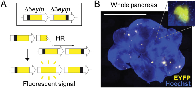
Fig. 1.
The FYDR mouse detects HR in situ in intact pancreas tissue. (A) Reconstitution of full-length EYFP coding sequence from two truncated copies through replication fork restart by HR. Note that the appearance of fluorescent signal indicates the gain of one repeat unit. Arrows represent expression constructs. EYFP coding sequences are in yellow, promoter and polyadenylation signal sequences are in white, and deleted sequences are in black. Drawing is not to scale. (B) Representative image of a pancreas from a FYDR mouse showing fluorescent foci in situ in intact tissue. Freshly harvested, unfixed whole pancreas was counterstained with Hoechst, compressed to 0.5mm and imaged under a fluorescent microscope. Fluorescence is pseudocolored. Original magnification, ×1. Scale bar = 1cm. Inset: individual focus at ×40 original magnification.

