Fig. 3.
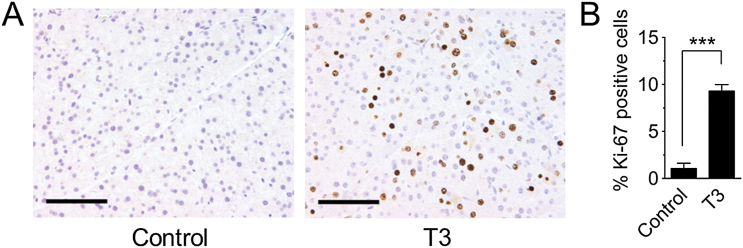
Hormone-induced cell proliferation in the pancreas. Mice were fed a diet containing 4 ppm thyroid hormone (T3). Pancreata were harvested at peak T3-induced proliferation, after treatment for 3 days (female mice) or 4 days (male mice). (A) Representative images of histological sections stained for the proliferation marker Ki-67. Original magnification, ×20. Scale bar = 100 μm. (B) Quantification of Ki-67 labeling in control mice (n = 24) and in T3-treated mice (n = 28) shows significantly higher labeling index after T3 treatment. Data are mean ± SEM. ***P < 0.001 (Student’s t-test).

