Abstract
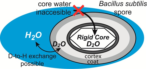
Dormant bacterial spores are able to survive long periods of time without nutrients, withstand harsh environmental conditions, and germinate into metabolically active bacteria when conditions are favorable. Numerous factors influence this hardiness, including the spore structure and the presence of compounds to protect DNA from damage. It is known that the water content of the spore core plays a role in resistance to degradation, but the exact state of water inside the core is a subject of discussion. Two main theories present themselves: either the water in the spore core is mostly immobile and the core and its components are in a glassy state, or the core is a gel with mobile water around components which themselves have limited mobility. Using deuterium solid-state NMR experiments, we examine the nature of the water in the spore core. Our data show the presence of unbound water, bound water, and deuterated biomolecules that also contain labile deuterons. Deuterium–hydrogen exchange experiments show that most of these deuterons are inaccessible by external water. We believe that these unreachable deuterons are in a chemical bonding state that prevents exchange. Variable-temperature NMR results suggest that the spore core is more rigid than would be expected for a gel-like state. However, our rigid core interpretation may only apply to dried spores whereas a gel core may exist in aqueous suspension. Nonetheless, the gel core, if present, is inaccessible to external water.
Introduction
Gram-positive bacteria, particularly of the Bacillus and Clostridium genera, have a sporulation mechanism by which they can protect their DNA and become dormant under harsh conditions that would otherwise kill them.1 This survival mechanism allows for the bacteria to survive in a metabolically inactive state for long periods of time until conditions, such as temperature or nutrient content, allow for germination and outgrowth. Some spores have been recovered and cultured after storage in amber for millions of years.2 The structure of the spore is well-understood, separated into distinct regions: an outer spore coat which is laminar and serves as a protective barrier;3−7 the cortex, composed of cross-linked peptidoglycan distinct from that found in the germinated bacterial cell wall and implicated in the heat resistance of spores;8−11 and the core, containing the bacterial DNA as well as protective molecules such as small acid-soluble proteins (SASPs)12−14 and an aquo-coordination complex between Ca2+ and dipicolinic acid (pyridine-2,6-dicarboxylic acid).15−18 The spore core has a lowered water content compared to that of vegetative cells, which is thought to be a major factor in spore heat resistance.19−21
The exact nature of the water in the spore core is not well understood. Two main paradigms have formed around this question. One theory is that the core water is in an amorphous (glassy) state. This was suggested by Sapru and Labuza in 199322 by predicting glass transition temperatures for spores based on inactivation kinetics and finding that more heat-resistant spores had higher predicted glass transition temperatures. Ablett in 199923 used differential scanning calorimetry to identify features of B. subtilis spores that were indicative of low moisture content even in an excess water environment and using 13C NMR on calcium dipicolinate concluded it was likely in an amorphous solid state indicative of a core with low water content consistent with a glassy state. Amorphous calcium dipicolinate was also seen using Raman spectroscopy of B. cereus spores.24,25 The other theory is that the core water is in a gel-like state. This hypothesis was put forward initially by Black and Gerhardt26 as a means of explaining their results on water uptake in spores using tritium-labeled H2O; they proposed that the spore core is an insoluble gel whose components are cross-linked but water-permeable. More recently, some27,28 have argued for the gel-like core theory based on deuterium relaxation times recorded from D2O-exchanged spores. Proponents of the gel core theory assert that the gel provides thermal heat resistance during dormancy by protecting proteins against thermal denaturation, assisted by the lower water content of the core.27 Sunde et al.27 asserted that, were the core in a glassy state, there would be a broad signal apparent in a 2H NMR spectrum of the spore; however, such a signal would be undetectable using the magnetic relaxation dispersion experiments as performed. Kaieda et al.28 analyzed a quadrupolar-echo spectrum of hydrated spores and concluded that its line shape represents mobile water and labile deuterons from proteins, nucleic acids, and peptidoglycan. Thus, no immobilized water could be detected in the 2H NMR spectrum of fully hydrated spores.28
Here, we report 2H NMR spectra of deuterated spores of a wild-type Bacillus subtilis (ATCC 6051) and the chloramphenicol-resistant strain Bacillus subtilis 1A578.29 Deuterium–hydrogen exchange experiments were conducted to examine the permeability of water into the spore and to compare them against calcium dipicolinate trihydrate (a known spore core component) and bovine serum albumin (as a spore protein analogue). In addition, variable-temperature (VT) NMR experiments were performed to determine how the deuteron mobility varies as the spores are heated and cooled. Our results suggest that there is immobile water in the spore core that can be detected using quadrupolar echo deuterium NMR. Variable-temperature NMR data does not show a gradual increase in molecular mobility proportional to temperature as would be expected for a spore core containing a gel of highly mobile water. Instead, our VT NMR data indicate that deuterated biomolecules and deuterated water retain their anisotropic motion properties at 50 °C. Heating to 100 °C does not affect viability, and the NMR spectra show that the proteins remain immobile at this temperature. However, 100 °C causes a transition of some deuterons from anisotropic to isotropic motion which we attribute to immobile water associated with protein hydration. These observations support the glassy-state core theory for our samples of dried spore powders. Nonetheless, we also observe mobile water in the core as recently reported27,28 and it remains possible that an aqueous spore suspension could have a gel-like core.
Materials and Methods
Spore Preparation
Bacillus subtilis 1A578, a chloramphenicol-resistant strain,29 was obtained from Bacillus Genetic Stock Center (BGSC, Department of Chemistry at the Ohio State University). Spores were prepared from stock Bacillus subtilis ATCC 6051 and 1A578. Frozen stock was inoculated overnight in 20 mL aliquots of LB medium, which was used to inoculate 200 mL of LB medium at 1% in a 1 L Erlenmeyer flask. The culture was allowed to grow (200 rpm @ 37 °C) for 24 h, after which it was centrifuged at 10000g at 4 °C for 20 min, the supernatant decanted, and the pellet resuspended in a separate 1 L flask with 200 mL of chemically defined sporulation medium (CDSM, composition included in the Supporting Information)30 and allowed to grow (200 rpm @ 37 °C) until the culture exhibited at least 80% spores via phase contrast microscopy (between 3 and 10 days). For partially deuterated spores, cultures were inoculated into 25% D2O LB medium and resuspended in 25% D2O CDSM for sporulation. Once the sample had sufficient spores, the culture was spun down, washed 1× with sterile Milli-Q water, spun down, and resuspended in a centrifuge tube in 20 mL of sterile 50% v/v EtOH:H2O. The resuspended spores were shaken at 200 rpm at 25 °C for 2 h to kill any remaining vegetative cells. The spores were then centrifuged 3X (10000g, 4 °C, 20 min); the supernatant was decanted and the spores were washed 3X with 20 mL aliquots of sterile Milli-Q water between each centrifugation. After washing, the spores were resuspended in a fresh 20 mL aliquot of sterile water and stored overnight at 4 °C; the following morning the culture was spun down and resuspended in fresh sterile water. The final step of purification involved delivery of the spores into a sterile centrifuge tube via syringe filtration using 3.1 and 1.2 μm glass fiber filters connected in series. The purified spores were centrifuged (10000g, 4 °C, 20 min), the supernatant was decanted, and the pellet was resuspended and transferred to a glass vial with 99% D2O (total volume 8 mL). The spore suspension was frozen with liquid nitrogen and lyophilized for 2 days to form a fluffy, charged white or off-white powder, which was stored at −20 °C until use. Spores were packed into 5 mm ceramic magic-angle-spinning rotors and sealed with O-ring caps for NMR experiments.
For deuterium–hydrogen exchange experiments, the lyophilized deuterated spores were transferred to a sterile 15 mL Falcon tube and resuspended in sterile Milli-Q water. The tube was sealed with a plastic cap, covered in parafilm, and stored at 4 °C with occasional shaking for 1 week. The exchanged spores were transferred to a centrifuge tube and spun at 10000g (4 °C, 20 min). Water was decanted, and the wet spores were resuspended with sterile Milli-Q water to aid their transfer into a glass vial prior to lyophilization as described above. Spores for hydrogen–deuterium exchange experiments were prepared in a similar manner. Lyophilized spores grown in protonated media were suspended in sterile D2O for 1 week before drying.
Synthesis of Amorphous and Crystalline Calcium Dipicolinate (CaDP) and Deuteration of BSA
Deuterated amorphous calcium dipicolinate trihydrate was prepared by modifying the procedure of Johnson et al.31 to use 99% D2O. After dissolving the dipicolinic acid and adding calcium hydroxide to a pH between 9 and 10, the heated solution was allowed to cool to room temperature and then stored in a desiccator for 4 days. Subsequently, the solution was further dried via lyophilization for 24 h. The resulting powder was packed into an NMR rotor to collect 2H NMR data.
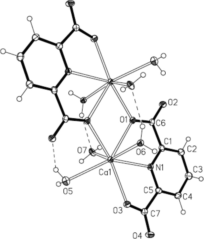
Crystalline deuterated CaDP was prepared using a modification of the procedure by Bailey et al.,32 replacing deionized water with 99% D2O. Slight heating was necessary to dissolve the dipicolinic acid. After addition of Ca(OH)2, the solution was transferred into several 13 mm OD test tubes and stored in a desiccator for ∼1 month. Needle-shaped crystals began to form within 1–2 weeks. The crystal structure was determined by X-ray crystallography and found to have the same space group and similar dimensions as previously reported data.33 Details of the X-ray crystallography procedures and results are provided as Supporting Information.
Bovine serum albumin (BSA, Sigma-Aldrich, Inc.) was dissolved in 99% D2O (100 mg/8.0 mL), stirred for 24 h at 4 °C, and lyophilized for 5 days before packing in a 5 mm rotor and collecting a deuterium solid-state echo (SSECHO) spectrum. A second sample was prepared by dissolving 100 mg of BSA in 8 mL of D2O, mixing for 24 h, and drying via lyophilization for 19 days.
NMR Experiments
Quadrupolar echo 2H NMR experiments were collected by placing the filled ceramic rotor into a Varian 5 mm wide-line NMR probe and inserting the probe into an Oxford 400 MHz (9.4 T) magnet (2H frequency = 61.424 MHz). Data was collected using a Varian Unityplus console and VNMR 6.1C software on a Sun workstation. The pulse sequence (SSECHO, 90x–τ1–90y–τ2–acquire) used a pulse width of 6 μs, recycle delay of 120 s, τ1 of 60 μs, and τ2 of 50 μs. The acquisition time was 40 ms. Deuterium NMR calibrations were performed with malonic acid-d4 and pure D2O samples to optimize the 90° pulse width and verify the appropriate spectral appearance of our calibration standards. The spore samples were studied at room temperature. The 2H T1 for immobilized water was found to be 24 s using a sample of frozen D2O (−40 °C) cooled by a stream of nitrogen gas chilled by a XRII FTS refrigeration unit. Data analysis was accomplished using VNMR 6.1C provided by Varian Inc.
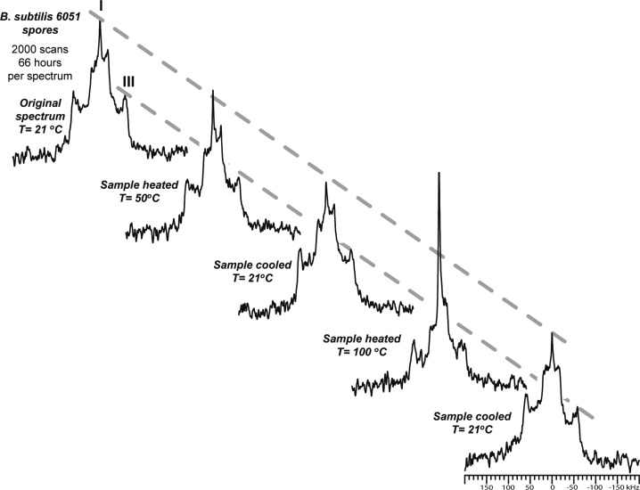
Variable-temperature 2H NMR experiments were performed by delivering heated dry air to the sample. The sample was held at the desired temperature for 15 min prior to data acquisition. Data was collected with the SSECHO pulse sequence using a delay time of 120 s for each of the 2000 transients (66 h data collection time). An initial spectrum was collected at 21 °C followed by heating to 50 °C. A spectrum collected at this temperature was compared to the 21 °C spectrum, and the sample was then cooled back to 21 °C. Another spectrum was collected to see whether changes that occurred, if any, at 50 °C were reversible. Subsequent spectra were also collected at 100 and 21 °C, respectively.
Results
Deuterium–Hydrogen Exchange
The deuterium quadrupolar echo NMR spectrum of partially deuterated B. subtilis ATCC 6051 spores is shown in Figure 1A. There are three distinct features that are present in this spectrum: feature I, a sharp singlet peak at 0 Hz referenced to a liquid D2O standard; feature II, a Pake doublet with a separation of approximately 32 kHz; and feature III, another Pake doublet with a 120 kHz separation. A second deuterium NMR spectrum was collected after the B. subtilis ATCC 6051 spores were soaked in H2O (Figure 1B). Here, signals for features I, II, and III are present, although the intensity is lower due to either replacing labile deuterons with protons or sample loss after repacking the postexchange spores. After correcting for the latter through scaling of the spectrum by the same proportion as sample lost, an overlay of the pre- and postexchange spectra (Figure 1C) shows a diminution of features I and II while feature III is unchanged.
Figure 1.
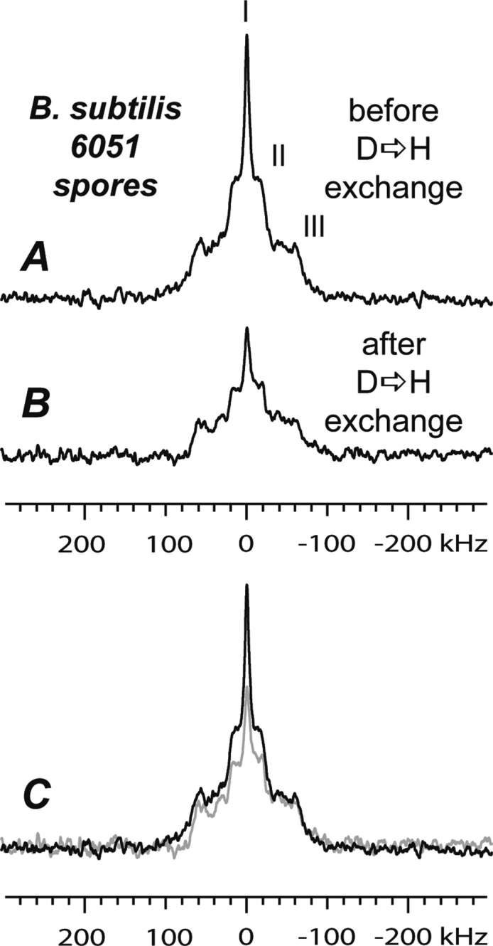
2H solid-state NMR spectrum of 25% D2O-labeled B. subtilis ATCC 6051 spores (A); of the same sample after one-week’s exchange with H2O (B); A and B overlaid on the same axis and scaled for sample loss (C). Three main features (I, II, III) are apparent in the spectra, with feature I decreasing most after exchange. Spectra were collected at room temperature over 10,000 scans with a delay time of 120 s.
Using the same methodology, spores of B. subtilis 1A578 were also studied using deuterium–hydrogen exchange via quadrupolar 2H NMR. Figure 2 shows the spectra for spores of B. subtilis 1A578 grown in 25% deuterated media before (A) and after (B) a one-week exchange with H2O, and a comparison of the two spectra on the same axis (C) corrected for mass loss during sample preparation. The 1A578 spores appear to have sharper features II and III than those of ATCC 6051. Likewise, the comparison in Figure 2C shows that feature III is not affected by soaking in H2O whereas features I and II become smaller.
Figure 2.
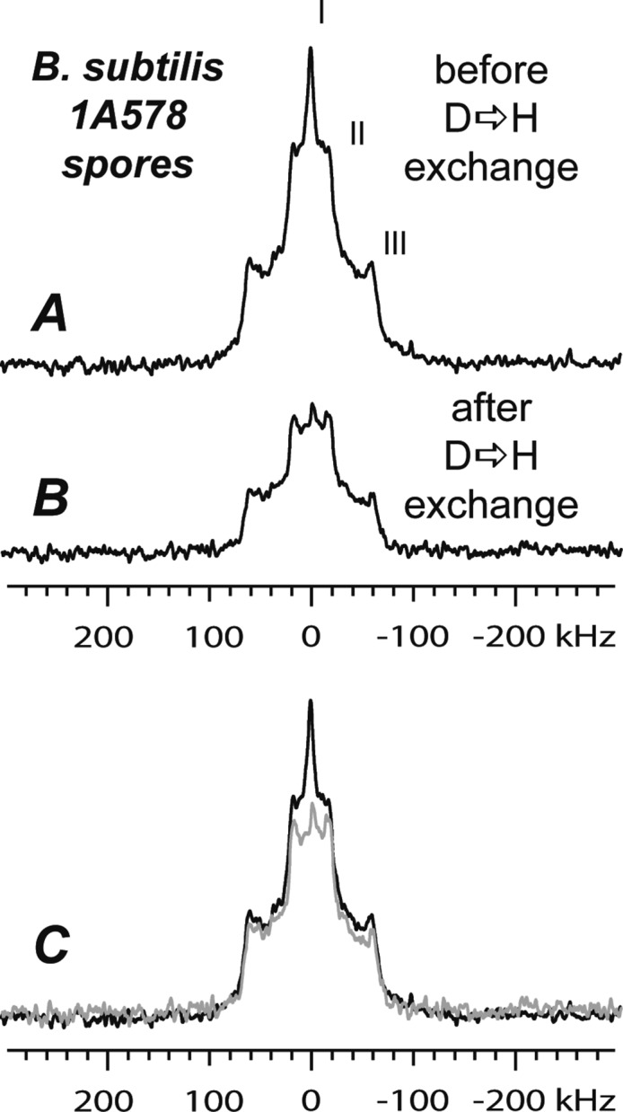
Deuterium quadrupolar echo NMR spectra of B. subtilis 1A578 spores: grown in 25% D2O media (A); same sample after one-week exchange with H2O (B); A and B shown on the same axis and scaled for mass (C). Three main features (I, II, III) are apparent indicating signals arising from different deuterons in the spore; feature I exhibits the largest decrease after exchange. Spectra were collected at room temperature over 10,000 scans with a delay time of 120 s.
Hydrogen—Exchange Deuterium NMR of Spores
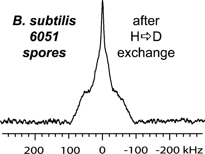
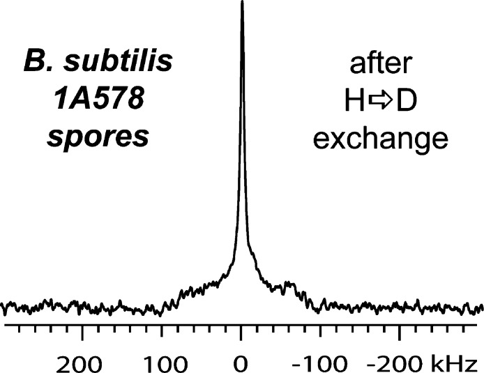
Spores of both strains of B. subtilis, ATCC 6051 and 1A578, grown in protonated media, were soaked in 99% D2O for 1 week and lyophilized, and quadrupolar echo 2H NMR spectra were collected. These spectra are shown as Figures 3 and 4, respectively. The spectrum of wild-type (ATCC 6051) spores shows a strong central feature I, an area of intermediate line broadening, and significant broadening at the baseline while features II and III are not observed. The chloramphenicol-resistant (1A578) spores produce a deuterium spectrum that is dominated by feature I and does not have features II and III, and broadening of the baseline is present but less intense.
Figure 3.
Deuterium solid-state spectrum of B. subtilis ATCC 6051 spores grown in protonated medium and soaked in D2O for one week’s exchange (room temperature, 120 s delay, 10,000 transients). Features II and III are not apparent, leaving only feature I and a broad signal.
Figure 4.
Deuterium solid-state NMR spectrum of spores of B. subtilis 1A578; this strain exhibits a different line shape after H-to-D exchange than B. subtilis ATCC 6051. Although the same overall changes (no features II or III and broad signal) are observed, the broad feature is much less intense. The spectrum was collected at room temperature with a 120 s delay time and a total of 7178 transients.
Bovine Serum Albumin Spectra
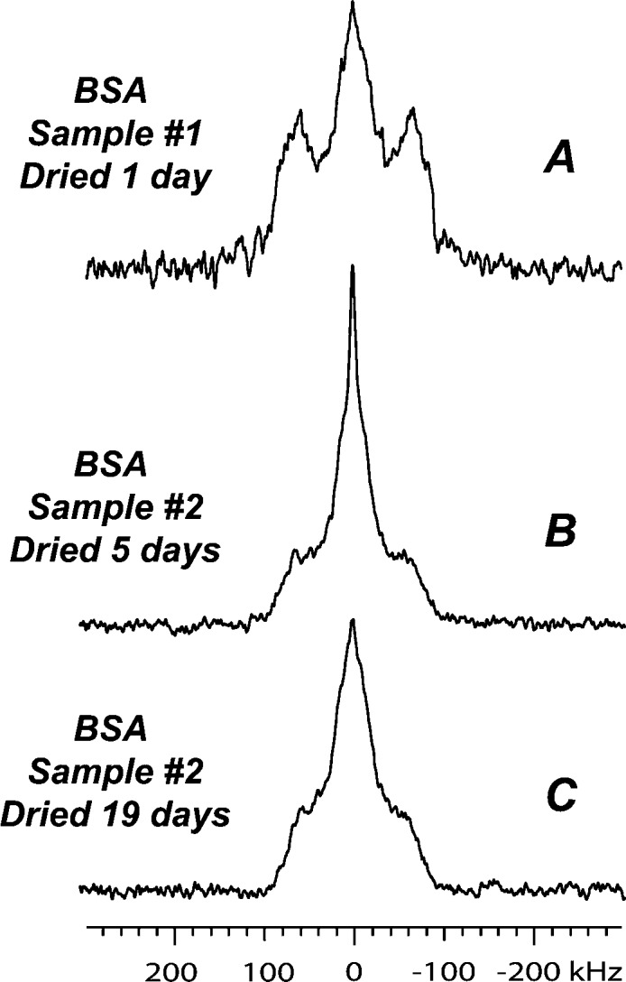
The spectra of BSA lyophilized for 5 days and for 19 days are shown in Figures 5B and 5C, respectively. The mobile D2O peak is reduced in the 19-day spectrum versus the 5-day spectrum, with little to no difference in the broader signal. These spectra are identical to previously reported spectra of a deuterated enzyme, subtilisin.34 However, we previously published a spectrum of deuterated BSA prepared under different conditions.35 This spectrum is shown in Figure 5A and shows a Pake doublet with a large separation, similar in width to feature III in Figures 1 and 2. For this sample, the protein:water ratio (500 mg:10 mL) and drying (1 day) were insufficient to achieve complete deuterium exchange and removal of excess water. Hence, Figure 5A has a lower SNR and the additional set of Pake doublets are not seen in the samples with extended drying (Figure 5B and 5C).
Figure 5.
A comparison of one sample of deuterated lyophilized bovine serum albumin lyophilized 1 day (A) with a second sample (BSA) dried for 5 days (B) and 19 days (C). The Pake doublet observed in A is attributed to bound water that vanishes when the sample is dried for longer periods of time (B, C). Spectra were collected at room temperature with a delay time of 120 s over 4364 transients (A) or 10,000 transients (B, C).
Deuterium NMR Spectra of Calcium Dipicolinate
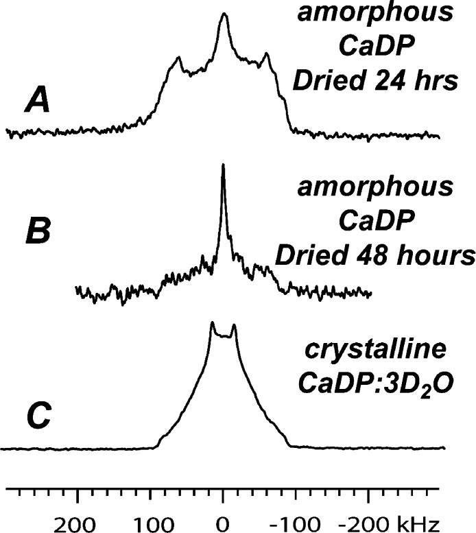
Figure 6 shows the quadrupolar echo deuterium NMR spectra of the amorphous and crystalline forms of calcium dipicolinate (CaDP) respectively. The amorphous sample presents itself as having a Pake doublet with a peak splitting of 116 kHz, with a central peak that is broader than that observed in the B. subtilis spores (Figure 6A). When a smaller sample of the amorphous CaDP is subjected to additional drying (Figure 6B), the Pake doublet disappears and the spectrum resembles that of the deuterated BSA samples (Figures 5B and 5C). The crystal form exists as a trihydrate, and, when isolated from deuterated water, crystalline CaDP·3D2O does not show a central peak (feature I), instead revealing a Pake doublet with a peak splitting of 31 kHz that broadens as it nears the baseline (Figure 6C). The crystal structure determined using a small portion of the CaDP·3D2O NMR sample is shown in Figure 7.
Figure 6.
2H NMR quadrupolar echo spectrum of amorphous deuterated calcium dipicolinate trihydrate dried for 24 h (A), a separate amorphous CaDP sample dried for 48 h (B), and crystalline calcium dipicolinate trihydrate (C). A Pake doublet surrounds a broad mobile D2O peak in A, which is not evident with longer drying on the other amorphous sample (B); crystalline CaDP shows a Pake doublet at ∼31 kHz indicative of limited motion (C). Spectrum A was collected at room temperature with a delay time of 120 s over 7768 transients; B over ∼2200 transients; C over 10,000 transients.
Figure 7.
Crystal structure of deuterated calcium dipicolinate trihydrate (CaDP·3D2O), as shown in a thermal ellipsoid plot, showing that it forms a dimer as a crystal.
Variable-Temperature Deuterium NMR
Figure 8 shows various spectra collected during the VT NMR experiment involving B. subtilis ATCC 6051. The first spectrum collected (top left) is similar to that of Figure 1A, with variations caused by lower signal-to-noise ratio in these spectra collected with 2000 scans (66 h) versus 10,000 scans (333 h). When heated to 50 °C there are only minor differences observed, mostly a slight decrease in the Pake doublet features II and III. This change appears to reverse itself when the sample is cooled back to 21 °C. Feature I becomes dominant and features II and III become reduced when the sample is heated to 100 °C. As before, the spectrum resumes its previous appearance when cooled back to 21 °C (bottom right).
Figure 8.
Spectra obtained for the variable-temperature deuterium NMR of 25% deuterated B. subtilis ATCC 6051 spores. Very slight changes are seen when the spores are heated to 50 °C and cooled; larger changes to the line shape are apparent when heated to 100 °C but appear to revert themselves when cooled to the original temperature. These changes and their reversion to previous line shape are suggestive of glass-like transitions inside a rigid spore core. Spectra were collected with a delay time of 120 s and 2000 transients at the temperatures indicated.
Discussion
Assignment of Deuterium NMR Spectra
The distinct features (I, III, and III) in the deuterium quadrupolar echo NMR spectrum of partially deuterated B. subtilis ATCC 6051 spores (Figure 1A) are assigned by comparison to samples of known composition. Feature I is attributed to deuterons with isotropic motion, possibly from water that is mobile or very dynamic in its environment; future experiments will examine feature I in more detail to study the T1 of this peak. Feature II has a Pake doublet separation (32 kHz) similar to that seen in the crystalline calcium dipicolinate sample, whose crystal structure shows water coordinated to the metal ion, carboxylic acids, and nearby water molecules. However, feature II could also be due to nonlabile deuterons incorporated in biomolecules. Using a growth medium with 25% D2O, our samples will have partial deuteration of every molecule. Thus, methyl groups will be found as −CH2D and give a Pake pattern with a 38 kHz peak separation.36 The Pake pattern is reduced from the typical 140–220 kHz separation found in organic compounds37 due to free rotation. It is well-known that −CD3 molecules in proteins and peptides present a similar NMR spectrum.38 Labile water associated with the lipid bilayers, such as the spore membrane, gives a peak separation of a few kilohertz while deuterons in the lipid itself produce a larger separation, on the order of 30 kHz.39,40 Feature II could also be interpreted as the result of C2 symmetry jumps; this is common in crystalline hydrates41 and, if applicable to calcium dipicolinate trihydrate, would tie feature II to the spore core as well through the CaDP·3H2O.
Attribution of feature III (peak separation of 120 kHz) to immobilized water molecules has been disputed and was suggested to arise from labile deuterons in biomolecules.28 We have prepared and studied labile deuterons in the BSA protein sample, and the 2H NMR spectrum does not have feature III present after extended drying (Figures 5B and 5C). Spectra similar to Figure 5B have been reported for hydrated subtilisin enzyme isolated from a D2O solution. This sample contains labile O–D and N–D groups along with a layer of hydration water strongly associated with the enzyme.34 For a sample of BSA dried for 1 day, the spectrum shows a Pake doublet with a separation similar to that of feature III. We attribute the Pake doublet in Figure 5A to water molecules (HOD) bound to the protein sample. These HOD molecules are not firmly attached to the protein as they can be removed with vacuum drying (Figures 5B and 5C). The outer edges of the Pake doublet in Figure 5A are less sharp than the Pake doublet of frozen water (Figure 9C). This indicates that excess water associated with BSA is immobilized in a heterogeneous fashion. The variation in chemical environment can be due to localized differences in numbers of neighboring water molecules and the hydrogen bonding network. Similar observations are made with CaDP samples with a wide Pake doublet (Figure 6A) that is removed with further drying (Figure 6B).
Figure 9.
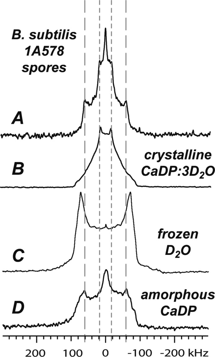
A comparison of the Pake doublets of B. subtilis 1A578 (A) versus crystalline CaDP·3D2O (B), bulk frozen D2O (C), and amorphous CaDP·3D2O (D), indicating the likelihood of the spore Pake doublets arising from immobilized core components. In this model feature III arises from bound water similar to that of amorphous CaDP but unlike bulk water; feature II may arise from methyl rotations or from the crystalline CaDP·3D2O signal.
The shape of feature III (Figures 1 and 2) shows that these deuterons are in an environment of substantial homogeneity compared to water associated with amorphous proteins or CaDP. If these deuterons are protein-associated water, they would be very susceptible to a temperature-induced increase in isotropic motion. These dynamics should be reversible and would not require the protein itself to undergo isotropic motion. Such behavior is shown after heating the spore to 100 °C and cooling it back down to 21 °C (Figure 8). The dramatic change of the anisotropic feature III into the isotropic signal of feature I supports our view that these deuterons are in rigid water associated with biomolecules, and their increased motion does not require a diffuse liquid-like state. Within a few degrees of the D2O melting point (3.8 °C), water molecules flip between ice crystal sites yielding a narrow isotropic peak. Wittebort et al.42 used variable temperature 1H and 2H NMR studies to examine molecular dynamics in ice. In the hexagonal form of ice, Ih, the central water molecule is bound to 2 neighboring H atoms (via the oxygen) and 2 neighboring oxygen atoms (via the protons). These four bonds form a tetrahedron around the central water molecule. In Wittebort’s sample, a narrow line is seen at −6 °C and attributed to isotropic reorientation within the ice lattice. At −24 °C, the narrow line is very small due to a reduction in dynamics. Our spore samples were analyzed at room temperature, and thus bulk ice is not expected, although the water could be dynamic within a rigid lattice; the relatively small size of the narrow line at −6 °C in Wittebort’s sample implies that its contribution to feature I would be negligible. Observation of immobile water at room temperature is not without precedence. A mixture of mobile and solid water has been reported for hydrated minerals. The sodium-silicate kanemite has three bound water molecules which move between tetrahedral binding sites within the mineral structure, though some water is found in between layers of the mineral.41,43 Rigid water in the kanemite sample produces a Pake pattern separation >190 kHz due to Na–O–D and Si–O–D bonds at −120 °C. At room temperature, a weak powder pattern is observed symmetric to an isotropic peak, indicating less mobile water undergoing jumps. Thus, water in nonbulk environments generates Pake patterns with varying quadrupolar coupling constants. Li et al. report that water in corn starch samples produces Pake pattern separations less than 144 kHz.44 Thus, it is not unexpected that excess water bound to proteins could give a Pake pattern splitting of 120 kHz compared to that of 144 kHz for bulk ice.
Dimers of α-truxillic acid form with two carboxylic acid groups arranged in a linear fashion. The result is a pair of O–H···O=C hydrogen bonds and replacement with deuterium producing a sharp Pake doublet with a 117 kHz splitting,45 but only when those carboxylic acids are in the linear arrangement. We do not believe a homogeneous chemical environment such as that found in α-truxillic acid, however, produces feature III in bacterial spores. There are numerous carboxylic acid groups in the spore proteins, but they form hydrogen bonds with amide groups. Carboxylic acid groups in the peptidoglycan are arranged in a random disordered fashion and would not be expected to adopt a homogeneous linear conformation. The crystalline calcium dipicolinate unit cell shows one water bond to a carboxylic acid group, yet we do not see feature III in Figure 6C. Most of the carboxyl groups are involved in direct chelation of the Ca2+ ions, reducing the occurrence of O–D···O=C hydrogen bonding.
Another possible source of feature III is nonlabile deuterons found as rigid C–D groups.36,46 This labeling would be widespread among the various proteins, DNA, peptidoglycan, and membranes found in a mature spore. These deuterons could be responsible for the remaining portion of feature III at 100 °C not converted to the isotropic feature I. Thus, we believe that feature III is due to both nonlabile deuterons in biomolecules and rigid water associated with biomolecules. Due to the complexity of the spore sample and the heterogeneous nature of the deuterium spectrum, with many components, we were unable to generate a satisfactory quantitative estimate of each component in the spore. As seen with previous reports,28 quantitative assignment would be dependent on the level of hydration. Nonetheless, our qualitative study provides additional insight in the spore core water environment. Future studies will attempt quantification through line shape simulation and comparison of simulated spectra with the spectra reported here.
Sites Accessible to Exchange
The exchange of accessible deuterons with H2O is demonstrated in Figure 1 using spores of B. subtilis ATCC 6051. The decrease in the mobile D2O peak (feature I) is expected; mobile D2O would exchange freely with exogenous water. However, a significant fraction of mobile water is unreachable by external H2O. These deuterons can be both core and noncore water. In the latter case, deuterons are protected from exchange by protein conformations that trap water in a rigid matrix. Dormant spores, even hydrated, would maintain proteins in tight conformations which would limit access and exchange as a means of hindering chemical attack. If water can cross the membrane, some core-associated deuterons must be protected from exchange by protein conformations or the transport and exchange of water is very slow compared to the 7-day exchange period. As the exchange was carried out in a water suspension, excess water would cause full hydration and swelling of the spore samples, although in a state of dormancy active water transport would cease to function. A rigid core model would protect water from exchange, but it is also possible that a rigid core could transition into a gel state with excess hydration. If this were the case, some mobile deuterons in protein/DNA/CaDP environments must be inaccessible to gel water, or the core is a mixture of gel and rigid domains restricting access to exchange sites. Nevertheless, this analysis does not preclude the opportunity for gel core water to be present with dynamics different than bulk water.28 However, experiments targeted to study core-water dynamics after H-to-D exchange would report only accessible water while trapped water would be invisible and not contribute to relaxation rate analysis. From the data in Figures 1 and 2, this trapped water has a noticeable contribution to feature I. Thus, Kaieda et al.28 do not report all possible water in hydrated spores.
However, it is also possible that the membrane prevents water diffusion. In this scenario, the mobile water peak observed here and in previous work would be due to noncore water molecules protected from exchange by protein conformations. Unfortunately, changes to feature I after exchange cannot be used to evaluate whether the mobile water peak is due to core or noncore water. It is not unreasonable that the lipid bilayer would undergo significant structural and/or biochemical changes during sporulation to protect the core from dehydration or attack by sporicidal agents through the removal or closure of transmembrane channels and transport proteins, or structural changes to the phospholipids themselves creating a tighter network. Deuterium NMR is commonly used to examine the motion of lipid membranes and dynamics within the hydrophobic tails47−53 as well as other relevant biomolecules such as DNA.54 If the lipid bilayer deuteron signals could be isolated from other components in the spore, these experiments could be used in the future to elucidate such changes. After lyophilization, the samples are exposed to atmospheric moisture during the 2-week time period required to collect a deuterium NMR spectrum. The rotor, approximately half to three-quarters full, allows for water vapor in the headspace to penetrate the spore and continue exchange processes. We do not detect changes in the spectrum over time or after recollection of prior data with the same sample. Thus, atmospheric H2O is not able to access exchangeable deuterons. Drying may cause the spore to acquire hydrophobic properties in the coat protein layers or membrane or change from a gel to rigid core.
The inner Pake doublet (feature II) also decreases, indicating some of these deuterons are in an environment susceptible to exchange. No change would be seen for nonlabile deuterons in biomolecules created in the culture and sporulation media. If the spore core is impenetrable, water in the calcium dipicolinate trihydrate would remain after exchange and contribute to feature II, if the core CaDP has a structure similar to the crystalline material produced in our laboratory. It is also possible that some of the water associated with CaDP is subject to exchange, perhaps on the exterior of crystallites, whereas most of the water in CaDP is protected. Kaieda et al.28 proposed that these signals could be due to deuterated methyl groups; similar line shapes were seen in magic-angle-spinning (MAS) spectra of deuterated valines inside of glycophorin A.38
Interestingly, feature III is relatively unchanged after soaking in H2O. If this Pake doublet were, as suggested, due solely to labile deuterons,28 the expected result of a deuterium–hydrogen exchange would be a loss of deuterium signal in the spectrum, or at least a decrease in intensity proportional to that seen for features I and II. Thus, these deuterons are either nonlabile or nonaccessible to external water. As described above, a glassy core would protect labile water deuterons in the core. Additionally, if linear hydrogen bonds such as in α-truxillic acid dimers45 are responsible for feature III, they may persist in water if the biomolecules or peptidoglycan do not rearrange in response to swelling from excess hydration. This seems unlikely as Bacillus spores change shape/size in response to external water.55 If these hydrogen bonds are susceptible to exchange, and responsible for feature III, then they would be nonexistent in the postexchange spectrum. Thus, the persistence of feature III after the D-to-H exchange process supports either bound water molecules in the spore or the partial deuteration of carbon atoms found in various biomolecules. As described above, we believe that both species are present.
Similar behavior was observed in deuterium–hydrogen exchange experiments with a sample of B. subtilis 1A578 spores, as seen in Figure 2. This indicates that changes to features I and II, and the persistence of feature III, are not a property confined to the ATCC 6051 strain. Nevertheless, the spores of the chloramphenicol-resistant and wild-type strains show dramatic differences when subjected to hydrogen-to-deuterium exchange experiments. In these experiments, spores were produced using protonated growth and sporulation media. Labile, and accessible, protons were replaced with deuterium by soaking in sterile D2O.
The postexchange spectra for ATCC 6051 spores show a line shape (Figure 3) lacking the sharp features II and III of Figure 1A. Hydrocarbons of biomolecules and lipid groups would not contribute to the post H-to-D exchange spectra. The lack of a strong feature II reinforces the conclusion that deuterons responsible for features II and III are not labile (perhaps in C–D or CHD groups) or not easily accessible for exchange (located in the core and/or protected by proteins). However, the 1A578 spores produce a spectrum (Figure 4) dominated by mobile water (feature I). The broadened baseline has a shape similar to that of the wild-type spores (Figure 3) but lower in intensity. Proteins external to the core may have a structural conformation that protects labile protons from exchange with deuterons. A proteomics study with the chloramphenicol-resistant 1A578 found 85 proteins already identified from Bacillus species and 69 novel proteins.56 Thus, unique protein structures may exist that resist exchange with deuterium, a possibility that warrants further study. Both spore samples were handled in a similar fashion, thus comparison of the spectra indicates that 1A578 has a greater fraction of mobile water. This could result from the chloramphenicol-resistant spores retaining a greater mass of mobile water, having a mobile water fraction with greater resistance to drying, or the labile deuterons being predominately found as mobile water. These deuterons obviously have rapid isotropic motion, but it is not clear if they are located in the core, noncore regions, or both.
If feature III of the spore deuterium NMR spectrum in Figure 1A was indicative of labile deuterons, asserted by Kaieda et al.,28 the spectrum observed in Figure 3 should look identical (or nearly so) to that seen in Figure 1A. Instead, Figure 3 shows a feature II that is practically indistinguishable from the central mobile deuterium peak and broad shoulders instead of the sharp Pake doublet seen in feature III of Figure 1A. Likewise, if feature III were the result of O–D···O=C hydrogen bonds susceptible to exchange, the H-to-D experiment should produce a sharp Pake doublet. Instead, we speculate that the broad signals in Figures 3 and 4 are from labile deuterons of proteins. When the spectra in Figures 3 and 4 are compared to a spectrum of BSA, the result is a nearly identical overlap for the wild-type 6051 spores (Figure 10). However, with its large fraction of mobile water, the resistant 1A578 spores (Figure 11) do not overlap with the BSA spectrum unless the protein spectrum is reduced in height. While the scaling factor was 3.3, this number is somewhat meaningless as we are not convinced that labile deuterons in the coat proteins of 1A578 spores would be exchanged to a similar degree as labile deuterons in a homogeneous solution of BSA.
Figure 10.
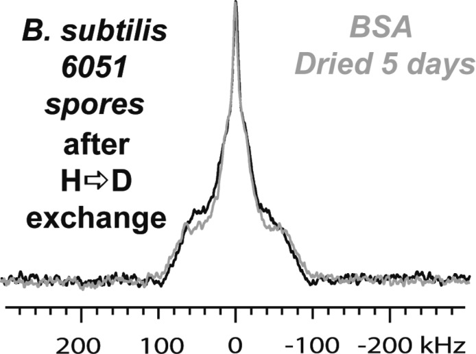
Postexchange B. subtilis ATCC 6051 spore spectrum (Figure 3) to which the five-day-dried BSA spectrum (Figure 5B) has been overlaid. BSA’s spectrum fits very well to the spectrum of the whole spore.
Figure 11.
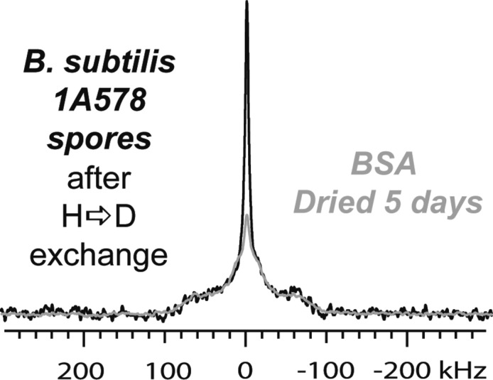
Postexchange B. subtilis 1A578 spore spectrum (Figure 4) overlaid on the same spectrum as the five-day lyophilized deuterated BSA (Figure 5B). BSA’s spectrum fits well to the broad peak feature in 1A578 but less so to the mobile water peak feature. The difference in line shape between this and Figure 10 is attributed to structural changes in B. subtilis 1A578 resulting in higher protein conformational homogeneity and may be responsible for the differences in line shape overall between B. subtilis 1A578 and B. subtilis ATCC 6051.
Rigid Spore Core from Variable-Temperature Deuterium Solid-State NMR
We speculate that the spore core is more a solid than a gel-like state, but exchange experiments alone do not provide enough evidence to distinguish the physical state of the core. Instead, we base our analysis on variable-temperature data collected to provide this distinction. Figure 8 shows the results of the VT NMR experiments with B. subtilis ATCC 6051 spores. It is evident that the mobile D2O peak does not increase perceptibly when the sample is heated from 21 to 50 °C. However, when cooled back to 21 °C and heated to 100 °C, the central peak intensity increases dramatically. The high temperature causes significant portions of the spore to undergo isotropic, or nearly isotropic, motion. Other portions of the spore retain anisotropic behavior as evidenced by the persistence of features II and III after heating. The lack of a dramatic change in features II and III at 50 °C suggests that components able to undergo isotropic motion are not affected by the moderate heating. This analysis does not support a gel-like core with a high mobile water content, which would be expected to show gradual changes as the temperature is increased. The rigid core analysis is similar to the predictions made by Sapru22 indicating glass-transition temperatures in between 50 and 100 °C for various Bacillus strains.
The analysis of our VT NMR data builds upon our assignment of spectra to different components, drying effects on the spectra, and changes caused by H/D or D/H exchange. As discussed below, we suggest a spore model with a rigid core where CaDP and biomolecules are rigid (features II and III) with a smaller fraction of deuterons existing as nonrigid, perhaps mobile, water (feature I). Data collected at 100 °C suggests that there is a phase change between 50 and 100 °C responsible for the large increase in the isotropic deuteron signal. The higher fraction of mobile deuterons could arise from faster dynamics of nonlabile deuterons in biomolecules. Nevertheless, 100 °C is insufficient to convert every deuteron of features II and III into an isotropic state. Deuterated peptidoglycan, contributing to feature III, is a highly cross-linked network and not expected to have large degrees of freedom, even with heating. Also, these deuterons may exist in proteins that resist conformation changes at high temperature. Crystalline CaDP may generate feature II, and work in our lab shows that crystalline CaDP does not melt in boiling water.
When the sample is cooled back to 21 °C (from 100 °C), NMR data show that the sample characteristics which generate the room temperature spectra are nearly identical to those of the initial spectra collected at 21 °C prior to heating. This suggests that changes in the deuteron environment due to heating are reversible. Deuterated biomolecules would return to an immobile state, but we do not know if these biomolecules have been denatured to render the spore inert. Previous work by the Setlow Lab has shown that the viability of B. subtilis spores is relatively unaffected by dry heating to 100 °C.20 The viability of these spores also suggests that the protein denaturation is modest and would not generate the isotropic signal at 100 °C. Thus, changes to feature III in Figure 8 are attributed to bound water molecules where heating increases their motion to generate an isotropic peak, yet cooling allows them to revert to the bound state without affecting the viability of the spore itself. From this viewpoint, the VT NMR data suggest the presence of bound water contributing to feature III in contrast to the assertions promoted by Kaieda et al.
Proton NMR has been used to examine water in spores; however, the combination of heterogeneous samples and narrow chemical shift dispersion creates difficulties in assigning spectra. Nevertheless, the water will generate a very strong signal, and its chemical environment will influence spin relaxation rates. Bradbury et al. reported equal amounts of water in core and cortex + coat regions. Water was found to be both mobile and immobile. After separating the coat proteins and PG cortex, the water in these samples was found to be immobile; thus, mobile water was assigned to the core.57 Using solid-state NMR spectroscopy, the characteristic 13C resonance for dipicolinic acid (DPA)58 was detected with cross-polarization in freeze-dried and rehydrated spores of B. subtilis.55 CPMAS data reflect those 13C spins that maintain strong dipolar coupling with nearby 1H nuclei, and thus indicate limited or absent molecular motion. In contrast, single-pulse MAS (SPMAS) signals are strongly affected by mobile species where strong spin–lattice effects cause fast T1 relaxation. Leuschner and Lillford could not detect DPA with 13C SPMAS, demonstrating that the DPA is in a firm rigid lattice, and rehydration of the core does not increase the molecular motion of DPA. From these prior studies, the core-specific DPA molecules may exist in a glassy core with water around the DPA also in an immobile state. We have shown in Figure 9B that the trihydrate form of crystalline CaDP produces a deuterium spectrum with a Pake doublet similar to feature II. This does not prove that CaDP in the core is crystalline, only that it is possible that CaDP could adopt a structural arrangement similar to the crystal structure. It is entirely possible that the CaDP is found as microcrystals in a glassy matrix of water, DNA, and proteins. The D-to-H exchange experiments show slight reduction of feature II; this reduction could be due to exchangeable waters around the CaDP crystallites.
Kaieda et al.28 reported a quadrupolar echo 2H NMR spectrum of fully hydrated Mn2+ depleted B. subtilis PS533 spores and concluded that the broad ∼130 kHz feature was entirely the result of labile deuterons in immobilized proteins and other biomolecules in the spore. Unfortunately, it is hard to understand how Kaieda et al. could state that there was no evidence for immobilized water in the core if their data and experimental techniques could not detect immobile core-associated D2O. Their protocol is essentially a hydrogen-to-deuterium exchange experiment introducing D2O after growing spores in deuterium-free media; however, instead of lyophilization, the authors used the wet spore pellet for their NMR analysis. The line shape observed in Figures 10 and 11 (particularly Figure 11) is similar to that shown in Kaieda et al.28 for their quadrupolar echo 2H NMR results. In their paper, the wet cell pellet creates a massive signal for mobile water. The authors claim that the wet pellet spectrum demonstrates swollen spores with a gel-like core and that their relaxation rate measurements reflect the dynamics of water within the core region. We do not dispute the validity of their data collection or their observation of water fractions with bulk and nonbulk behavior. We assert that their measurements are not representative of all water molecules in the spore sample. The persistence of feature I after D-to-H exchange suggests that water remains trapped, perhaps in a rigid core as suggested by our VT NMR data. Samples of 1A578 spores generated from protonated media and followed by deuterium exchange produce a spectrum (Figure 11) similar to that in Kaieda’s work. We believe that our spectrum reflects the mobile water content (core and noncore) of a dried spore sample while Kaieda’s data originates with fully hydrated samples. Upon closer inspection, their spectrum shows a broadened baseline similar to that seen with BSA protein reported here, and we agree that this baseline broadening is due to labile deuterons in biomolecules. Lastly, Sunde et al.27 assert that, were the core in a glassy state, there would be a broad signal apparent on a 2H NMR spectrum of the spore; however, such a signal would not be detectable using the magnetic relaxation dispersion experiments as performed. We have observed this signal in different spore strains, and its width of ∼120 kHz is consistent with excess water bound to BSA and CaDP.
The reason why the spectra reported in Kaieda et al.28 are similar to that in Figure 11 (the B. subtilis 1A578), and why there are differences in the line shape between B. subtilis ATCC 6051 and 1A578, may have an explanation. While ATCC 6051 is a wild-type strain, 1A578 is a variant of B. subtilis 168 that has resistance to chloramphenicol.29 It is possible that the mechanism of resistance causes structural changes in the vegetative cells of this strain. Such changes might manifest themselves in subtle alterations in the spore structure resulting in the differences in the deuterium NMR spectra of Figures 1–4. The strain used both in Kaieda et al.’s work28 and in the paper by Sunde et al.27 is B. subtilis PS533, noted as being resistant to kanamycin.59 Thus, the similarity of the deuterium NMR spectra reported by Kaieda et al. to B. subtilis 1A578 spectra shown in Figure 4 may result from introducing a resistance locus to B. subtilis. From this viewpoint, it is possible that the data reported by Sunde et al. are also influenced by a similar phenomenon. It is also noteworthy that features II and III of the wild-type B. subtilis ATCC 6051 spores (Figure 1) show greater broadening and heterogeneity compared with the sharper lines seen in Figure 2, data from chloramphenicol resistant strain. Sharper lines result from enhanced structural homogeneity, and thus the resistance locus appears to have caused an observable reduction in variation of proteins/biomolecule conformation. This observation merits additional study with other bacterial spore strains. It may be possible to apply other sophisticated biomolecular NMR experiments to explore structural biology via 13C and 15N labeling.
Conclusions
In this work, we studied deuterium NMR spectra of B. subtilis ATCC 6051 and 1A578 with an aim of determining the state of water inside of the spore core. This was aided by variable-temperature NMR studies of these species and 2H NMR spectra of calcium dipicolinate and bovine serum albumin. The overall NMR line shape of spectra for spores of both B. subtilis strains indicates that water is in multiple distinct states in the spore. The wide Pake doublet and the maintenance of its signal through deuterium–hydrogen exchange indicates that it is bound water, likely associated with the core. This is also demonstrated through the hydrogen–deuterium exchange spectra where the only signals that appear on the spectrum are those similar to bulk proteins such as those seen in deuterated bovine serum albumin. The difference between the spore spectrum shown in Kaieda et al.28 and that of B. subtilis ATCC 6051 is postulated to be due to structural differences perhaps attributable to different proteins or antibiotic resistances, similar to B. subtilis 1A578. The crystalline calcium dipicolinate spectrum, when compared to spore spectra, suggests that the inner Pake doublet may be attributable to immobile core components. In addition, the lack of proportional increase in the mobile deuterium signal during the variable-temperature NMR experiments, and the return of the signal to its original line shape upon cooling, suggest a narrow transition along the lines predicted by Sapru22 and Ablett23 and thus a glassy core.
The existence of bound water has come under recent scrutiny27 from the measurement of deuterium relaxation rates. These samples were dispersed in deuterated water, and deuterium relaxation rates were measured with solution-state NMR methods at different magnetic field strengths. The spin–lattice relaxation could be explained by molecular motions on the order of 50 ps, which implies a mobile water fraction within the core. As described by the authors, this correlation time is too fast for water to be in a “rigid lattice” environment that would produce a 200 kHz wide spectrum. However, these water molecules are invisible in their deuterium relaxation rate measurements. As shown in this work, deuterium wide line NMR spectroscopy is able to detect both mobile and rigid lattice waters simultaneously. Our data demonstrate the existence of rigid lattice water molecules in bacterial spores and that these molecules are in a different chemical environment than water molecules within the tetrahedral bonding network of frozen water. Additional data show that immobilized water in spores is resilient and unable to undergo chemical exchange with water molecules in exogenous solution.
Acknowledgments
This work is supported by the National Institutes of Health (1R01GM090064-01), a NASA EPSCoR Research Infrastructure Development (RID) grant (NN07AL49A), and the University of Oklahoma. We also wish to express our gratitude to Dr. Kasthuri Venkateswaran, California Institute of Technology, Jet Propulsion Laboratory, NASA, for insights and helpful discussions.
Supporting Information Available
The X-ray crystallographic unit cell properties and other crystallographic data of calcium dipicolinate trihydrate. This material is available free of charge via the Internet at http://pubs.acs.org.
The authors declare no competing financial interest.
Funding Statement
National Institutes of Health, United States
Supplementary Material
References
- Higgins D.; Dworkin J. Recent Progress in Bacillus subtilis Sporulation. FEMS Microbiol. Rev. 2012, 36, 131–148. [DOI] [PMC free article] [PubMed] [Google Scholar]
- Cano R. J.; Borucki M. K. Revival and Identification of Bacterial Spores in 25- to 40-Million-Year-Old Dominican Amber. Science 1995, 268, 1060–1064. [DOI] [PubMed] [Google Scholar]
- Driks A. Bacillus subtilis Spore Coat. Microbiol. Mol. Biol. Rev. 1999, 63, 1–20. [DOI] [PMC free article] [PubMed] [Google Scholar]
- Driks A. The Bacillus Spore Coat. Phytopathology 2004, 94, 1249–1251. [DOI] [PubMed] [Google Scholar]
- Driks A. Maximum Shields: the Assembly and Function of the Bacterial Spore Coat. Trends Microbiol. 2002, 10, 251–254. [DOI] [PubMed] [Google Scholar]
- Takamatsu H.; Watabe K. Assembly and Genetics of Spore Protective Structures. Cell. Mol. Life Sci. 2002, 59, 434–444. [DOI] [PMC free article] [PubMed] [Google Scholar]
- Riesenman P. J.; Nicholson W. L. Role of the Spore Coat Layers in Bacillus subtilis Spore Resistance to Hydrogen Peroxide, Artificial UV-C, UV-B, and Solar UV Radiation. Appl. Environ. Microbiol. 2000, 66, 620–626. [DOI] [PMC free article] [PubMed] [Google Scholar]
- Warth A. D.; Strominger J. L. Structure of the Peptidoglycan from Spores of Bacillus subtilis. Biochemistry 1972, 11, 1389–1396. [DOI] [PubMed] [Google Scholar]
- Atrih A.; Foster S. J. The Role of Peptidoglycan Structure and Structural Dynamics during Endospore Dormancy and Germination. Antonie van Leeuwenhoek 1999, 75, 299–307. [DOI] [PubMed] [Google Scholar]
- Mallidis C. G.; Scholefield J. Relation of the Heat Resistance of Bacterial Spores to Chemical Composition and Structure. II. Relation to Cortex and Structure. J. Appl. Bacteriol. 1987, 63, 207–215. [DOI] [PubMed] [Google Scholar]
- Atrih A.; Zöllner P.; Allmaier G.; Foster S. J. Structural Analysis of Bacillus subtilis 168 Endospore Peptidoglycan and Its Role during Differentiation. J. Bacteriol. 1996, 178, 6173–6183. [DOI] [PMC free article] [PubMed] [Google Scholar]
- Setlow P. Small, Acid-Soluble Spore Proteins of Bacillus Species: Structure, Synthesis, Genetics, Function, and Degradation. Annu. Rev. Microbiol. 1988, 42, 319–338. [DOI] [PubMed] [Google Scholar]
- Moeller R.; Setlow P.; Reitz G.; Nicholson W. L. Roles of Small, Acid-Soluble Spore Proteins and Core Water Content in Survival of Bacillus subtilis Spores Exposed to Environmental Solar UV Radiation. Appl. Environ. Microbiol. 2009, 75, 5202–5208. [DOI] [PMC free article] [PubMed] [Google Scholar]
- Setlow B.; Setlow P. Small, Acid-Soluble Proteins Bound to DNA Protect Bacillus subtilis Spores from Killing by Dry Heat. Appl. Environ. Microbiol. 1995, 61, 2787–2790. [DOI] [PMC free article] [PubMed] [Google Scholar]
- Paidhungat M.; Ragkousi K.; Setlow P. Genetic Requirements for Induction of Germination of Spores of Bacillus subtilis by Ca2+-Dipicolinate. J. Bacteriol. 2001, 183, 4886–4893. [DOI] [PMC free article] [PubMed] [Google Scholar]
- Ghiamati E.; Manoharan R.; Nelson W. H.; Sperry J. F. UV Resonance Raman Spectra of Bacillus Spores. Appl. Spectrosc. 1992, 46, 357–364. [Google Scholar]
- Setlow B.; Atluri S.; Kitchel R.; Koziol-Dube K.; Setlow P. Role of Dipicolinic Acid in Resistance and Stability of Spores of Bacillus subtilis with or without DNA-Protective α/β-Type Small Acid-Soluble Proteins. J. Bacteriol. 2006, 188, 3740–3747. [DOI] [PMC free article] [PubMed] [Google Scholar]
- Magge A.; Granger A. C.; Wahome P. G.; Setlow B.; Vepachedu V. R.; Loshon C. A.; Peng L.; Chen D.; Li Y.-q.; Setlow P. Role of Dipicolinic Acid in the Germination, Stability, and Viability of Spore of Bacillus subtilis. J. Bacteriol. 2008, 190, 4798–4807. [DOI] [PMC free article] [PubMed] [Google Scholar]
- Setlow P. Spores of Bacillus subtilis: Their Resistance to and Killing by Radiation, Heat and Chemicals. J. Appl. Microbiol. 2006, 101, 514–525. [DOI] [PubMed] [Google Scholar]
- Nicholson W. L.; Munakata N.; Horneck G.; Melosh H. J.; Setlow P. Resistance of Bacillus Endospores to Extreme Terrestrial and Extraterrestrial Environments. Microbiol. Mol. Biol. Rev. 2000, 64, 548–572. [DOI] [PMC free article] [PubMed] [Google Scholar]
- Beaman T. C.; Gerhardt P. Heat Resistance of Bacterial Spores Correlated with Protoplast Dehydration, Mineralization, and Thermal Adaptation. Appl. Environ. Microbiol. 1986, 52, 1242–1246. [DOI] [PMC free article] [PubMed] [Google Scholar]
- Sapru V.; Labuza T. P. Glassy State in Bacterial Spores Predicted by Polymer Glass-Transition Theory. J. Food Sci. 1993, 58, 445–448. [Google Scholar]
- Ablett S.; Darke A. H.; Lillford P. J.; Martin D. R. Glass Formation and Dormancy in Bacterial Spores. Int. J. Food Sci. Technol. 1999, 34, 59–69. [Google Scholar]
- Kong L.; Setlow P.; Li Y.-q. Analysis of the Raman Spectra of Ca2+-Dipicolinic Acid Alone and in the Bacterial Spore Core in Both Aqueous and Dehydrated Environments. Analyst 2012, 137, 3683–3689. [DOI] [PubMed] [Google Scholar]
- Huang S.-s.; Chen D.; Pelczar P. L.; Vepachedu V. R.; Setlow P.; Li Y.-q. Levels of Ca2+-Dipicolinic Acid in Individual Bacillus Spores Determined Using Microfluidic Raman Tweezers. J. Bacteriol. 2007, 189, 4681–4687. [DOI] [PMC free article] [PubMed] [Google Scholar]
- Black S. H.; Gerhardt P. Permeability of Bacterial Spores. IV. Water Content, Uptake, and Distribution. J. Bacteriol. 1962, 83, 960–967. [DOI] [PMC free article] [PubMed] [Google Scholar]
- Sunde E. P.; Setlow P.; Hederstedt L.; Halle B. The Physical State of Water in Bacterial Spores. Proc. Natl. Acad. Sci. U.S.A. 2009, 106, 19334–19339. [DOI] [PMC free article] [PubMed] [Google Scholar]
- Kaieda S.; Setlow B.; Setlow P.; Halle B. Mobility of Core Water in Bacillus subtilis Spores by 2H NMR. Biophys. J. 2013, 105, 2016–2023. [DOI] [PMC free article] [PubMed] [Google Scholar]
- Anderson L. M.; Henkin T. M.; Chambliss G. H.; Bott K. F. New Chloramphenicol Resistance Locus in Bacillus subtilis. J. Bacteriol. 1984, 158, 386–388. [DOI] [PMC free article] [PubMed] [Google Scholar]
- Hageman J. H.; Shankweiler G. W.; Wall P. R.; Franich K.; McCowan G. W.; Cauble S. M.; Grajeda J.; Quinones C. Single, Chemically Defined Sporulation Medium for Bacillus subtilis: Growth, Sporulation, and Extracellular Protease Production. J. Bacteriol. 1984, 160, 438–441. [DOI] [PMC free article] [PubMed] [Google Scholar]
- Johnson T. J.; Williams S. D.; Valentine N. B.; Su Y.-F. The Infrared Spectra of Bacillus Bacteria Part II: Sporulated Bacillus—The Effect of Vegetative Cells and Contributions of Calcium Dipicolinate Trihydrate, CaDP·3H2O. Appl. Spectrosc. 2009, 63, 908–915. [DOI] [PubMed] [Google Scholar]
- Bailey G. F.; Karp S.; Sacks L. E. Ultraviolet-Absorption Spectra of Dry Bacterial Spores. J. Bacteirol 1965, 89, 984–987. [DOI] [PMC free article] [PubMed] [Google Scholar]
- Strahs G.; Dickerson R. E. The Crystal Structure of Calcium Dipicolinate Trihydrate (A Bacterial Spore Metabolite). Acta Crystallogr. B 1968, 24, 571–578. [DOI] [PubMed] [Google Scholar]
- Lee C. S.; Ru M. T.; Haake M.; Dordick J. S.; Reimer J. A.; Clark D. S. Multinuclear NMR Study of Enzyme Hydration in an Organic Solvent. Biotechnol. Bioeng. 1998, 57, 686–693. [PubMed] [Google Scholar]
- Rice C. V.; Friedline A.; Johnson K.; Zachariah M.; Thomas K. J. III The Nature of Water within Bacterial Spores: Protecting Life in Extreme Environments. Proc. SPIE 2011, 8152, 81520V.(7 pp). [Google Scholar]
- Stepanov A. G.; Luzgin M. V.; Romannikov V. N; Zamaraev K. I. Carbenium Ion Properties of Octane-1 Adsorbed on Zeolite H-ZSM-5. Catal. Lett. 1994, 24, 271–284. [Google Scholar]
- Duer M.Introduction to Solid-State NMR Spectroscopy; Blackwell Publishing: Malden, MA, 2004. [Google Scholar]
- Liu W.; Crocker E.; Siminovitch D. J.; Smith S. O. Role of Side-Chain Conformational Entropy in Transmembrane Helix Dimerization of Glycophorin A. Biophys. J. 2003, 84, 1263–1271. [DOI] [PMC free article] [PubMed] [Google Scholar]
- Bechinger B.; Seelig J. Interaction of Electric Dipoles with Phospholipid Head Groups. A 2H and 31P NMR Study of Phloretin and Phloretin Analogues in Phosphatidylcholine Membranes. Biochemistry 1991, 30, 3923–3929. [DOI] [PubMed] [Google Scholar]
- Lee D.-K.; Kwon B. S.; Ramamoorthy A. Freezing Point Depression of Water in Phospholipid Membranes—A Solid-State NMR Study. Langmuir 2008, 24, 13598–13604. [DOI] [PMC free article] [PubMed] [Google Scholar]
- O’Hare B.; Grutzeck M. W.; Kim S. H.; Asay D. B.; Benesi A. J. Solid State Water Motions Revealed by Deuterium Relaxation in 2H2O-Synthesized Kanemite and 2H2O-Hydrated Na+-Zeolite A. J. Magn. Reson. 2008, 195, 85–102. [DOI] [PubMed] [Google Scholar]
- Wittebort R. J.; Usha M. G.; Ruben D. J.; Wemmer D. E.; Pines A. Observation of Molecular Reorientation in Ice by Proton and Deuterium Magnetic Resonance. J. Am. Chem. Soc. 1988, 110, 5668–5671. [Google Scholar]
- Benesi A. J.; Grutzeck M. W.; O’Hare B.; Phair J. W. Room-Temperature Icelike Water in Kanemite Detected by 2H NMR T1 Relaxation. Langmuir 2005, 21, 527–529. [DOI] [PubMed] [Google Scholar]
- Li S.; Dickinson L. C.; Chinachoti P. Mobility of ‘Unfreezable’ and ‘Freezable’ Water in Waxy Corn Starch by 2H and 1H NMR. J. Agric. Food Chem. 1998, 46, 62–71. [DOI] [PubMed] [Google Scholar]
- Khan M.; Brunklaus G.; Enkelmann V.; Spiess H.-W. Transient States in [2 + 2] Photodimerization of Cinnamic Acid: Correlation of Solid-State NMR and X-ray Analysis. J. Am. Chem. Soc. 2008, 130, 1741–1748. [DOI] [PubMed] [Google Scholar]
- Jelinski L. W. Solid State Deuterium NMR Studies of Polymer Chain Dynamics. Annu. Rev. Mater. Sci. 1985, 15, 359–377. [Google Scholar]
- Seelig A.; Seelig J. The Dynamic Structure of Fatty Acyl Chains in a Phospholipid Bilayer Measured by Deuterium Magnetic Resonance. Biochemistry 1974, 13, 4839–4845. [DOI] [PubMed] [Google Scholar]
- Seelig J.; Macdonald P. M. Phospholipids and Proteins in Biological Membranes. 2H NMR as a Method To Study Structure, Dynamics, and Interactions. Acc. Chem. Res. 1987, 20, 221–228. [Google Scholar]
- Brown M. F.; Nevzorov A. A. 2H-NMR in Liquid Crystals and Membranes. Colloids Surf., A 1999, 158, 281–298. [Google Scholar]
- Smith R. L.; Oldfield E. Dynamic Structure of Membranes by Deuterium NMR. Science 1984, 225, 280–288. [DOI] [PubMed] [Google Scholar]
- Kintanar A.; Kunwar A. C.; Oldfield E. Deuterium Nuclear Magnetic Resonance Spectroscopic Study of the Fluorescent Probe Diphenylhexatriene in Model Membrane Systems. Biochemistry 1986, 25, 6517–6524. [DOI] [PubMed] [Google Scholar]
- Davis J. H. The Description of Membrane Lipid Conformation, Order and Dynamics by 2H-NMR. Biochim. Biophys. Acta 1983, 737, 117–171. [DOI] [PubMed] [Google Scholar]
- Petrache H. I.; Dodd S. W.; Brown M. F. Area per Lipid and Acyl Length Distributions in Fluid Phosphatidylcholines Determined by 2H NMR Spectroscopy. Biophys. J. 2000, 79, 3172–3192. [DOI] [PMC free article] [PubMed] [Google Scholar]
- Robinson B. H.; Mailer C.; Drobny G. Site-Specific Dynamics in DNA: Experiments. Annu. Rev. Biophys. Biomol. Struct. 1997, 26, 629–658. [DOI] [PubMed] [Google Scholar]
- Leuschner R. G. K.; Lillford P. J. Effects of Hydration on Molecular Mobility in Phase-Bright Bacillus subtilis Spores. Microbiology 2000, 146, 49–55. [DOI] [PubMed] [Google Scholar]
- Kuwana R.; Kasahara Y.; Fujibayashi M.; Takamatsu H.; Ogasawara N.; Watabe K. Proteomics Characterization of Novel Spore Proteins of Bacillus subtilis. Microbiology 2002, 148, 3971–3982. [DOI] [PubMed] [Google Scholar]
- Bradbury J. H.; Foster J. R.; Hammer B.; Lindsay J.; Murrell W. G. The Source of the Heat Resistance of Bacterial Spores. Study of Water in Spores by NMR. Biochim. Biophys. Acta 1981, 678, 157–164. [DOI] [PubMed] [Google Scholar]
- Lundin R. E.; Sacks L. E. High-Resolution Solid-State 13C Nuclear Magnetic Resonance of Bacterial Spores: Identification of the Alpha-Carbon Signal of Dipicolinic Acid. Appl. Environ. Microbiol. 1988, 54, 923–928. [DOI] [PMC free article] [PubMed] [Google Scholar]
- Setlow B.; Setlow P. Role of DNA Repair in Bacillus subtilis Spore Resistance. J. Bacteriol. 1996, 178, 3486–3495. [DOI] [PMC free article] [PubMed] [Google Scholar]
Associated Data
This section collects any data citations, data availability statements, or supplementary materials included in this article.


