

Abstract
Triceps ruptures are less common injuries presenting to the orthopaedic or emergency department setting compared with other musculoskeletal injuries. This to some extent reduces the level of index of suspicion or chances of considering the triceps rupture as one of the differential diagnosis while examining a patient following upper limb injury. The literature search shows that a significant proportion of patient diagnosis has been missed during initial presentation, leading to a delay in diagnosis and in providing definitive treatment, ranging from 6 to 18 months. The triceps are the primary extensor of the elbow and are supplied with the radial nerve. Any injury to the triceps can adversely affect the functioning of the limb and influence the ability to work and return to employment. We share our experience of treating a patient with a triceps rupture, in whom the diagnosis was made 6 months after injury; the patient was able to return to manual work 3 months after surgical repair.
Background
The triceps brachii muscle has three distinct muscle heads—the long, medial and lateral head—inserting onto the olecranon process of the ulna. The long head arises from the infraglenoid tubercle of the scapula while the lateral head arises from a ridge over the posterior aspect of the humerus above the radial groove and partly forms the lateral intermuscular septum. The medial head arises from the posterior aspect of the humerus below the radial groove, partly from the medial intermuscular septum and with a few fibres from the lateral intermuscular septum.1 All the three heads are innervated by the radial nerve. Triceps ruptures are not common.
Incidence of triceps ruptures is less than 2% of total tendon-related injuries and less than 1% of tendon-related injuries in the upper extremity.2 We found limited reports of isolated medial head of triceps rupture in the literature search using PubMed, CINHAL and EMBASE, and scant mention of it in the major orthopaedic journal web portals. However, the common factor among those reported was that they were cases of young active males aged between 23 and 42 years. There are few reports describing full thickness tear or avulsion of the distal triceps including the medial head.
The diagnosis is made based on mechanism of injury, clinical presentation with bruising and swelling along the posterior aspect of arm with limited or lack of extension. In some cases a palpable gap at the rupture site can be noted. It is reported that a significant proportion of these cases have missed the diagnosis on initial assessment. Investigations in the form of MRI and ultrasound scan will help in identifying and describing the location and extent of tear.
Case presentation
A 44-year-old man presented with limited and weak extension in his non-dominant left elbow of 4 months duration. He had slipped on an icy driveway and fallen with an outstretched arm in an attempt to save himself. At the time he had presented to the hospital's emergency department and had later been reviewed by an orthopaedic surgeon at the same hospital. Owing to persistent symptoms he was then referred to the upper limb unit.
On reviewing his initial assessment notes, he had presented with bruising over the posteromedial aspect of his left arm associated with swelling and painful limited elbow extension. Four months later, he was unable to extend his elbow against gravity (figure 1), and had no clinically palpable gap along the distal triceps. He had wasting of the triceps muscle (figures 2 and 3) and loss of contour of the muscle mass (figures 4 and 5) on the affected side with no signs of radial nerve impairment.
Figure 1.
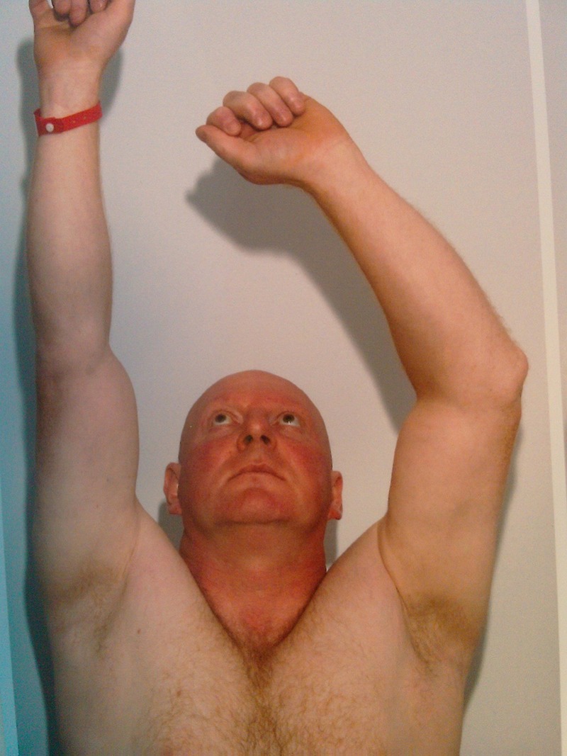
Loss of extension against gravity.
Figure 2.
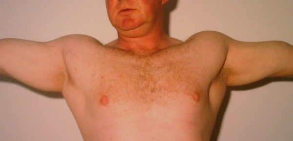
Muscle wasting with loss of contour.
Figure 3.
Muscle wasting with loss of contour.
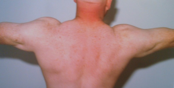
Figure 4.
Difference in contour in both arms.
Figure 5.
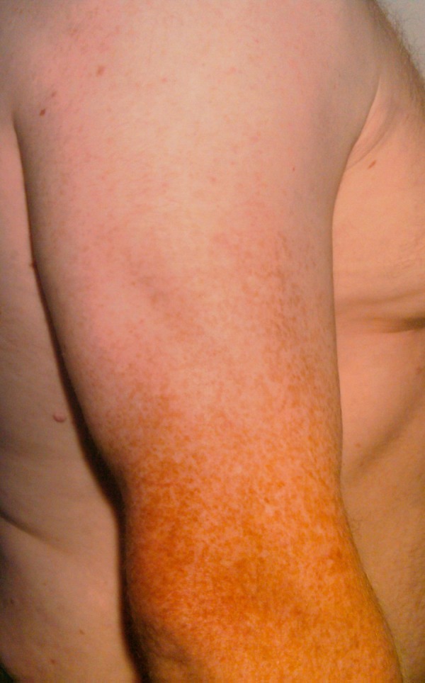
Difference in contour in both arms.
Investigations
Our patient was referred nearly 5 months after his injury. During that period he had undergone imaging in the form of MRI and was diagnosed to have partial triceps rupture. Since his symptoms had not improved with conservative treatment, MRI were reviewed by a different radiologist with a special interest in musculoskeletal imaging, and the scan was repeated, confirming the suspicion of medial head of triceps rupture.
Differential diagnosis
Triceps rupture
Radial nerve neuropraxia
Elbow joint intra-articular pathology
Treatment
The triceps were explored using a midline incision over the olecranon process extending proximally. The medial head rupture was confirmed and repaired using drill holes and anchor sutures followed by Kessler’s box sutures. The elbow was immobilised for 2 weeks postoperatively and mobilised by active-assisted physiotherapy.
Outcome and follow-up
The patient was closely monitored during and after rehabilitation. He had regained full extension with good power at his 3, 5 and 9 months follow-up appointments. He was able to return to his job of painting and decorating without any further incidence.
Discussion
Owing to the fact that the incidence of triceps ruptures is very low, it can be easily missed during initial assessment and diagnosis. Delay in diagnosis and treatment can have functional and financial implications. In the above-described case, the patient was unable to return to his job as painter and decorator for nearly 6 months following his injury. A good understanding of the anatomy and functions, with a high index of suspicion would have helped in obtaining an early accurate diagnosis.
Following increasing incidence of triceps insufficiency after total elbow arthroplasty and other surgical procedures involving triceps dissection and repair, a few cadaveric studies have been made to better understand the anatomy of the triceps and their attachments. Keener et al3 have analysed 36 fresh-frozen cadaveric specimens with particular attention to the insertion. They concluded that the triceps have broad medial to lateral insertions correlating to the size of the olecranon and expand in proximal-to-distal dimension. In their study they noted the medial tendon to have a mean width of 12.1 mm and a confluence with the rest of the triceps tendon. Whereas Madsen et al4 described the triceps as having two separate components at insertion, with the superficial component arising from the long and lateral head and the deep component arising from the medial head. This was based on a study of eight fresh-frozen cadaveric specimens. This finding is similar to Belentani et al,5 whose findings were based on 12 cadaveric specimen MRIs and histological study.
Athwal et al6 analysed 15 cadaveric upper extremities, 8 of which had separate medial head insertion posterior and deep to the common long and lateral head, and 7 had three heads in a combined common insertion. In addition to reporting the anatomical variation they also described an arthroscopic technique to repair medial head rupture using double-loaded metallic suture anchors.
Van Riett et al7 reported their outcome of surgical repair of triceps rupture on 23 patients. Among them two had rupture of only the medial head. Ten of 23 injuries were initially misdiagnosed or missed. In their study, the average time from injury to primary repair was 63 days.
Medial head of triceps ruptures are often missed, leading to significant delay in treatment and regaining of full function. Detailed clinical assessment is required with a high index of suspicion. Risk factors include repeated local steroid injections, chronic renal impairment with secondary hyperparathyroidism, attrition from osteophytes and olecranon bursitis.8 Imaging in the form of MRI can help with confirmation and can localise the tear.
Various techniques are described in open and arthroscopic repairs for tendon injuries; the choice should be based on the surgeon’s experience and preferences. At this stage, there is limited literature with reported outcomes from robust trials of different surgical techniques for medial head ruptures repair to make an evidence-based decision on a particular type of repair.
Learning points.
Thorough assessment including complete physical examination helps with diagnosis.
MRI aid confirming the diagnosis.
Surgical repair gives good results even if performed at a later stage due to delay in diagnosis.
It is important to be aware of normal anatomical variations in triceps insertion and to have a high index of suspicion.
Acknowledgments
The authors thank Mr Leo Jacobs (primary surgeon) for providing guidance and support with this case report.
Footnotes
Contributors: The literature search was performed by both the authors, the case report writing was done by RMG and the editing and formatting by NK.
Competing interests: None.
Patient consent: Obtained.
Provenance and peer review: Not commissioned; externally peer reviewed.
References
- 1.Johnson D. Gray's anatomy: the anatomical basis of clinical practice. 40th edn. Churchill Livingstone: Eslsevier, 2008 [Google Scholar]
- 2.Rafael JS, Nicholas G, Michael S, et al. Acute triceps rupture: case report and retrospective chart review. J Shoulder Elbow Surg 2006;15:130–4 [DOI] [PubMed] [Google Scholar]
- 3.Keener J, Chafik D, Kim M, et al. Insertional anatomy of the triceps brachii tendon. J Shoulder Elbow Surg 2010;19:399–405 [DOI] [PubMed] [Google Scholar]
- 4.Madsen M, Marx R, Millett P, et al. Surgical anatomy of the triceps Brachii tendon insertion: anatomic study and clinical correlation. Am J Sports Med 2006;34:1839–43 [DOI] [PubMed] [Google Scholar]
- 5.Belentani C, Pastore D, Wangwinyuvirat M, et al. Triceps brachii tendon: anatomic-MR imaging study in cadavers with histologic correlation. Skeletal Radiol 2009;38:171–5 [DOI] [PubMed] [Google Scholar]
- 6.Athwal GS, Mcgill R, Rispoli D. Isolated avulsion of the medial head of the triceps tendon: an anatomic study and arthroscopic repair in 2 cases. Arthroscopy 2009;25:983–8 [DOI] [PubMed] [Google Scholar]
- 7.Van Riett RP, Morrey BF, Ho E, et al. Surgical treatment of distal triceps ruptures. J Bone Joint Surg Am 2003;85:1961–7 [DOI] [PubMed] [Google Scholar]
- 8.Sing RK, Pooney J. Complete rupture of the triceps brachii muscle. Br J Sports Med 2002;36:467–9 [DOI] [PMC free article] [PubMed] [Google Scholar]