Abstract
Jejunal lipomata are an unusual cause of adult intussusception. We report a case of a 44-year-old Chinese woman who presented with a 3-day history of abdominal pain and nausea; she had a 5-year history of similar episodic symptoms after meals. Contrast-enhanced CT revealed a fat-density lead point in the jejunum resulting in intussusception. Single port laparoscopic surgery was performed with reduction of the intussusception, bowel resection and primary anastamosis. Histology confirmed a benign submucosal lipoma. We discuss the recent published literature on this rare entity and show CT images and intraoperative pictures.
Background
Gastrointestinal lipomata are a rare cause of intussusception resulting in partial chronic intestinal obstruction. This condition is often diagnosed late due to its non-specific symptoms, which usually spontaneously resolve. Fortunately, diagnosis can be easily made with a CT scan being the most useful modality. It is important for clinicians to have a high index of suspicion in such instances so as to arrange for appropriate investigation. On diagnosis, surgical resection is straightforward and offers full relief of symptoms.
Case presentation
A 44-year-old Chinese woman presented with a 3-day history of central abdominal pain which was colicky in nature. There was associated nausea but no vomiting. Her bowel habits were normal. She reported similar episodes of discomfort over the past 5 years, which tended to occur after meals and spontaneously resolved thereafter. She had previously consulted multiple general practitioners who had attributed her symptoms to gastritis. No previous gastroscopy had been performed. She had a medical history of hypertension as well as three previous caesarean sections and tubal ligation. Physical examination revealed a mild fullness in the lower abdomen without other significant findings.
Investigations
Blood tests were performed, with a haemoglobin level of 11.9 g/dL and total white cell count of 6.09×10⁹/L. Contrast-enhanced CT of the abdomen showed a 6 cm segment of jejunojejunal intussusception in the left flank, with a fat-density lead point (figures 1 and 2). There was no significant proximal bowel dilation and no additional lesions were found.
Figure 1.
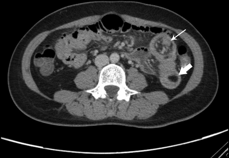
Axial view contrast-enhanced CT of the abdomen showing a 6 cm segment of jejunojejunal intussusception in the left flank secondary to a fat-density lead point (bold arrow). Small bowel along with its mesentery (thin arrow) is pulled.
Figure 2.
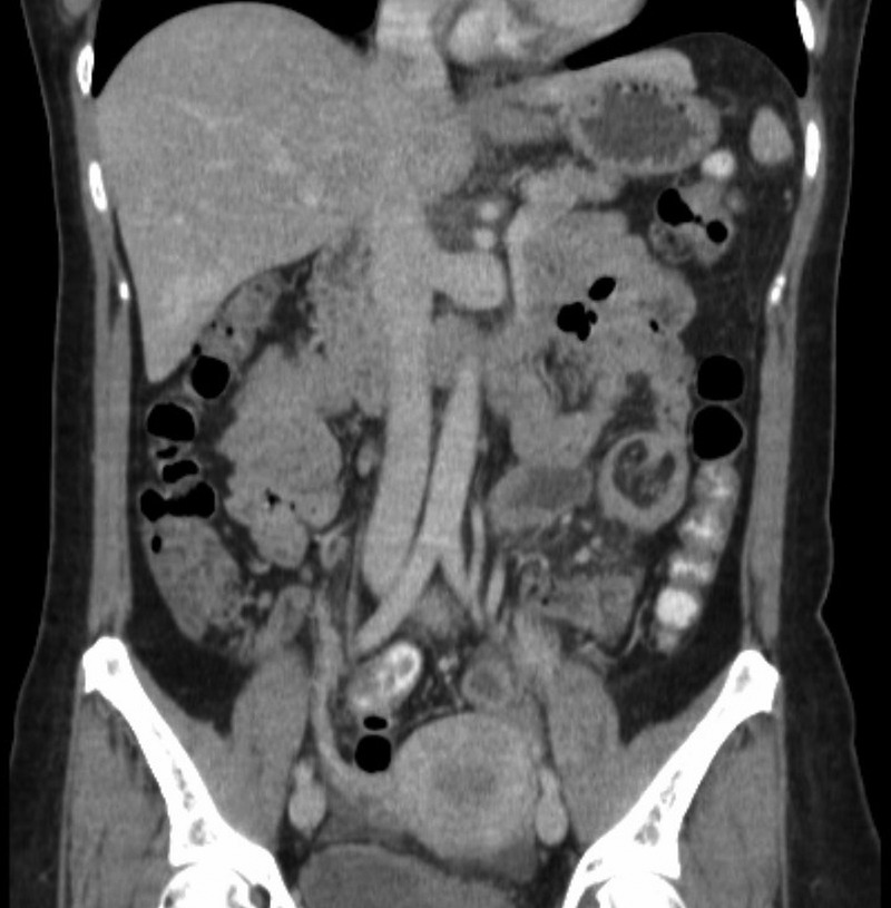
Coronal view.
Differential diagnosis
The impression based on CT findings was that of a jejunal lipoma causing intussusception.
Treatment
The patient underwent laparoscopic-assisted small bowel resection using a single umbilical glove port. The jejunal intussusception was reduced laparoscopically and delivered via the wound (Figures 3 and 4). Small bowel resection and functional end-to-end anastamosis were performed using a linear cutting stapler device. Examination of the specimen revealed a 3×2 cm lipomatous tumour (figures 5 and 6). The rest of the bowel appeared normal.
Figure 3.
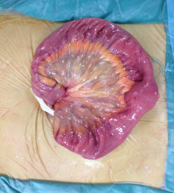
Jejunal loop containing the lipoma delivered via a single umbilical port incision.
Figure 4.
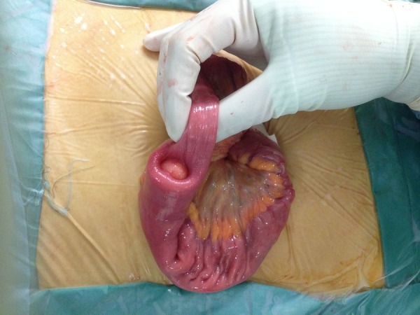
Jejunal loop containing the lipoma delivered via a single umbilical port incision.
Figure 5.
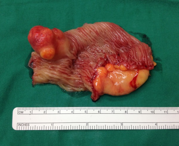
Intraluminal component of resected specimen showing a 3×2 cm lipomatous tumour.
Figure 6.
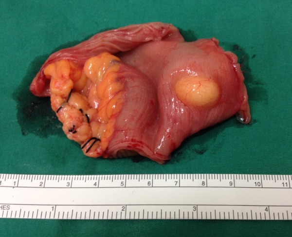
Serosal extension of submucosal lipoma.
Outcome and follow-up
The patient had an uneventful recovery period and was discharged on postoperative day 4. She has remained symptom free on follow-up. Histology confirmed a submucosal lipoma. No malignant features were seen.
Discussion
Adult intussusception is rare with the majority of cases having a neoplastic lead point. Malignancy is the cause in 70–80% of colonic intussusception and 25–35% of small bowel intussusception.1 2 Almost all malignant causes of colonic intussusception are primary adenocarcinoma while metastatic tumours account for the majority of malignant lead points in the small bowel, most often from melanoma, sarcoma and lung cancer.2
Among benign causes of small bowel intussusception, polyps of various origins (Peutz-Jeghers, hamartomatous, fibrous, inflammatory), diverticula (Meckel's) and small bowel lipomata have been described.2 Gastrointestinal lipomata are an unusual entity, representing only 1–2% of all gastrointestinal tumours, and most commonly occur in the colon, followed by small bowel, and occasionally in the stomach.3 They most commonly present with symptoms of intestinal obstruction or acute bleeding from mucosal ulceration.4 Obstructive symptoms are often non-specific, partial or episodic from intermittent intussusception and spontaneous reduction, resulting in delayed diagnosis and treatment.5 Of 51 reported cases of gastrointestinal lipomata causing adult intussusceptions, 26 were of small bowel origin and only 5 arose from the jejunum.3
CT scan is the best modality for diagnosis of intestinal intussusception,1 2 and may also be used for staging purposes should malignancy be suspected. Other useful modalities for small bowel evaluation are push or double balloon enteroscopy and video capsule endoscopy,6 but have limited usage in cases of obstruction. Barium studies and abdominal ultrasound may show classic signs of intussusception but have limited sensitivity for diagnosing intussusception when compared to CT scan. It is therefore important to have a high index of suspicion so as to order the appropriate test when encountering a patient with a similar history.
Bowel resection and primary anastamosis is the treatment of choice. In contrast to colonic intussusceptions, where all should be resected en block without reduction, a selective approach can be applied for small bowel cases. Reduction should not be attempted where signs of bowel ischaemia and inflammation are present or malignancy is suspected due to fears of tumour seeding.6 Small bowel lipomata are usually solitary but may be multiple in up to 5% of cases;3 careful examination of the rest of the bowel is prudent to rule out other lesions not detected on preoperative imaging.
Learning points.
Jejunojejunal intussusception secondary to lipomata is a rare cause of intestinal obstruction, and should be considered as a differential in cases of partial chronic obstruction.
A CT scan is the best modality for diagnosis of this condition; showing a target sign with a fat-density lead point.
Surgery is simple and straightforward, however, all possible diagnoses should be considered; operative management may differ in cases of suspected malignancy.
Footnotes
Contributors: IS-E wrote the manuscript, assisted in the surgery and postoperative care of the patient. FJF edited the final manuscript and participated in postoperative care. CLT was the lead surgeon in charge who performed the surgery.
Competing interests: None.
Patient consent: Obtained.
Provenance and peer review: Not commissioned; externally peer reviewed.
References
- 1.Goh BK, Quah HM, Chow PK, et al. Predictive factors of malignancy in adults with intussusception. World J Surg 2006;30:1300–4 [DOI] [PubMed] [Google Scholar]
- 2.Eisen LK, Cunningham JD, Aufses AH Jr. Intussusception in adults: institutional review. J Am Coll Surg 1999;188:390–5 [DOI] [PubMed] [Google Scholar]
- 3.Mouaqit O, Hasnai H, Chbani L, et al. Adult intussusceptions caused by a lipoma in the jejunum: report of a case and review of the literature. World J Emerg Surg 2012;7:28. [DOI] [PMC free article] [PubMed] [Google Scholar]
- 4.Wardi J, Langer P, Shimonov M. Unusual cause of upper gastrointestinal bleeding. J Surg Case Rep 2013;2013:pii: rjt013. [DOI] [PMC free article] [PubMed] [Google Scholar]
- 5.Jai SR, Bensardi F, Chehab F, et al. Jejunal lipoma with intermittent intussusception revealed by partial obstructive syndrome. Saudi J Gastroenterol 2008;14:206–7 [DOI] [PMC free article] [PubMed] [Google Scholar]
- 6.Manouras A, Lagoudianakis EE, Dardamanis D, et al. Lipoma induced jejunojejunal intussusception. World J Gastroenterol 2007;13:3641–4 [DOI] [PMC free article] [PubMed] [Google Scholar]


