
FIGURE 2.
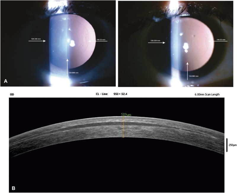
A, Serial digital clinical photographs (16×) of 32-year-old woman operated for +6.5 D hyperopia in the right eye with FILI. Photographs were taken on day 15 and 6 months after the operation. Distance from edge of the lenticule to limbus was measured at 3 points and verified at every visit to check for centering and any shift in position. B, Six-month postoperative anterior segment optical coherence tomography of an eye treated for +6.5 D hyperopia showing a clear and well-centered lenticule.