Abstract
Incomplete DNA methylation reprogramming in cloned embryos leads to low cloning efficiency. Our previous studies showed that the epigenetic modification agents 5-aza-2’-deoxycytidine (5-aza-dC) or trichostatin A (TSA) could enhance the developmental competence of porcine cloned embryos. Here, we investigated genomic methylation dynamics and specific gene expression levels during early embryonic development in pigs. In this study, our results showed that there was a typical wave of DNA demethylation and remethylation of centromeric satellite repeat (CenRep) in fertilized embryos, whereas in cloned embryos, delayed demethylation and a lack of remethylation were observed. When cloned embryos were treated with 5-aza-dC or TSA, CenRep methylation reprogramming was improved, and this was similar to that detected in fertilized counterparts. Furthermore, we found that the epigenetic modification agents, especially TSA, effectively promoted silencing of tissue specific genes and transcription of early embryo development-related genes in porcine cloned embryos. In conclusion, our results showed that the epigenetic modification agent 5-aza-dC or TSA could improve genomic methylation reprogramming in porcine cloned embryos and regulate the appropriate expression levels of genes related to early embryonic development, thereby resulting in high developmental competence.
Keywords: DNA methylation, Epigenetic modification agents, Pig, Reprogramming, Somatic cell nuclear transfer
Though somatic cell nuclear transfer (SCNT) has been achieved in many species, overall cloning efficiency is still low, and this limits the applications of cloning technology in agriculture, medicine and basic research [1,2,3].
It is generally believed that low cloning efficiency is mainly due to incomplete epigenetic reprogramming [4, 5]. To improve epigenetic reprogramming in cloned embryos, various strategies have been used, and epigenetic modification agents, such as 5-aza-dC, TSA, scriptaid and valproic acid, are usually applied and have enhanced the developmental competence of cloned embryos [6,7,8,9]. Our previous results also show that 5-aza-dC or TSA could improve cloning efficiency [10, 11]. However, the mechanism underlying the developmental improvement of cloned embryos induced by epigenetic modification agents is still poorly understood.
As the most studied epigenetic modification, DNA methylation could reflect the epigenetic reprogramming degree in cloned embryos; therefore, the mechanism of epigenetic reprogramming induced by SCNT mainly focuses on DNA methylation [12,13,14]. Previous studies have shown that compared with that of in vivo or in vitro fertilized embryos, the genome of cloned embryos is usually highly methylated, leading to poor cloning efficiency [13, 15]. Since epigenetic modification agents could improve the development of cloned embryos, it is thought that DNA methylation reprogramming must be improved in treated embryos. At present, some studies have shown that epigenetic modification agents could rescue the disrupted methylation of imprinting genes [6, 16]. However, the effect of epigenetic modification agents on global methylation reprogramming during early embryonic development has been unclear.
Previous studies have shown that the centromeric satellite repeat (CenRep) methylation level could represent the genomic methylation status [17]; thus, CenRep was selected to test genomic methylation reprogramming during early embryonic development. In this study, we first treated porcine cloned embryos with 5-aza-dC or TSA to enhance their development, then investigated genomic methylation dynamics during early embryonic development and finally tested the transcripts of DNA methyltransferase, tissue specificity, pluripotency, zygotic genome activation and blastocyst quality-related genes in embryos.
Materials and Methods
Chemicals were purchased from Sigma-Aldrich (St. Louis, MO, USA), and disposable and sterile plasticware was obtained from Nunclon (Roskilde, Denmark), unless otherwise stated. All experiments were approved by the Animal Care Commission of Shandong Academy of Agricultural Sciences according to animal welfare laws, guidelines and policies.
Porcine fetal fibroblast cell (PFF) culture
PFF culture has been described previously [11]. Briefly, PFFs were isolated from a 35-day-old fetus. After removal of the head, internal organs and limbs, the remaining tissues were finely minced into pieces, digested with 0.25% trypsin-0.04% ethylenediaminetetraacetic acid solution (GIBCO) and then dispersed in high glucose enriched Dulbecco’s modified Eagle’s medium (DMEM; GIBCO) containing 10% fetal bovine serum (FBS; GIBCO) and 1% penicillin-streptomycin (GIBCO). The dispersed cells were centrifuged, resuspended and cultured in DMEM. Until confluence, PFFs were digested, centrifuged, resuspended in FBS containing 10% dimethyl sulfoxide, and they were then stored in liquid nitrogen until use. Prior to SCNT, PFFs were thawed and cultured, and they were subsequently used in 3 to 5 passages.
Oocyte collection and in vitro maturation (IVM)
Oocyte maturation has been described previously [11]. Briefly, porcine ovaries were collected from a local slaughterhouse and transported to the laboratory. Follicles were aspirated, and follicular contents were washed with HEPES-buffered Tyrode’s lactate. Cumulus-oocyte complexes (COCs) with at least three uniform layers of compact cumulus cells and a uniform cytoplasm were recovered, washed and cultured in maturation medium under mineral oil at 38.5 C in a 5% CO2 atmosphere and saturated humidity. After 42 h, COCs were vortexed in 1 mg/ml hyaluronidase to remove cumulus cells. Only oocytes with a visible polar body, regular morphology and a homogenous cytoplasm were used.
IVF and SCNT embryo culture, treatment and collection
The procedures for porcine IVF and SCNT have been described in one of our previous reports [18]. Briefly, for IVF, the semen was incubated, resuspended and washed in DPBS supplemented with 0.1% (w/v) BSA. The spermatozoa were diluted with modified Tris-buffered medium (mTBM) to the appropriate concentration. Matured oocytes were washed in mTBM, transferred into fertilization medium and co-incubated with spermatozoa. Then the embryos were washed and cultured in porcine zygote medium-3 (PZM-3) for subsequent development. For SCNT, matured oocytes and PFFs were placed in manipulation medium. After enucleation, donor cells were placed into the perivitelline space. Fusion and activation of the cell-cytoplast complexes were induced by electroporation, and the fusion rate was confirmed by microscopic examination. Then reconstructed embryos were cultured in PZM-3 for subsequent development. The cleavage and blastocyst rates of IVF and SCNT embryos were evaluated at 48 h and 156 h, respectively.
For 5-aza-dC or TSA treatment [10, 11], cloned embryos were cultured in PZM-3 supplemented with 25 nM (optimized) 5-aza-dC (NT-AZA) or 40 nM (optimized) TSA (NT-TSA) for 24 h, washed and then transferred into PZM-3 for further culture.
For embryo collection, the 1-cell, 2-cell, 4-cell, 8-cell and blastocyst stage embryos in the IVF, NT-CON (cloned), NT-AZA and NT-TSA groups were collected at 6 h, 24 h, 48 h, 72 h and 156 h, respectively.
Bisulfite sequencing
Bisulfite sequencing has been reported [18]. Briefly, pooled samples were digested with Proteinase K and then treated with sodium bisulfite to convert all unmethylated cytosine to uracil using an EZ DNA Methylation-GoldTM Kit (Zymo Research) according to the manufacturer’s protocol. For semen, the sperm was collected by centrifugation, washed in SMB solution (10 mM Tris-HCl, 10 mM EDTA, 50 mM NaCl and 2% SDS, pH 7.2) and then incubated in SMB solution supplemented with 40 mM dithiothreitol and 0.3 mg/ml Proteinase K at 56 C for 1 h. For samples of 103 PFFs, 200 MII oocytes and 150, 80, 30, 20 and 10 pooled zona pellucida-removed embryos at the 1-cell, 2-cell, 4-cell, 8-cell and blastocyst stages, respectively, digestion was performed in M-Digestion Buffer supplemented with Proteinase K at 50 C for 20 min. After digestion, a CT (cytosine to thymine) conversion reagent was added to purified genomic DNA at 98 C for 10 min and 64 C for 2.5 h. Then the samples were desalted, purified and diluted with M-Elution Buffer. Subsequently, PCR was carried out to amplify CenRep (Z75640) using the reported primers [17] and Hot Start TaqTM Polymerase (TaKaRa) with a profile of 94 C for 5 min and 45 cycles of 94 C for 30 sec, 55 C for 30 sec and 72 C for 1 min, followed by 72 C for 10 min. Then the amplified products were verified by electrophoresis and purified using an Agarose Gel DNA Purification Kit (TaKaRa). The purified fragments were cloned into pMD18-T Vector (TaKaRa) and subjected to sequence analysis.
Quantitative real-time PCR
Measurement of gene expression with quantitative real-time PCR has been applied in our previous studies [11, 18]. Briefly, total RNA was extracted from 30 pooled embryos at each stage using an RNeasy Mini Kit (Qiagen) according to the manufacturer’s instructions. Reverse transcription was performed using a PrimeScript® RT Reagent Kit (TaKaRa) with the following parameters: 37 C for 15 min and 85 C for 5 sec, and the cDNA was stored at –20 C until use. For quantitative real-time PCR, reactions were performed in 96-well optical reaction plates (Applied Biosystems) using SYBR® Premix ExTaqTM II (TaKaRa) and a 7500 Real-Time PCR System (Applied Biosystems) with the following conditions: 95 C for 30 sec and 40 two-step cycles of 95 C for 5 sec and 60 C for 34 sec, followed by a dissociation stage consisting of 95 C for 15 sec, 60 C for 1 min and 95 C for 15 sec. For every sample, the cycle threshold (CT) values were obtained from three replicates. The primers used for amplification of target and internal reference genes are presented in Supplementary Table 1 (on-line only). The relative expression levels of target genes were analyzed using the 2−ΔΔCT method.
Statistical analysis
Differences in data (mean ± SEM) were analyzed with the SPSS statistical software. Statistical analysis of data concerning genomic methylation and gene expression levels was performed with one-way analysis of variance (ANOVA). For all analyses, differences were considered to be statistically significant when P<0.05.
Results
Delayed and incomplete genomic methylation reprogramming in porcine cloned embryos
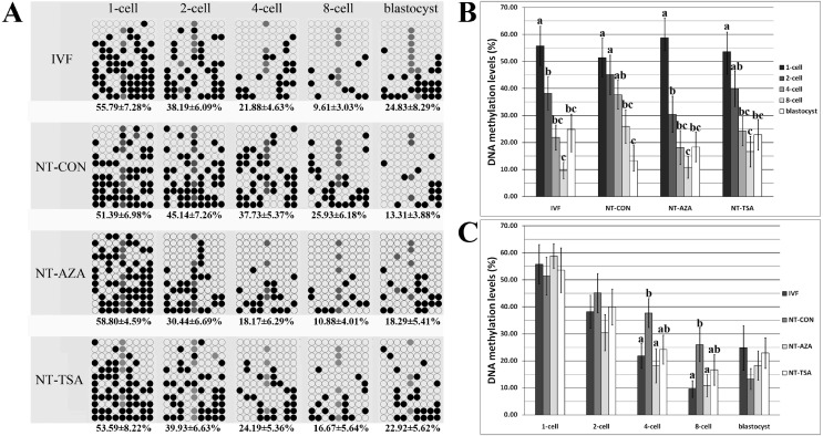
The CenRep methylation statuses of sperm and MII oocytes were examined, and a significant difference was observed (P<0.05), with sperm showing 64.58% methylation and MII oocytes showing 28.87% methylation (Suppl Fig. 1: on-line only). After fertilization, genomic demethylation was not observed in the 1-cell stage embryos in comparison with the mean methylation of sperm and oocytes (Fig. 1 and Supplementary Fig. 1). In the cleavage stage embryos, CenRep methylation displayed a continuous decrease from the 1-cell to 8-cell stage, and significant differences were observed between the 1-cell and 2-cell stages or the 4-cell and 8-cell stages (Fig. 1, P<0.05). In blastocysts, the CenRep methylation level was higher than that in the 8-cell stage (Fig. 1), indicating that genomic remethylation occurred in blastocysts. Over all, IVF embryos displayed a typical wave of genomic demethylation and remethylation.
Fig. 1.
CenRep methylation status. A: CenRep methylation status at the 1-cell, 2-cell, 4-cell, 8-cell and blastocyst stages of IVF, NT-CON, NT-AZA and NT-TSA embryos; B: dynamic CenRep methylation profiles in the IVF, NT-CON, NT-AZA and NT-TSA groups, respectively; C: CenRep methylation status at the 1-cell, 2-cell, 4-cell, 8-cell and blastocyst stages of IVF, NT-CON, NT-AZA and NT-TSA embryos. Black and white circles indicate methylated and unmethylated CpG sites, respectively, and gray circles represent mutated and/or single nucleotide polymorphism (SNP) variation at certain CpG sites. The data are expressed as means ± SEM. a–c Values for a given group in columns with different superscripts differ significantly (P < 0.05).
The CenRep methylation level in PFFs was 52.28%, and after SCNT, no significant differences were found between PFFs and the 1-cell stage embryos (Fig. 1 and Supplementary Fig. 1). In cloned embryos, gradual demethylation from the 1-cell stage to blastocyst stage was observed, suggesting that remethylation did not take place in the blastocyst stage (Fig. 1). When comparing the methylation levels within individual developmental stages between cloned and IVF embryos, the levels in the 4-cell and 8-cell stage cloned embryos were significantly higher than those in their fertilized counterparts (P<0.05). These results strongly suggested that genomic methylation reprogramming in cloned embryos was delayed and incomplete.
Epigenetic modification agents improved genomic methylation reprogramming in porcine cloned embryos
The epigenetic modification agents 5-aza-dC or TSA could significantly enhance the developmental competence of porcine cloned embryos (Table 1 and Supplementary Fig. 2: on-line only, P<0.05). Here, the alterations of genomic methylation levels in these treated cloned embryos were investigated. In the NT-AZA group, the 1-cell stage embryos did not significantly differ from PFFs, the 1-cell to 8-cell stage embryos underwent DNA demethylation in a gradual fashion, with the level of methylation in the 2-cell stage in particular significantly lower than that in the 1-cell stage (P<0.05), and blastocysts displayed remethylation in comparison with 8-cell stage embryos, which was similar to the pattern in the IVF group (Fig. 1 and Supplementary Fig. 1). When comparing the methylation status within individual developmental stages between the NT-AZA and NT-CON or IVF groups, genomic demethylation was shifted earlier in 2-cell stage, 4-cell and 8-cell stage embryos in the NT-AZA group, with the levels of methylation being significantly lower than those in the NT-CON group (P<0.05), and no significant differences were observed between the NT-AZA and IVF group during early embryonic development. These results indicated that 5-aza-dC could improve genomic methylation reprogramming in cloned embryos.
Table 1. Development of cloned embryos treated with 5-aza-dC or TSA.
| Groups | No. embryos (Rep.) |
No. embryos cleaved (% ± SEM) |
No. blastocysts (% ± SEM) |
Blastocyst cell numbers (mean ± SEM) & |
| NT-CON | 242 (5) | 209 (85.79 ± 0.95)a | 50 (20.50 ± 0.70)a | 37 ± 3 (n=49) |
| NT-AZA | 247 (5) | 223 (89.88 ± 1.14)b | 67 (27.30 ± 1.24)b | 37 ± 2 (n=55) |
| NT-TSA | 238 (5) | 210 (88.82 ± 1.12)ab | 118 (50.71 ± 2.21)c | 38 ± 2 (n=76) |
& Blastocyst cell numbers of less than 16 or blastocysts used for molecular analysis (10 or 40 blastocysts in the NT-AZA or NT-TSA group, respectively) were not included. a–c Values in the same column with different superscripts differ significantly (P < 0.05).
In the NT-TSA group, similar DNA methylation dynamics to the NT-AZA or IVF group were observed (Fig. 1). When comparing the differences between the NT-TSA and NT-AZA groups, the NT-TSA group showed a methylation pattern that was much more similar to that of the IVF group, as the NT-AZA group underwent faster genomic demethylation and slower genomic remethylation, though no significant differences were observed. Overall, our results showed that the epigenetic modification agents 5-aza-dC and TSA rescued the disrupted genomic methylation reprogramming in cloned embryos.
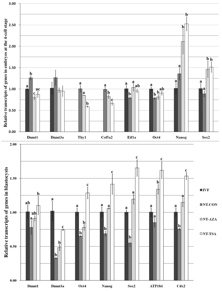
Epigenetic modification agents improved the expression levels of genes related to early embryo development
To further explore the mechanism underlying the improved development of cloned embryos treated with 5-aza-dC or TSA, the transcript levels of early embryonic development-related genes in the zygotic genome activation (ZGA) and blastocyst stages were investigated (Fig. 2). Compared with the IVF group, the NT-CON group displayed a significantly higher transcript level of Dnmt1 and lower expression levels of Oct4 and Eif1a at the ZGA stage (P<0.05), and significantly lower transcripts of Dnmt3a, Oct4, Nanog, Sox2 and Cdx2 at the blastocyst stage (P<0.05), suggesting that zygotic genes were not effectively activated and that the blastocyst quality was poor in cloned embryos.
Fig. 2.
Transcript levels of early embryo development-related genes at the zygotic genome activation and blastocyst stages of IVF, NT-CON, NT-AZA and NT-TSA embryos. The transcript abundance for each gene (Thy1 and Col5a2 in cloned embryos) in IVF embryos was considered the control. The data are expressed as means ± SEM. a–c Values for a given gene at a certain stage in columns with different superscripts differ significantly (P < 0.05).
In the NT-AZA group, the mRNA expression levels of early embryonic development-related genes was improved in comparison to those in the NT-CON group, showing significantly higher transcripts of Eif1a, Nanog and Sox2 and lower expression of Dnmt1 and Col5a2 at the ZGA stage (P<0.05), and significantly higher mRNA expression of Nanog, Sox2, ATP1b1 and Cdx2 at the blastocyst stage (P<0.05). When compared with the IVF group, the NT-AZA group displayed no significant alterations of Eif1a and Cdx2, suggesting that 5-aza-dC could improve ZGA and blastocyst quality, though significantly lower expression levels of Oct4 at the ZGA stage and Dnmt3a and Oct4 at the blastocyst stage were still observed (P<0.05).
In the NT-TSA group, significant downregulation of Dnmt1 and tissue-specific gene transcripts at the ZGA stage and upregulation of Nanog and Sox2 expression levels at the ZGA stage and DNA methyltransferase, pluripotency and blastocyst quality-related gene transcripts at the blastocyst stage were observed in comparison with the NT-CON group (P<0.05). When compared with 5-aza-dC, TSA was more effective for gene expression regulation, showing significant silencing of the tissue-specific genes at the ZGA stage and activation of Dnmt3a, pluripotency and blastocyst quality-related genes at the blastocyst stage (P<0.05). Furthermore, in comparison to those in the IVF group, significant upregulation of pluripotency and blastocyst quality-related gene transcripts was observed in the NT-TSA group (P<0.05), though the Dnmt3a transcript level at the blastocyst stage was still significantly lower (P<0.05). Thus, these above results showed that treating cloned embryos with 5-aza-dC or TSA improved the transcription levels of genes related to early embryonic development.
Discussion
Our study showed that treating porcine cloned embryos with 5-aza-dC or TSA could enhance genomic methylation reprogramming and regulate the appropriate transcript levels of early embryonic development-related genes in cloned embryos, thereby resulting in improvement of the development of porcine cloned embryos.
It is generally believed that epigenetic modification agents could improve nuclear reprogramming, and previous studies have shown that 5-aza-dC or TSA could enhance the developmental competence of cloned embryos [6, 16, 19]. However, the mechanism underlying the improvement of development is poorly studied. In this study, we investigated genomic methylation reprogramming in cloned embryos. In comparison with IVF embryos, cloned embryos took on a process of delayed demethylation without remethylation, suggesting that incomplete methylation reprogramming may be one cause of the developmental block or lethality of cloned embryos [13]. As for the reason for incomplete methylation reprogramming in cloned embryos, it is possible that there is a mechanism that causes the donor cell nucleus to preserve its methylation pattern, making oocyte-specific factors incompletely reprogram its nucleus [20]. When 5-aza-dC or TSA were applied, genomic methylation reprogramming was improved and was similar to that in IVF counterparts, showing a typical pattern of demethylation and remethylation. These findings may provide explanations for the observations that 5-aza-dC or TSA enhanced the developmental competence of cloned embryos. As for the improvement of genomic methylation reprogramming, it is possible that 5-aza-dC was incorporated into the genome and that TSA modified the chromatin structure [4]. Of course, other mechanisms also exist [4, 11]. In regard to the differences in genomic methylation reprogramming between the NT-AZA and NT-TSA group, one possible explanation is that the manners of genomic methylation regulation induced by 5-aza-dC or TSA are different, and regulation of histone modification possibly fits better with genomic demethylation and remethylation in cloned embryos [6, 21]. The results concerning embryonic development and the numbers of born and live piglets per surrogate (data not shown) could confirm this explanation. As to how genomic methylation is processed to achieve unimpaired reprogramming in the NT-AZA or NT-TSA group, more information is needed to clarify the mechanism.
In view of the genomic methylation dynamics during early embryonic development, our results also suggest that the partially progressive demethylation possibly results from replication-related passive demethylation and that active demethylation may not occur, even though traditional bisulfite sequencing could not distinguish between 5-methylcytosine and 5-hydroxymethylcytosine [17, 22]. Due to the important role of 5-hydroxymethylcytosine in somatic nuclear reprogramming [23], new technologies such as oxidative bisulfite sequencing will be applied to clarify the genomic demethylation mechanism during embryonic development.
DNA methylation reprogramming is thought to be possibly associated with gene transcription regulation [14, 24]. Our study showed that the transcription level of Dnmt1 at the ZGA stage in the NT-CON group was significantly higher than that in the IVF group, while the Dnmt3a transcript level was significantly downregulated at the blastocyst stage, possibly explaining the cause of failure of DNA demethylation and remethylation in the NT-CON group [13]. Incomplete genomic methylation reprogramming would lead to the disturbed expression levels of genes related to early embryonic development, showing continuous expression of tissue-specific genes, no effective activation of pluripotent genes and downregulation of blastocyst quality-related gene expression in cloned embryos, thereby resulting in low cloning efficiency [25, 26]. When cloned embryos were treated with 5-aza-dC or TSA, the expression levels of Dnmt1 and Dnmt3a in the treatment groups, especially the NT-TSA group, were improved and were much closer to those in the IVF group (Supplementary Fig. 3: on-line only), suggesting that DNA methylation reprogramming in the NT-AZA and NT-TSA groups would be facilitated [27], and our results showed that genomic methylation reprogramming was improved in the NT-AZA and NT-TSA groups. The results of gene transcription showed that the gene expression patterns in the NT-AZA and NT-TSA groups were also appropriate, which was strongly consistent with the improved genomic methylation reprogramming. Previous studies reported that appropriate transcription of these early embryonic development-related genes is essential for cloned embryo development [28]. Thus, we speculate that the improvement of developmental competence of cloned embryos is probably due to the rescued genomic methylation reprogramming enhancing the restoration of the expression levels of early embryonic development-related genes. Certainly, not all the gene transcription levels were consistent with the overall DNA methylation status during embryonic development, as each gene has its own specific methylation pattern. At present, these gene methylation patterns in cloned embryos have not been well elucidated, and they are very worthy of investigation.
In conclusion, our results showed that treating porcine cloned embryos with 5-aza-dC or TSA improved genomic methylation reprogramming and regulated the appropriate transcripts of genes related to early embryonic development, thereby resulting in improvement of the development of porcine cloned embryos.
Supplementary Material
Acknowledgments
This work was supported by grants from the Taishan Scholar and Distinguished Experts from Overseas program (HH), the earmarked fund for the China Agriculture Research System (CARs-37 to HH), the National Natural Science Fund of China (31272586 to HH), the National Major Breeding Program of Genetically Modified Organisms (2011ZX08008-004), the Shandong Agricultural Significant Application and Technological Innovation Fund (HH), the Pioneering Fund for Overseas Students in Jinan (20120203 to HH) and the China Postdoctoral Science Foundation (2014M551943). The authors declare that no conflicting financial interests exist.
References
- 1.Lee K, Prather RS. Advancements in somatic cell nuclear transfer and future perspectives. Anim Front 2013; 3: 56–61. [Google Scholar]
- 2.Rodriguez-Osorio N, Urrego R, Cibelli JB, Eilertsen K, Memili E. Reprogramming mammalian somatic cells. Theriogenology 2012; 78: 1869–1886. [DOI] [PubMed] [Google Scholar]
- 3.Galli C, Lagutina I, Perota A, Colleoni S, Duchi R, Lucchini F, Lazzari G. Somatic cell nuclear transfer and transgenesis in large animals: current and future insights. Reprod Domest Anim 2012; 47(Suppl 3): 2–11. [DOI] [PubMed] [Google Scholar]
- 4.Zhao J, Whyte J, Prather RS. Effect of epigenetic regulation during swine embryogenesis and on cloning by nuclear transfer. Cell Tissue Res 2010; 341: 13–21. [DOI] [PubMed] [Google Scholar]
- 5.Palini S, De Stefani S, Scala V, Dusi L, Bulletti C. Epigenetic regulatory mechanisms during preimplantation embryo development. Ann N Y Acad Sci 2011; 1221: 54–60. [DOI] [PubMed] [Google Scholar]
- 6.Xu W, Li Z, Yu B, He X, Shi J, Zhou R, Liu D, Wu Z. Effects of DNMT1 and HDAC inhibitors on gene-specific methylation reprogramming during porcine somatic cell nuclear transfer. PLoS ONE 2013; 8: e64705. [DOI] [PMC free article] [PubMed] [Google Scholar]
- 7.Zhao J, Hao Y, Ross JW, Spate LD, Walters EM, Samuel MS, Rieke A, Murphy CN, Prather RS. Histone deacetylase inhibitors improve in vitro and in vivo developmental competence of somatic cell nuclear transfer porcine embryos. Cell Reprogram 2010; 12: 75–83. [DOI] [PMC free article] [PubMed] [Google Scholar]
- 8.Costa-Borges N, Santaló J, Ibáñez E. Comparison between the effects of valproic acid and trichostatin A on the in vitro development, blastocyst quality, and full-term development of mouse somatic cell nuclear transfer embryos. Cell Reprogram 2010; 12: 437–446. [DOI] [PubMed] [Google Scholar]
- 9.Cervera RP, Martí-Gutiérrez N, Escorihuela E, Moreno R, Stojkovic M. Trichostatin A affects histone acetylation and gene expression in porcine somatic cell nucleus transfer embryos. Theriogenology 2009; 72: 1097–1110. [DOI] [PubMed] [Google Scholar]
- 10.Kong QR, Zhu J, Huang B, Huan YJ, Wang F, Shi YQ, Liu ZF, Wu ML, Liu ZH. TSA improve transgenic porcine cloned embryo development and transgene expression. Yi Chuan 2011; 33: 749–756 (In Chinese). [DOI] [PubMed] [Google Scholar]
- 11.Huan YJ, Zhu J, Xie BT, Wang JY, Liu SC, Zhou Y, Kong QR, He HB, Liu ZH. Treating cloned embryos, but not donor cells, with 5-aza-2′-deoxycytidine enhances the developmental competence of porcine cloned embryos. J Reprod Dev 2013; 59: 442–449. [DOI] [PMC free article] [PubMed] [Google Scholar]
- 12.Li E, Zhang Y. DNA methylation in mammals. Cold Spring Harb Perspect Biol 2014; 6: a019133. [DOI] [PMC free article] [PubMed] [Google Scholar]
- 13.Peat JR, Reik W. Incomplete methylation reprogramming in SCNT embryos. Nat Genet 2012; 44: 965–966. [DOI] [PubMed] [Google Scholar]
- 14.Smith ZD, Meissner A. DNA methylation: roles in mammalian development. Nat Rev Genet 2013; 14: 204–220. [DOI] [PubMed] [Google Scholar]
- 15.Deshmukh RS, Østrup O, Østrup E, Vejlsted M, Niemann H, Lucas-Hahn A, Petersen B, Li J, Callesen H, Hyttel P. DNA methylation in porcine preimplantation embryos developed in vivo and produced by in vitro fertilization, parthenogenetic activation and somatic cell nuclear transfer. Epigenetics 2011; 6: 177–187. [DOI] [PubMed] [Google Scholar]
- 16.Wang Y, Su J, Wang L, Xu W, Quan F, Liu J, Zhang Y. The effects of 5-aza-2′- deoxycytidine and trichostatin A on gene expression and DNA methylation status in cloned bovine blastocysts. Cell Reprogram 2011; 13: 297–306. [DOI] [PMC free article] [PubMed] [Google Scholar]
- 17.Zhao MT, Rivera RM, Prather RS. Locus-specific DNA methylation reprogramming during early porcine embryogenesis. Biol Reprod 2013; 88: 48. [DOI] [PMC free article] [PubMed] [Google Scholar]
- 18.Wei Y, Huan Y, Shi Y, Liu Z, Bou G, Luo Y, Zhang L, Yang C, Kong Q, Tian J, Xia P, Sun Q-Y, Liu Z. Unfaithful maintenance of methylation imprints due to loss of maternal nuclear Dnmt1 during somatic cell nuclear transfer. PLoS ONE 2011; 6: e20154. [DOI] [PMC free article] [PubMed] [Google Scholar]
- 19.Li J, Svarcova O, Villemoes K, Kragh PM, Schmidt M, Bøgh IB, Zhang Y, Du Y, Lin L, Purup S, Xue Q, Bolund L, Yang H, Maddox-Hyttel P, Vajta G. High in vitro development after somatic cell nuclear transfer and trichostatin A treatment of reconstructed porcine embryos. Theriogenology 2008; 70: 800–808. [DOI] [PubMed] [Google Scholar]
- 20.Yamanaka K, Sakatani M, Kubota K, Balboula AZ, Sawai K, Takahashi M. Effects of downregulating DNA methyltransferase 1 transcript by RNA interference on DNA methylation status of the satellite I region and in vitro development of bovine somatic cell nuclear transfer embryos. J Reprod Dev 2011; 57: 393–402. [DOI] [PubMed] [Google Scholar]
- 21.Jafarpour F, Hosseini SM, Hajian M, Forouzanfar M, Ostadhosseini S, Abedi P, Gholami S, Ghaedi K, Gourabi H, Shahverdi AH, Vosough AD, Nasr-Esfahani MH. Somatic cell-induced hyperacetylation, but not hypomethylation, positively and reversibly affects the efficiency of in vitro cloned blastocyst production in cattle. Cell Reprogram 2011; 13: 483–493. [DOI] [PMC free article] [PubMed] [Google Scholar]
- 22.Huang Y, Pastor WA, Shen Y, Tahiliani M, Liu DR, Rao A. The behaviour of 5-hydroxymethylcytosine in bisulfite sequencing. PLoS ONE 2010; 5: e8888. [DOI] [PMC free article] [PubMed] [Google Scholar]
- 23.Wossidlo M, Nakamura T, Lepikhov K, Marques CJ, Zakhartchenko V, Boiani M, Arand J, Nakano T, Reik W, Walter J. 5-Hydroxymethylcytosine in the mammalian zygote is linked with epigenetic reprogramming. Nat Commun 2011; 2: 241. [DOI] [PubMed] [Google Scholar]
- 24.Muers M. Gene expression: Disentangling DNA methylation. Nat Rev Genet 2013; 14: 519. [DOI] [PubMed] [Google Scholar]
- 25.Yin LJ, Zhang Y, Lv PP, He WH, Wu YT, Liu AX, Ding GL, Dong MY, Qu F, Xu CM, Zhu XM, Huang HF. Insufficient maintenance DNA methylation is associated with abnormal embryonic development. BMC Med 2012; 10: 26. [DOI] [PMC free article] [PubMed] [Google Scholar]
- 26.Cao S, Han J, Wu J, Li Q, Liu S, Zhang W, Pei Y, Ruan X, Liu Z, Wang X, Lim B, Li N. Specific gene-regulation networks during the pre-implantation development of the pig embryo as revealed by deep sequencing. BMC Genomics 2014; 15: 4. [DOI] [PMC free article] [PubMed] [Google Scholar]
- 27.Chung YG, Ratnam S, Chaillet JR, Latham KE. Abnormal regulation of DNA methyltransferase expression in cloned mouse embryos. Biol Reprod 2003; 69: 146–153. [DOI] [PubMed] [Google Scholar]
- 28.Wrenzycki C, Herrmann D, Gebert C, Carnwath JW, Niemann H. Gene expression and methylation patterns in cloned embryos. Methods Mol Biol 2006; 348: 285–304. [DOI] [PubMed] [Google Scholar]
Associated Data
This section collects any data citations, data availability statements, or supplementary materials included in this article.


