

Abstract
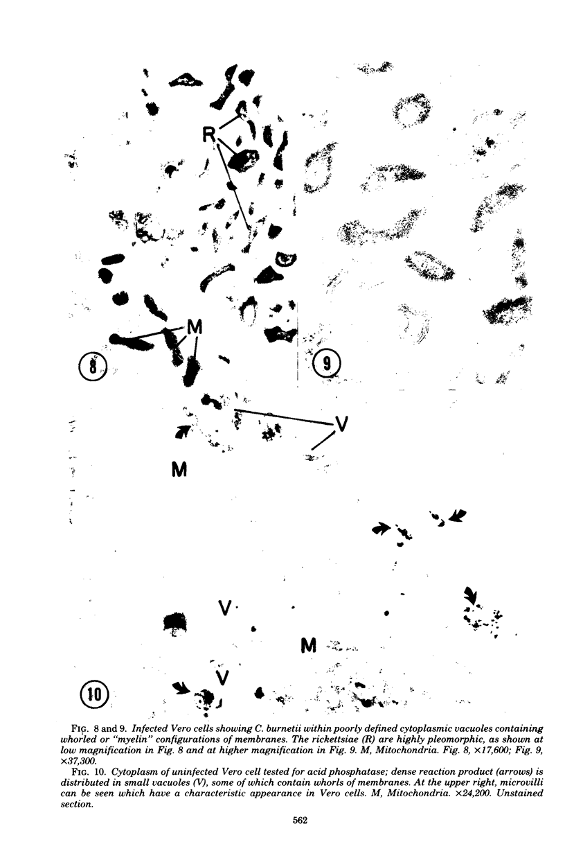
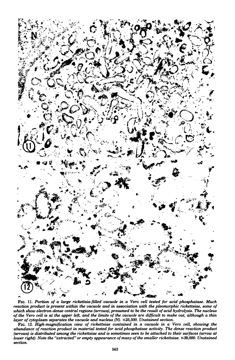
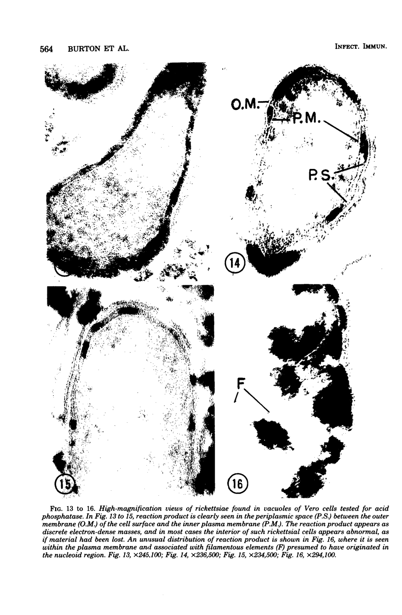
Mouse fibroblasts (L-929) and Vero (green monkey kidney) cells were infected with the rickettsia Coxiella burnetti, and persistent infections developed and were studied over a 6- to 10-month period. Ultrastructural comparisons were made between the two infected cell types, and both were tested cytochemically for the presence of acid phosphatase, a marker enzyme of lysozymes. Rickettsiae were always observed within vacuoles, and some infected L cells showed flattened endoplasmic reticulum as compared with uninfected cells. Rickettsiae in Vero cells were most often seen in vacuoles containing whorls of membranes ("myelin configurations") which were also seen in uninfected cells. Rickettsiae in Vero cells were pleomorphic, with acid phosphatase reaction product in their periplasmic space. This suggests either rickettsial degradation by lysosomal enzymes which penetrated the cell envelope or a penetration after the rickettsiae were dead. Vacuoles of infected Vero cells showed much more reaction product than that in infected L cells, and most rickettsiae in L cells had a normal appearance and showed no reaction product in their periplasmic space.
Full text
PDF


Images in this article


Selected References
These references are in PubMed. This may not be the complete list of references from this article.
- BOZEMAN F. M., HOPPS H. E., DANAUSKAS J. X., JACKSON E. B., SMADEL J. E. Study on the growth of Rickettsiae. I. A tissue culture system for quantitative estimations of Rickettsia tsutsugamushi. J Immunol. 1956 Jun;76(6):475–488. [PubMed] [Google Scholar]
- BREZINA R., KAZAR J. PHAGOCYTOSIS OF COXIELLA BURNETI AND THE PHASE VARIATION PHENOMENON. Acta Virol. 1963 Sep;7:476–476. [PubMed] [Google Scholar]
- Burton P. R., Kordová N., Paretsky D. Electron microscopic studies of the rickettsia Coxiella burneti: entry, lysosomal response, and fate of rickettsial DNA in L-cells. Can J Microbiol. 1971 Feb;17(2):143–150. doi: 10.1139/m71-025. [DOI] [PubMed] [Google Scholar]
- Downs C. M. Phagocytosis of coxiella burneti, phase I and phase II by peritoneal monocytes from normal and immune guinea pigs and mice. Zentralbl Bakteriol Orig. 1968 Apr;206(3):329–343. [PubMed] [Google Scholar]
- Kindig D. A., Kirsten W. H. Virus-like particles in established murine cell lines: electron-microscopic observations. Science. 1967 Mar 24;155(3769):1543–1545. doi: 10.1126/science.155.3769.1543. [DOI] [PubMed] [Google Scholar]
- Kordová N., Burton P. R., Downs C. M., Paretsky D., Kovácová E. The interaction of Coxiella burnetti phase I and phase II in Earle's cells. Can J Microbiol. 1970 Feb;16(2):125–133. doi: 10.1139/m70-021. [DOI] [PubMed] [Google Scholar]
- PIETRYK H. C., WEISS E. Growth of Coxiella burnetii in monolayer cultures of chick embryo entodermal cells. J Bacteriol. 1956 Aug;72(2):235–241. doi: 10.1128/jb.72.2.235-241.1956. [DOI] [PMC free article] [PubMed] [Google Scholar]
- REYNOLDS E. S. The use of lead citrate at high pH as an electron-opaque stain in electron microscopy. J Cell Biol. 1963 Apr;17:208–212. doi: 10.1083/jcb.17.1.208. [DOI] [PMC free article] [PubMed] [Google Scholar]
- ROBERTS A. N., DOWNS C. M. Study on the growth of Coxiella burnetii in the L strain mouse fibroblast and the chick fibroblast. J Bacteriol. 1959 Feb;77(2):194–204. doi: 10.1128/jb.77.2.194-204.1959. [DOI] [PMC free article] [PubMed] [Google Scholar]
- SCHAECHTER M., BOZEMAN F. M., SMADEL J. E. Study on the growth of Rickettsiae. II. Morphologic observations of living Rickettsiae in tissue culture cells. Virology. 1957 Feb;3(1):160–172. doi: 10.1016/0042-6822(57)90030-2. [DOI] [PubMed] [Google Scholar]
- Weiss E. Growth and physiology of rickettsiae. Bacteriol Rev. 1973 Sep;37(3):259–283. doi: 10.1128/br.37.3.259-283.1973. [DOI] [PMC free article] [PubMed] [Google Scholar]
- Wisseman C. L., Jr, Waddell A. D., Silverman D. J. In vitro studies on Rickettsia-host cell interactions: lag phase in intracellular growth cycle as a function of stage of growth of infecting Rickettsia prowazeki, with preliminary observations on inhibition of rickettsial uptake by host cell fragments. Infect Immun. 1976 Jun;13(6):1749–1760. doi: 10.1128/iai.13.6.1749-1760.1976. [DOI] [PMC free article] [PubMed] [Google Scholar]