Abstract
Investigations of fear conditioning in rodents and humans have illuminated the neural mechanisms of fear acquisition and extinction. However, the neural mechanism of memory consolidation of fear conditioning is not well understood. To address this question, we measured brain activity and the changes in functional connectivity following fear acquisition using resting-state functional magnetic resonance imaging. The amygdala–dorsal anterior cingulate cortex (dACC) and hippocampus–insula functional connectivity were enhanced, whereas the amygdala–medial prefrontal cortex (mPFC) functional coupling was decreased during fear memory consolidation. Furthermore, the amygdala–mPFC functional connectivity was negatively correlated with the subjective fear ratings. These findings suggest the amygdala functional connectivity with dACC and mPFC may play an important role in memory consolidation of fear conditioning. The change of amygdala-mPFC functional connectivity could predict the subjective fear. Accordingly, this study provides a new perspective for understanding fear memory consolidation.
Keywords: fear memory consolidation, functional connectivity, amygdala, resting-state fMRI
INTRODUCTION
Learning about potential dangers in the environment is critical for adaptive function, however, when experienced over a long period of time it can have a devastating effect. Some people, for instance, continue to suffer chronically from stress symptoms, may develop a wide range of psychopathologies including anxiety, phobia and post-traumatic stress disorder (PTSD). Researchers have argued that dysregulation mechanisms underlying the development and maintenance of ‘conditioned’ fear responses may provide an explanation (Schiller et al., 2009; Indovina et al., 2011). Understanding the neural mechanism of fear process is an important step in translating basic research to the treatment of fear-related disorders. Using the paradigm of classical fear conditioning, researchers have been able to map the pathways of the neural mechanisms of fear learning and extinction (LeDoux, 2003). Extensive imaging research has established several key regions involved in fear learning and expression, including the amygdala, insula, dorsal anterior cingulate, prefrontal cortex and temporal cortex. Specially, the amygdala is a brain structure that directly mediates aspects of fear learning and facilitates fear memory operations in other regions, including the hippocampus and prefrontal cortex (LaBar and Cabeza, 2006). The thickness of dorsal anterior cingulate, insula and temporal lobe were also positively correlated with conditioned fear responses (LaBar and Cabeza, 2006; Hartley et al., 2011; Linnman et al., 2011; Milad et al., 2007a, 2007b). The neural circuit involved in fear extinction included the amygdala, hippocampus (Hip), ventromedial prefrontal cortex (vmPFC) and dorsal anterior cingulated cortex (dACC) (Morgan and LeDoux, 1995; Milad and Quirk, 2002; Quirk, 2002). Moreover, the thickness of vmPFC and the dorsal anterior cingulate cortex (dACC) were closely associated with psychophysiological response during fear extinction (Milad et al., 2005; Hartley et al., 2011). Amygdala–hippocampal, amygdala–vmPFC, dACC–vmPFC and hippocampal–vmPFC functional coupling were also closely correlated with the fear acquisition and extinction process (Kalisch et al., 2006; Milad et al., 2007b; Lang et al., 2009). From fear acquisition to extinction, there are another two steps of fear process: consolidation and reconsolidation.
Resting-state functional magnetic resonance imaging (rs-fMRI) was regarded as important tools to examine spontaneous brain activity and the integrity of inter-regional functional coupling (Friston et al., 1997; Raichle et al., 2001; Greicius et al., 2003; Buckner and Vincent, 2007). Using rs-fMRI, previous researchers have indicated that spontaneous brain activity served key role in memory consolidation(Daselaar et al., 2010; Stevens et al., 2010). These results supported the system consolidation theory, which hypothesized that the medial temporal lobe (MTL) (mainly hippocampus and parahippocampus) is required for initial storage and recall and that the neo-cortex is considered as the area where remote memory is stored (Wang et al., 2012). Specifically, one useful strategy was to use the resting activity of one brain region of interest (ROI) to identify other brain regions that are functionally connected (i.e. seed-based approach). For example, in a recent investigation, Roy et al. (2009) used a seed-based approach to show that fluctuations in amygdala activity are positively coupled with the vmPFC but negatively coupled with the dorsal mPFC (dmPFC) at rest. Moreover, Sripada et al. (2012) found that veterans with PTSD showed greater positive connectivity between the amygdala and insula, reduced positive connectivity between the amygdala and hippocampus and reduced anticorrelation between the amygdala and dACC and rostral ACC during resting state. Using resting-state functional connectivity (RSFC), recent memory consolidation studies also suggested that the functional connectivity between brain areas played an important role in memory consolidation (fronto-parietal network, hippocampal-lateral occipital network) (Albert et al., 2009; Tambini et al., 2010). With respect to fear memory consolidation, Van Marle et al. (2010) found that enhanced functional connectivity between amygdala and dACC, insula and brainstem in a resting-state period directly following experimentally induced, moderate psychological stress. Moreover, Schultz et al. (2012) found that the amygdala–dmPFC functional coupling was increased following fear acquisition.
Interestingly, recent studies have demonstrated the utility for using RS-fMRI data to predict behavioral outcomes. Specially, individuals with high anxiety were characterized by negatively correlated amygdala–vmPFC functional connectivity, whereas low anxious subjects showed positively correlated activity. Further, high anxious subjects showed amygdala–dmPFC activity that was uncorrelated, whereas low anxious subjects showed negatively correlated activity at rest (Kim et al., 2011). Using DTI, Kim and Whalen (2009) observed that a stronger structural integrity in a pathway linking the amygdala and vmPFC also predicted lower anxiety levels. Moreover, Schultz et al. (2012) found that behavioral measure of explicit memory performance and implicit autonomic measure of conditioning were significantly correlated with the change in amygdala connectivity with superior frontal gyrus and ACC, respectively by using the RSFC. However, little is known about brain regions and functional networks of memory consolidation of fear conditioning when the brain is ‘at rest’ following fear acquisition. To address this question, we measured brain activity and the changes in functional connectivity following fear acquisition using rs-fMRI. Moreover, we also performed the correlation between individual’s change in functional coupling and subjective fear and trait anxiety score.
Based on these researches, we expected the inter-regional functional coupling would change over time between after acquisition and before acquisition. More specifically, the amygdala enhanced its positive coupling with the dACC, whereas the amygdala decreased its positive coupling with the mPFC. Furthermore, the change of the inter-regional functional coupling could be predicted by individual differences of subjective fear and trait anxiety scores.
MATERIALS AND METHODS
Participants
In total, 56 right-handed college students were recruited for the study (28 females; meanage = 21.68, s.d. = 1.70) and they were paid for their participation. All subjects completed the State-Trait Anxiety Inventory (Spielberger et al., 1983) prior to the start of the experiment. There was no significant difference in trait anxiety between the experimental group (mean = 41.2, s.d. = 1.46) and the control group (mean = 40.6, s.d. = 1.85), t(54) = 0.26, P = 0.83. Subjects were pre-assessed to exclude those with a previous history of neurological or psychiatric illness. All subjects were recruited from a Chinese university, gave informed consent and the study was approved by the Institutional Review Board of the Southwest University. They were divided into two groups. The experimental group consisted of 36 college students from a Chinese university (18 females) for task analysis and seven participants were removed in the resting analysis due to excessive head movement. Another group of 20 college students (10 females) in control group was recruited from the same university and four participants were removed from the resting analysis due to excessive head movement.
Stimuli
Another 50 participants rated 533 pictures chosen from the Internet and International Affective Picture System which can be used in the fear conditioning task (Lang et al., 1999). In accordance with the previous literature, we used a dimensional model for measuring pictures along three dimensions, ‘valence’, ‘arousal’ and ‘the degree of fear’. They rated the respective dimensions on a 7-point Likert scale. Finally, we chose 60 fear pictures and 160 neutral pictures, in which the disparity of the degree of fear and the valence was as large as possible [the fear picture (fear): mean = 5.72, s.d. = 0.51, the neutral picture (fear): mean = 1.85, s.d. = 0.55, t(49) = 33.38, P < 0.001; the fear picture (valence): mean = 6.08, s.d. = 1.09, the neutral picture(valence): mean = 2.86, s.d. = 0.50, t(49) = 18.35, P < 0.001]. To match the arousal degree between fear pictures and neutral pictures, we also rated and chose the neutral pictures. The arousal of fear pictures and neutral pictures was as follows: the fear picture (arousal): mean = 5.12, s.d. = 0.35; the neutral picture (arousal): mean = 4.74, s.d. = 0.27, t(49) = 1.36, P > 0.1. The conditioned stimulus (CS+, CS−) were yellow and blue squares and the unconditioned stimulus (US) were the fear pictures.
Design and procedure
The experiment began with a baseline rest condition (REST1, 10 min), then participants completed fear acquisition task (40 min) before the experimental rest condition (REST2, 10 min) (Figure 1). During acquisition, all experimental subjects underwent a Pavlovian discrimination fear-conditioning paradigm with partial reinforcement, whereas the control group underwent the same task without reinforcement. The conditioned stimulus (CS+, CS−) were yellow and blue squares (2 s) and the US was the fear picture (2 s) terminating with the CS+. The inter-trial-interval was 2–6 s. The CS+ was paired with the fear picture on a 62.5% partial reinforcement schedule and the CS− was always paired with neutral picture. Subjects were instructed to pay attention to the screen and try to figure out the relationship between the squares appearing on the screen and the fear picture. Moreover, when the CS+ appeared on screen, the subjects need to press key ‘1’, otherwise they should press ‘3’. Two orders were used to counterbalance for key and designations of colored squares (blue or yellow) as CS+ or CS−. In control group, all procedures were the same as the experimental group except for no reinforcement. In the control group, subjects performed a square-object processing task (‘square-object encoding’) or a square-scene processing task (‘square-scene encoding’). Both tasks required subjects to form an association between the color of square (blue or yellow) and the type of picture (object or scene). In other words, the subjects were instructed to predict the type of picture following the certain color of square. During the resting state, subjects were instructed to keep their eyes closed, relax their mind and remain motionless as much as possible. The resting scan lasted for 600 s. All participants informed that they had not fallen asleep during the scan. At last, subjective fear ratings (CS+ and CS–) were obtained immediately following REST2 using a 1–7 scale of fearfulness (on a 7-point Likert scale: 1, a little; 4, moderately; 7, extremely). Two-way mixed analysis of variance (ANOVA) on the group (experimental group vs control group) and the type of the conditioned stimulus (CS+ vs CS−) revealed that there was a significant interaction between two factors, F(1, 54) = 79.45, P < 0.001. To examine the effect of experiment treatment, we performed simple effect analysis. The following results were obtained: in the experimental group, the subjective fear ratings of CS+ and CS− was as follows: the CS+: mean = 5.42, s.d. = 1.52, the CS−: mean = 1.28, s.d. = 0.77, t(35) = 13.07, P < 0.001. In the control group, the subjective fear ratings of CS+ and CS− was as follows: the CS+: mean = 1.75, s.d. = 0.55, the CS−: mean = 1.6, s.d. = 0.60, t(19) = 1.14, P = 0.27. For another simple effect analysis, the following results were obtained: there was no significant difference between the subjective fear ratings of CS− in experimental group (mean = 1.28, s.d. = 0.77) and the subjective fear ratings in the control group (mean = 1.6, s.d. = 0.60), t(54) = 1.59, P = 0.12; however, participants in the experimental group have greater fear ratings of CS+ (mean = 5.42, s.d. = 1.52) than participants in the experimental group (mean = 1.75, s.d. = 0.55), t(54) = 10.34, P < 0.001. The behavioral results showed that the experimental group acquired the conditioned fear.
Fig. 1.
Schematic illustration of the paradigm for the fMRI experiment
Image acquisition and analysis
Structure MRI
T1-weighted images were recorded with a total of 176 slices at a thickness of 1 mm and in-plane resolution of 0.98 × 0.98 mm (TR = 1900 ms; TE = 2.52 ms; flip angle = 9°; FoV = 250 × 250 mm2).
Task-state functional MRI
Images were acquired with a Siemens 3T scanner (Siemens Magnetom Trio TIM, Erlangen, Germany). An Echo-Planar imaging (EPI) sequence was used for data collection, TR = 2000 ms; TE = 30 ms; flip angle = 90°; FoV = 192 × 192 mm2; matrix size = 64 × 64; voxel size = 3 × 3 × 3 mm3; interslice skip = 0.99 mm; Slices = 32.
We used SPM8 to analyze the functional data (Friston et al., 1994). For T2*-weighted images, slice order was corrected through slice timing and six parameters of head movement were estimated and removed in realign option; the first five images were discarded to achieve steady magnet state. The T1-weighted images were co-registered to the EPI mean images and segmented into white matter, gray matter and cerebrospinal fluid (CSF). The EPI images were then normalized to the MNI space with the structure information from co-registration and segmentation, the voxel size was 3 × 3 × 3 mm3 and spatially smoothing were taken with a Gaussian kernel at 8 × 8 × 8 mm3 in the full width at half maximum.
In the first-level specify, the four functional scanning runs were modeled in one GLM (general linear model). Five regressors (‘+’, CS+, CS−, fear picture and neutral picture) were created for each run after convolution with the canonical hemodynamic response function. These regressors further included six realignment parameters and the resulted design matrix was filtered with a high-band pass of 128 s. After these, we used the contrast of CS+ and CS− to explore the fear-related brain regions in the second-level specify and the threshold of P-value was 0.05 (FDR corrected, voxels ≥10). To investigate functional networks of automatic memory consolidation of fear conditioning when the brain is ‘at rest’ following fear acquisition, spherical ROIs were defined centered on the peak coordinates of the fear-related areas with the radius of 6 mm for our rs-fMRI data analysis. More specifically, ROIs identified were based on the group localization analyses (CS+ vs CS−) and FDR corrected was performed for multiple comparison in the voxel-level inference with corrected P < 0.05 and voxels ≥10 (voxel size threshold ≥10, corrected P-value < 0.05).
Resting-state functional MRI
The identical data acquisition parameter and preprocessing step were employed here as they were in task state. However, the spatially smoothing kernel was 6 × 6 × 6 mm3. The REST and DPARSF software were further used in rest-state analysis (Yan and Zang, 2010; Song et al., 2011). After preprocessing, the time series for each voxel was filtered (bandpass, 0.01–0.08 Hz) to remove the effects of very-low-frequency drift and high-frequency noise, e.g. respiratory and heart rhythms (Biswal et al., 1995; Lowe et al., 1998; Zang et al., 2007; Zhu et al., 2008).We calculated the voxel-wise functional connectivity with the ROIs of amygdala, which were defined above to find the regions that participated in the fear acquisition in the whole-brain level. Additionally, we also chose insula as seed for the key role in negative emotion processing (Van Marle et al., 2010). The functional connectivity was estimated based on the detrended, filtered and covariables removed images. The covariables included the six head motion parameters, global mean signal, white matter signal and CSF signal (Fox et al., 2005). Each participant’s time courses were obtained separately from activation maps of either before acquisition or after acquisition and were then used as regressors in a voxel-based whole-brain correlation analysis. Importantly, the time course from the same voxel was used as a regressor for both time points (before and after acquisition) for each participant. Change in the functional amygdala–dACC coupling was obtained by subtracting the individual time course correlation coefficient between amygdala and dACC after acquisition from the correlation coefficient before acquisition and converting it to normal distribution with Fisher’s z transformation. The same procedure was done for the change in the functional amygdala–mPFC, hippocampus–insula and vmPFC–insula coupling. The threshold was P < 0.01, corrected for multiple comparisons using the Bonferroni correction (Steiger, 2005; Lei et al., 2011). Finally, we computed the correlation between the change (Δcorrelation coefficient) in amygdala–mPFC functional connectivity for REST2–REST1 and subjective fear ratings to investigate whether the change of the amygdala–mPFC functional connectivity could predict subjective fear ratings. In addition, brain-behavior correlation analysis was also conducted between the change (Δcorrelation coefficient) in vmPFC–insula functional connectivity for REST2–REST1 and trait anxiety in order to examine whether the functional modification could be predicted by trait anxiety.
RESULTS
The neural circuits of fear acquisition
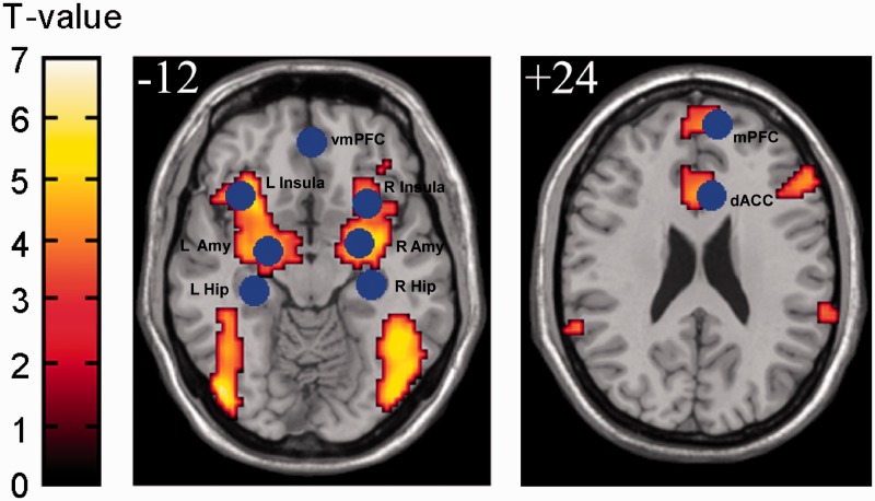
In order to investigate the brain activity related to fear acquisition, the neural correlates of differential fear learning were identified by comparing activity of the CS+ relative to the CS− [t(35) = 2.90, P < 0.05, FDR corrected], which revealed enhanced neural activity in the amygdala, dACC, mPFC, insula, thalamus and temporal lobe (Figure 2). To verify whether the fear matrix is only active in the experiment group, we performed the paired t-test between the CS+ and the CS− in the control group. The result revealed there was no significant difference of brain activity between CS+ and the CS− in the control group (P = 0.05, FDR corrected). The results suggested that the fear matrix is only active in the experiment group. These areas are commonly identified in human fMRI investigations of fear conditioning (Phelps and LeDoux, 2005; LaBar and Cabeza, 2006; Delgado et al., 2008a), indicating that these regions would be the network commonly labeled as the ‘fear acquisition matrix’. The whole-brain regions activated for CS+ vs CS− are displayed in Table 1.
Fig. 2.
Areas of brain activation in fear acquisition for the CS+ vs CS− condition. Based on functional result, we extract the ROI for resting analysis. The individual peak voxel within the group ROI was used for T-value and time course extraction for each subject.
Table 1.
Areas of brain activation for CS+ vs CS− in experimental group (Talairach coordinates)
| Region | Broadman's area (BA) | No. voxels | Peak t-value | x | y | z |
|---|---|---|---|---|---|---|
| Left inferior/middle frontal/superior temporal/parahippocampa gyrus | 47/45/13 | 311 | 5.07 | −36 | 19 | −8 |
| Right inferior/middle frontal/parahippocampa /superior temporal gyrus | 6/9/47 | 862 | 5.38 | 56 | 25 | 0 |
| Left superior temporal/parahippocampa /inferior frontal gyrus | 38/13 | 70 | 4.45 | −30 | 10 | −20 |
| Left superior temporal gyrus | 38 | 12 | 3.64 | −42 | 16 | −17 |
| Left middle/superior/inferior temporal /middle occipital gyrus | 39/22/37 | 306 | 5.50 | −53 | −59 | 9 |
| Right superior temporal/parahippocampa/inferior frontal gyrus | 38/13 | 69 | 5.12 | 33 | −10 | −9 |
| Right middle/superior/inferior temporal/fusiform /middle occipital /parahippocampa gyrus | 37/39/22 | 893 | 6.32 | 57 | −56 | 6 |
| Right middle /inferior occipital/inferior/middle temporal gyrus | 19/37/18 | 406 | 5.87 | 48 | −71 | −1 |
| Left fusiform /middle/inferior temporal/parahippocampa gyrus | 37/20 | 175 | 5.47 | −42 | −49 | −17 |
| Left middle/inferior occipital /middle/inferior temporal /fusiform gyrus | 19/18/37 | 373 | 6.48 | −45 | −77 | −6 |
| Left parahippocampa/thalamus/inferior frontal gyrus | 13/34/28 | 1512 | 6.42 | −9 | −7 | 4 |
Inter-regional functional coupling change over time (Voxel wise and ROI wise)
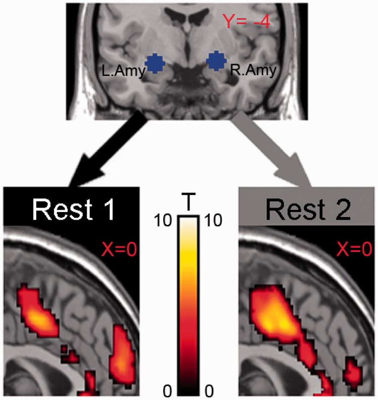
To further elucidate the network by which the amygdala exerts its differential effects, especially in regard to the medial aspect of the PFC (mPFC) and dACC, we performed a whole-brain voxel-based correlation using time courses obtained separately from the amygdala as seed (Voxel wise). ROIs were selected on the basis of activation of CS+ − CS− conditions in task functional MRI, that is, centering the ROI on the peak of activation (Talairach coordinate) (right amygdala, 24, −5, −12; left amygdala, −22, −8, −10; R Hip, 30, −25, −8; L Hip, −30, −28, −7; dACC, 6, 20, 19; mPFC, 9, 55, 20; vmPFC, 0, 45, −14; L insula, −36, 19, −8; R insula, 28, 15, −12) (Figure 2). Before acquisition, the amygdala was positively functionally coupled to the dACC and mPFC [t(28) = 3.07, P = 0.01, FDR corrected]. After acquisition, the amygdala exerted a similar pattern and strengthened positive functional coupling with dACC, whereas the amygdala decreased its positive coupling with the mPFC [t(28) = 3.09, P = 0.01, FDR corrected] (Figure 3).
Fig. 3.
Inter-regional functional coupling change over time. Sagittal views of inter-regional functional coupling maps, focused on the amygdala coupling with dACC and the mPFC separately obtained from before fear acquisition (left) and after fear acquisition (right) (P = 0.01, Bonferroni corrected).
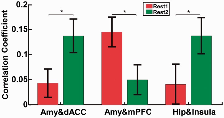
Next, we calculated the individual’s change in regional functional coupling using time courses obtained separately from the amygdala and insula as seed (ROI wise). In line with the voxel-wise analyses, functional connectivity analysis between REST1 and REST2, in which amygdala activation during fear acquisition was taken as the seed, revealed increased coupling between the amygdala and dACC [t(28) = 2.23, P = 0.03] and decreased coupling between the amygdala and the mPFC [t(28) = 2.18, P = 0.04]. In addition, we observed increased coupling between the hippocampus and insula [t(28) = 2.03, P = 0.05] (Figure 4). However, functional connectivity analysis revealed that there was no change of amygdala–dACC [t(15) = 0.51, P = 0.62], amygdala–mPFC [t(15) = 1.69, P = 0.51] and hippocampal–insula [t(15) = 1.24, P = 0.43] functional coupling in the control group (after acquisition vs before acquisition).
Fig. 4.
The change in amygdala–dACC, amygdala–mPFC and Hippocampus–insula, functional coupling change over time between REST1 and REST2; asterisk indicates P < 0.05.
Together, the voxel-wise and ROI-wise findings showed that demonstration of modifications over time in the strength of inter-regional functional coupling further characterizes the plasticity of fear-related neural networks. It also may determine the effect of the fear memory consolidation.
Brain–behavior correlation results
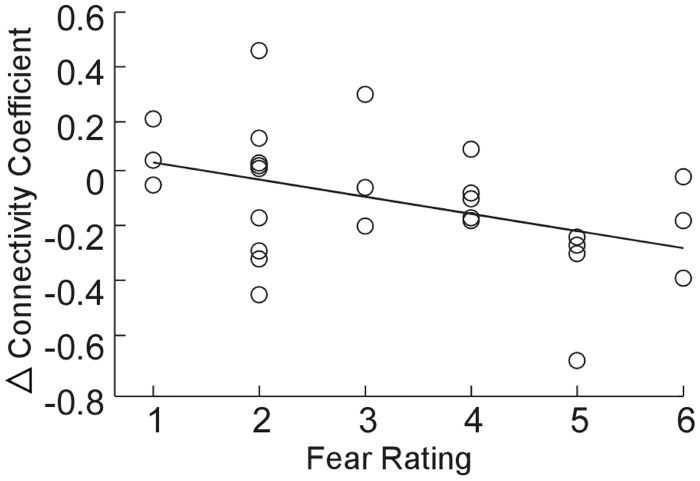
To investigate whether the change of the functional coupling could predict subjective fear ratings, we computed the correlation between the change (Δ) in amygdala–mPFC functional connectivity for REST2–REST1 and subjective fear ratings. The correlation analysis showed that the change (Δ) in amygdala–mPFC functional connectivity (REST2 vs REST1) was negatively correlated with the subjective fear ratings (r = −0.43, P = 0.03) (Figure 5).The findings suggested the changeof amygdala–mPFC functional connectivity could predict the subjective fear.
Fig. 5.
Individual difference in the change (Δcorrelation coefficient) of amgydala–mPFC functional connectivity predicted individuals’ subjective fear ratings, r = −0.43, P = 0.03
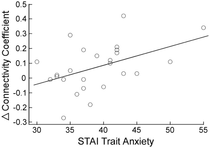
Furthermore, to determine whether individual differences in trait anxiety would correlate with the change in vmPFC–insula connectivity following fear learning, we performed a correlation analysis between the individual’s change in functional coupling and trait anxiety score. We found that individuals with higher trait anxiety scores showed significantly greater increase in vmPFC–insula functional coupling following fear conditioning (r = 0.46, P = 0.02) (Figure 6). No other fMRI results were moderated by trait anxiety. These findings indicated that vmPFC–insula functional modification could be predicted by individual differences in trait anxiety.
Fig. 6.
High-trait anxiety was associated with enhanced functional connectivity between vmPFC and insula during fear memory consolidation, r = 0.46, P = 0.02.
DISCUSSION
Using rs-fMRI, we investigated the neural circuits and changes of functional connectivity of automatic memory consolidation of fear conditioning. Our study yielded three main findings. First, in line with previous researches, several key regions (amygdala, dACC, mPFC, insula, thalamus and temporal lobe) which were labeled as the neural biomarkers of fear acquisition emerged in our task design. Second, the amygdala–dACC and hippocampus–insula functional connectivity was enhanced, whereas the amygdala–mPFC coupling was decreased during fear memory consolidation. Finally, the change in amygdala–mPFC functional connectivity (REST2 vs REST1) could predict the subjective fear. Besides, increased vmPFC–insula coupling was positively correlated with the level of trait anxiety. Combined, these results may provide a new perspective for exploration of the neural systems involved in fear memory consolidation.
Functional connectivity analysis revealed that amygdala–dACC and hippocampus–insula functional connectivity were increased during fear memory consolidation. With regard to rodent connectivity, physiological studies supported excitatory and inhibitory effects for prelimbic (PL) prefrontal cortex and infralimbic (IL) prefrontal cortex, respectively. Specifically, PL and IL can modulate fear expression through descending projections to the amygdala. PL targeted the basal nucleus of the amygdala, whereas IL targeted inhibitory areas such as the lateral division of the central nucleus and intercalated neurons (Royer and Pare, 2002; Vertes, 2004; Likhtik et al., 2005; Liang et al., 2011). Thus, via divergent projections, PL and IL can bi-directionally gate the expression of amygdala-dependent fear memories. Human imaging research has shown that the amygdala appeared to be vital for the rapid, automatic and non-conscious processing of emotional and social stimuli (Pessoa and Adolphs, 2010), emotional memory, specifically the modulation of encoding and consolidation of hippocampal-dependent memories (Phelps, 2004). Furthermore, the dACC has been proposed to modulate fear expression through excitation of the amygdala (Milad et al., 2007a), mediate the autonomic bodily arousal that normally accompanies vigilant states and promote the amygdala’s expression of the fear response and consolidation of fear memory. Both dACC and insula are densely and reciprocally connected with amygdala (Augustine 1996; Öngür and Price, 2000). Further, the dACC and the amygdala have synergistic roles in regulating purposive behavior, effected through bi-directional pathways (Schoenbaum et al., 2000) accounting for about half of all prefrontal projection neurons directed to the amygdala and receiving projections from the amygdala. (Ghashghaei et al., 2007). In mediating the autonomic arousal that accompanies vigilant states, the amygdala is additionally coupled to the dACC and anterior insula, key regions in autonomic interoceptive (Critchley, 2005; Seeley et al., 2007; Craig, 2009; Van Marle et al., 2010). This increase in connectivity could reflect the process of consolidating the memory of the CS-UCS contingency and the underlying strengthening of neural connections that support the permanent storage of fear memory. Several studies suggested that network level changes occur in order to support a memory after the initial learning event (Frankland and Bontempi, 2005). Specifically, using rs-fMRI, Van Marle et al. (2010) demonstrated that enhanced functional coupling of the amygdala with dACC in the immediate aftermath of acute psychological stress.The changes in amygdala connectivity with the dACC in this study may reflect the ongoing process of strengthening synapses between the amygdala which is critical for the long term storage of fear memory and the dACC which is important for the acquisition of fear conditioning. This pattern of co-activation may also indicate an extended state of hypervigilance that promotes sustained salience and mnemonic processing after stress. Additionally, the hippocampus serves a key role in the consolidation of long-term implicit memory and spatial navigation, e.g. episodic and semantic recollections of familiar objects and locations (Cowell et al., 2010; Squire and Wixted, 2011). Our findings of enhanced hippocampus–insula coupling following fear acquisition may be indicative of the immediate, prioritized memory consolidation of the fear/threaten stimuli. Alternatively, strengthened hippocampus–insula coupling after fear acquisition could be related to increased (ruminative) recollection of the fear/threaten material during rest. Collectively, enhanced amygdala–dACC and hippocampus–insula cooperation may boost fear memory consolidation processing. In particular, our data now suggested that homeostatic salience is persistently augmented in the immediate aftermath of acute stress.
However, the amygdala–mPFC coupling was decreased during fear memory consolidation. Humans with PTSD show that there is an increased activation of the amygdala in response to fear-related triggers that are accompanied by an abnormally low response in the mPFC that generally inhibits the amygdala (Lanius et al., 2003; Shin et al., 2005; Yehuda and LeDoux, 2007). Greater connectivity of the default network such as mPFC with the amygdala before acquisition may be particularly interesting in light of the suggestion that a function of the default network is to maintain the organism in a state of readiness for expected future events (Raichle and Gusnard, 2005). Interestingly, rapid activation of the amygdala in response to fear emotion escapes prefrontal cortex modulation (Dunsmoor et al., 2011). Specifically, studies in rodents have shown that prolonged stress alters mPFC and amygdala circuits, causing dendritic hypertrophy in mPFC (Radley et al., 2004) and hypertrophy in amygdala (Vyas et al., 2002). Thus, chronic exposures, in particularly, can lead to both a hyperactive amygdala-mediated fear response to threats and a weakened ability of mPFC to regulate these responses. Alternatively, however, persons with a hyperactive amygdala may be more likely to process neutral, unconscious or implicit threats, which would serve to even further weaken the ability of mPFC to regulate these responses. Indeed, even healthy persons elicit physiological responses to threatening stimuli that are processed unconsciously (Yehuda and LeDoux, 2007). Moreover, emotion activation studies in these individuals have shown hyperactivation in emotion-related regions, including the amygdala and insula and hypoactivation in emotion regulation regions, including the mPFC and ACC. This is consistent with our findings, in which the known role of the amygdala as a key region in threat detection (Adolphs et al., 1999), fear conditioning (Armony and LeDoux, 1997) and emotional salience (Whalen et al., 2001) and of the mPFC as a modulatory region interconnected with limbic structures (Price and Drevets, 2009) and involved in emotion regulation (Phan et al., 2002). But when humans confront threatening stimuli, the amygdala may escape the prefrontal cortex modulation. Ultimately, our findings make it reasonable to assume that the modifications in the strength of inter-regional functional coupling were strongly correlated with fear memory consolidation. Specifically, the amygdala and hippocampus were the two key regions which automatically encode and consolidate emotional information. The dACC and insula promote the amygdala’s and the hippocampus’s consolidation of fear memory, respectively. Conversely, the ability of mPFC to regulate the activation of the amygdala was weakened following fear acquisition. On the whole, these data suggest that while we spend critical moments engaging the environment to solve immediate tasks, we spend most of our time directed away from the environment in processing modes that consolidate the past, stabilize brain ensembles and prepare us for the future (Raichle, 2006; Buckner and Vincent, 2007).
Interestingly, our findings showed that the weaker amygdala–mPFC connectivity in functional coupling, the more subjective fear ratings following fear acquisition. The animal and human studies suggested that fear disorders may be related to a malfunction of the mPFC that makes it difficult to regulate fears that have been acquired. Thus, exaggerated fear and panic disorder may both involve heightened amygdala activity and weakened mPFC regulation (Morgan and LeDoux, 1995; LeDoux and Bemporad, 1997; Quirk and Beer, 2006). This was in line with our results, in which the ability of mPFC to regulate the activation of the amygdala was weakened following fear acquisition, when participants were susceptive to fear. The findings suggested that subjective fear ratings could be predicted by the change of amygdala–mPFC functional connectivity. Interestingly, Linnman et al. found that resting amygdala and medial prefrontal metabolism predicted functional activation of the fear extinction circuit. Specifically, higher resting amygdala metabolism predicted deactivation in the dACC and activation in the vmPFC during extinction training. These associations are consistent with the critical involvement of the amygdala in the extinction learning process as opposed to its separate roles in fear learning and fear expression. However, the predictive relationships were reversed during extinction recall (Linnman et al., 2012). This result is consistent with a role for the vmPFC in inhibiting the amygdala’s expression of the fear response during extinction recall and a role for the dACC in promoting it (Phelps et al., 2004; LaBar and Cabeza, 2006; Quirk et al., 2006; Admon et al., 2009; Albert et al., 2011). Our study indicated that the functional connectivity between mPFC and amygdala predicted the performance of fear extinction and functional activation of the fear extinction circuit in the future study. Moreover, the correlation analysis further indicated that high trait anxious individuals showed enhanced vmPFC–insula connectivity. The instructed use of emotion regulation techniques to reduce phasic fear responses has been demonstrated in non-anxious volunteers (Delgado et al., 2008b). Our data suggest that the vmPFC may be spontaneously regulating the activation of insula to downregulate fear, especially in high-trait anxious individuals. Moreover, as previous research suggested, anxiety levels could predict amygdala–mPFC connectivity and response magnitude during rest. Specifically, individuals with high anxiety were characterized by negatively correlated amygdala–vmPFC functional connectivity, whereas low anxious subjects showed positively correlated activity (Kim et al., 2011). Futhermore, using DTI, Kim and Whalen (2009) found that that a stronger structural integrity in a pathway linking the amygdala and vmPFC also predicted lower anxiety levels.” Additionally, our results may be that when the human confronted fear or threat stimuli, individuals of high-trait anxiety were more sensitive to solve the immediate threat and accordingly enhanced the strength of the functional connectivity between vmPFC and insula.
Using rs-fMRI, this study explored the neural mechanisms of the memory consolidation of fear conditioning, especially investigating the changes of functional connectivity which may be strongly associated with fear memory consolidation. The present study can help elucidate how humans consolidate from a fear acquisition episode. From a therapeutic point of view, these findings put forth a possible approach in which long-term treatment aims to downregulate the changes of amygdala–dACC functional connectivity and upregulate adaptive changes in the amygdala functional connectivity with the mPFC.
However, our study has several limitations. First, this study did not include objective behavioral data such as startle amplitude or SCR, so it may not be the stronger evidence that the participants acquired the conditioned fear. Second, we did not include more tests of fear memory after consolidation, so the correlations between behavioral measures of learning and changes of functional connectivity were not well understood. Specially, the correlations between SCR performance and changes of functional connectivity were not included in this study. Third, future studies should target the reconsolidation update mechanisms during the reconsolidation window, especially, brain plasticity in fear learning from the development perspective. Specifically, the retrieval–extinction conducted during the reconsolidation window of an old fear memory was demonstrated the best way to blockade the spontaneous recovery or the reinstatement of fear responses and prevent drug craving and relapse (Monfils et al., 2009; Schiller et al., 2009; Quirk and Milad, 2010; Xue et al., 2012). But less is known about the autonomic mechanism and functional connectivity in reconsolidation window. Collectively, these could be valuable areas of investigation for future research.
In summary, the amygdala–dACC and hippocampus–insula functional connectivity was enhanced, while the amygdala–mPFC coupling was decreased during fear memory consolidation. Furthermore, the change of amygdala–mPFC functional connectivity could predict the subjective fear. It was important to note here that the amygdala–dACC, amygdala–mPFC functional connectivity may be particularly valuable for automatic memory consolidation of fear conditioning. Characterization of the post-stress network changes in humans may represent a first step toward understanding the early phase of psychological trauma etiology.
Conflict of Interest
None declared.
Acknowledgments
This research was supported by National Natural Science Foundation of China (31271117, 31200857); the Fundamental Research Funds for the Central Universities (SWU1309002, SWU1209319); the National Key Discipline of Basic Psychology in Southwest University of China (TR201207-2, NSKD11047); Humanity and Social Science Youth foundation of Ministry of Education of China (12YJC190015).
REFERENCES
- Admon R, Lubin G, Stern O, et al. Human vulnerability to stress depends on amygdala's predisposition and hippocampal plasticity. Proceedings of the National Academy of Sciences United States of America. 2009;106(33):14120–5. doi: 10.1073/pnas.0903183106. [DOI] [PMC free article] [PubMed] [Google Scholar]
- Adolphs R, Tranel D, Hamann S, et al. Recognition of facial emotion in nine individuals with bilateral amygdala damage. Neuropsychologia. 1999;37(10):1111–7. doi: 10.1016/s0028-3932(99)00039-1. [DOI] [PubMed] [Google Scholar]
- Albert J, López-Martín S, Tapia M, Montoya D, Carretié L. The role of the anterior cingulate cortex in emotional response inhibition. Human Brain Mapping. 2011;33(9):2147–60. doi: 10.1002/hbm.21347. [DOI] [PMC free article] [PubMed] [Google Scholar]
- Albert NB, Robertson EM, Miall RC. The resting human brain and motor learning. Current Biology. 2009;19(12):1023. doi: 10.1016/j.cub.2009.04.028. [DOI] [PMC free article] [PubMed] [Google Scholar]
- Armony JL, LeDoux JE. How the brain processes emotional information. Annals of the New York Academy of Sciences. 1997;821(1):259–70. doi: 10.1111/j.1749-6632.1997.tb48285.x. [DOI] [PubMed] [Google Scholar]
- Augustine JR. Circuitry and functional aspects of the insular lobe in primates including humans. Brain Research Reviews. 1996;22(3):229–44. doi: 10.1016/s0165-0173(96)00011-2. [DOI] [PubMed] [Google Scholar]
- Biswal B, Zerrin Yetkin F, Haughton VM, Hyde JS. Functional connectivity in the motor cortex of resting human brain using echo-planar mri. Magnetic Resonance in Medicine. 1995;34(4):537–541. doi: 10.1002/mrm.1910340409. [DOI] [PubMed] [Google Scholar]
- Buckner RL, Vincent JL. Unrest at rest: default activity and spontaneous network correlations. Neuroimage. 2007;37(4):1091–96. doi: 10.1016/j.neuroimage.2007.01.010. [DOI] [PubMed] [Google Scholar]
- Cowell RA, Bussey TJ, Saksida LM. Components of recognition memory: Dissociable cognitive processes or just differences in representational complexity? Hippocampus. 2010;20(11):1245–62. doi: 10.1002/hipo.20865. [DOI] [PubMed] [Google Scholar]
- Craig A. How do you feel-now? The anterior insula and human awareness. Nature Reviews Neuroscience. 2009;10(1):59–70. doi: 10.1038/nrn2555. [DOI] [PubMed] [Google Scholar]
- Critchley HD. Neural mechanisms of autonomic, affective, and cognitive integration. The Journal of Comparative Neurology. 2005;493(1):154–66. doi: 10.1002/cne.20749. [DOI] [PubMed] [Google Scholar]
- Daselaar S, Huijbers W, de Jonge M, Goltstein P, Pennartz C. Experience-dependent alterations in conscious resting state activity following perceptuomotor learning. Neurobiology of Learning and Memory. 2010;93(3):422–7. doi: 10.1016/j.nlm.2009.12.009. [DOI] [PubMed] [Google Scholar]
- Delgado MR, Li J, Schiller D, Phelps EA. The role of the striatum in aversive learning and aversive prediction errors. Philosophical Transactions of the Royal Society B: Biological Sciences. 2008a;363(1511):3787–800. doi: 10.1098/rstb.2008.0161. [DOI] [PMC free article] [PubMed] [Google Scholar]
- Delgado MR, Nearing KI, LeDoux JE, Phelps EA. Neural circuitry underlying the regulation of conditioned fear and its relation to extinction. Neuron. 2008b;59(5):829–38. doi: 10.1016/j.neuron.2008.06.029. [DOI] [PMC free article] [PubMed] [Google Scholar]
- Dunsmoor JE, Prince SE, Murty VP, Kragel PA, LaBar KS. Neurobehavioral mechanisms of human fear generalization. Neuroimage. 2011;55(4):1878–88. doi: 10.1016/j.neuroimage.2011.01.041. [DOI] [PMC free article] [PubMed] [Google Scholar]
- Fox MD, Snyder AZ, Vincent JL, Corbetta M, Van Essen DC, Raichle ME. The human brain is intrinsically organized into dynamic, anticorrelated functional networks. Proceedings of the National Academy of Sciences of the United States of America. 2005;102(27):9673. doi: 10.1073/pnas.0504136102. [DOI] [PMC free article] [PubMed] [Google Scholar]
- Frankland PW, Bontempi B. The organization of recent and remote memories. Nature Reviews Neuroscience. 2005;6(2):119–30. doi: 10.1038/nrn1607. [DOI] [PubMed] [Google Scholar]
- Friston K, Buechel C, Fink G, Morris J, Rolls E, Dolan R. Psychophysiological and modulatory interactions in neuroimaging. Neuroimage. 1997;6(3):218–29. doi: 10.1006/nimg.1997.0291. [DOI] [PubMed] [Google Scholar]
- Friston KJ, Holmes AP, Worsley KJ, Poline JP, Frith CD, Frackowiak RSJ. Statistical parametric maps in functional imaging: a general linear approach. Human Brain Mapping. 1994;2(4):189–210. [Google Scholar]
- Ghashghaei H, Hilgetag C, Barbas H. Sequence of information processing for emotions based on the anatomic dialogue between prefrontal cortex and amygdala. Neuroimage. 2007;34(3):905–23. doi: 10.1016/j.neuroimage.2006.09.046. [DOI] [PMC free article] [PubMed] [Google Scholar]
- Greicius MD, Krasnow B, Reiss AL, Menon V. Functional connectivity in the resting brain: a network analysis of the default mode hypothesis. Proceedings of the National Academy of Sciences. 2003;100(1):253. doi: 10.1073/pnas.0135058100. [DOI] [PMC free article] [PubMed] [Google Scholar]
- Hartley CA, Fischl B, Phelps EA. Brain structure correlates of individual differences in the acquisition and inhibition of conditioned fear. Cerebral Cortex. 2011;21(9):1954–62. doi: 10.1093/cercor/bhq253. [DOI] [PMC free article] [PubMed] [Google Scholar]
- Indovina I, Robbins TW, Núñez-Elizalde AO, Dunn BD, Bishop SJ. Fear-conditioning mechanisms associated with trait vulnerability to anxiety in humans. Neuron. 2011;69(3):563–71. doi: 10.1016/j.neuron.2010.12.034. [DOI] [PMC free article] [PubMed] [Google Scholar]
- Kalisch R, Korenfeld E, Stephan KE, Weiskopf N, Seymour B, Dolan RJ. Context-dependent human extinction memory is mediated by a ventromedial prefrontal and hippocampal network. The Journal of Neuroscience. 2006;26(37):9503–11. doi: 10.1523/JNEUROSCI.2021-06.2006. [DOI] [PMC free article] [PubMed] [Google Scholar]
- Kim MJ, Gee DG, Loucks RA, Davis FC, Whalen PJ. Anxiety dissociates dorsal and ventral medial prefrontal cortex functional connectivity with the amygdala at rest. Cerebral Cortex. 2011;21(7):1667–73. doi: 10.1093/cercor/bhq237. [DOI] [PMC free article] [PubMed] [Google Scholar]
- Kim MJ, Whalen PJ. The structural integrity of an amygdala–prefrontal pathway predicts trait anxiety. The Journal of Neuroscience. 2009;29(37):11614–8. doi: 10.1523/JNEUROSCI.2335-09.2009. [DOI] [PMC free article] [PubMed] [Google Scholar]
- LaBar KS, Cabeza R. Cognitive neuroscience of emotional memory. Nature Reviews Neuroscience. 2006;7(1):54–64. doi: 10.1038/nrn1825. [DOI] [PubMed] [Google Scholar]
- Lang PJ, Bradley MM, Cuthbert BN. International Affective Picture System (IAPS): Technical Manual and Affective Ratings. Gainesville, FL: The Center for Research in Psychophysiology, University of Florida; 1999. [Google Scholar]
- Lang S, Kroll A, Lipinski SJ, et al. Context conditioning and extinction in humans: differential contribution of the hippocampus, amygdala and prefrontal cortex. European Journal of Neuroscience. 2009;29(4):823–32. doi: 10.1111/j.1460-9568.2009.06624.x. [DOI] [PMC free article] [PubMed] [Google Scholar]
- Lanius RA, Williamson PC, Hopper J, et al. Recall of emotional states in posttraumatic stress disorder: an fMRI investigation. Biological Psychiatry. 2003;53(3):204–10. doi: 10.1016/s0006-3223(02)01466-x. [DOI] [PubMed] [Google Scholar]
- LeDoux JE. Synaptic self: How our Brains Become Who We are. USA: Penguin Group; 2003. [Google Scholar]
- LeDoux J, Bemporad JR. The emotional brain. Journal of the American Academy of Psychoanalysis. 1997;25(3):525–8. [Google Scholar]
- Lei X, Ostwald D, Hu J, et al. Multimodal Functional Network Connectivity: An EEG-fMRI Fusion in Network Space. PloS One. 2011;6(9):e24642. doi: 10.1371/journal.pone.0024642. [DOI] [PMC free article] [PubMed] [Google Scholar]
- Liang Z, King J, Zhang N. Uncovering intrinsic connectional architecture of functional networks in awake rat brain. The Journal of Neuroscience. 2011;31(10):3776–83. doi: 10.1523/JNEUROSCI.4557-10.2011. [DOI] [PMC free article] [PubMed] [Google Scholar]
- Likhtik E, Pelletier JG, Paz R, Paré D. Prefrontal control of the amygdala. The Journal of Neuroscience. 2005;25(32):7429–37. doi: 10.1523/JNEUROSCI.2314-05.2005. [DOI] [PMC free article] [PubMed] [Google Scholar]
- Linnman C, Zeidan MA, Furtak SC, Pitman RK, Quirk GJ, Milad MR. Resting amygdala and medial prefrontal metabolism predicts functional activation of the fear extinction circuit. American Journal of Psychiatry. 2012;169(4):415–23. doi: 10.1176/appi.ajp.2011.10121780. [DOI] [PMC free article] [PubMed] [Google Scholar]
- Linnman C, Zeidan MA, Pitman RK, Milad MR. Resting cerebral metabolism correlates with skin conductance and functional brain activation during fear conditioning. Biological Psychology. 2011;89(2):450–9. doi: 10.1016/j.biopsycho.2011.12.012. [DOI] [PMC free article] [PubMed] [Google Scholar]
- Lowe M, Mock B, Sorenson J. Functional connectivity in single and multislice echoplanar imaging using resting-state fluctuations. Neuroimage. 1998;7(2):119–32. doi: 10.1006/nimg.1997.0315. [DOI] [PubMed] [Google Scholar]
- Milad MR, Quinn BT, Pitman RK, Orr SP, Fischl B, Rauch SL. Thickness of ventromedial prefrontal cortex in humans is correlated with extinction memory. Proceedings of the National Academy of Sciences of the United States of America. 2005;102(30):10706. doi: 10.1073/pnas.0502441102. [DOI] [PMC free article] [PubMed] [Google Scholar]
- Milad MR, Quirk GJ. Neurons in medial prefrontal cortex signal memory for fear extinction. Nature. 2002;420(6911):70–74. doi: 10.1038/nature01138. [DOI] [PubMed] [Google Scholar]
- Milad MR, Quirk GJ, Pitman RK, Orr SP, Fischl B, Rauch SL. A role for the human dorsal anterior cingulate cortex in fear expression. Biological Psychiatry. 2007a;62(10):1191–4. doi: 10.1016/j.biopsych.2007.04.032. [DOI] [PubMed] [Google Scholar]
- Milad MR, Wright CI, Orr SP, Pitman RK, Quirk GJ, Rauch SL. Recall of fear extinction in humans activates the ventromedial prefrontal cortex and hippocampus in concert. Biological Psychiatry. 2007b;62(5):446–54. doi: 10.1016/j.biopsych.2006.10.011. [DOI] [PubMed] [Google Scholar]
- Monfils MH, Cowansage KK, Klann E, LeDoux JE. Extinction-reconsolidation boundaries: key to persistent attenuation of fear memories. Science. 2009;324(5929):951–5. doi: 10.1126/science.1167975. [DOI] [PMC free article] [PubMed] [Google Scholar]
- Morgan MA, LeDoux JE. Differential contribution of dorsal and ventral medial prefrontal cortex to the acquisition and extinction of conditioned fear in rats. Behavioral Neuroscience. 1995;109(4):681. doi: 10.1037//0735-7044.109.4.681. [DOI] [PubMed] [Google Scholar]
- Öngür D, Price J. The organization of networks within the orbital and medial prefrontal cortex of rats, monkeys and humans. Cerebral Cortex. 2000;10(3):206–19. doi: 10.1093/cercor/10.3.206. [DOI] [PubMed] [Google Scholar]
- Pessoa L, Adolphs R. Emotion processing and the amygdala: from a'low road'to'many roads' of evaluating biological significance. Nature Reviews Neuroscience. 2010;11(11):773–83. doi: 10.1038/nrn2920. [DOI] [PMC free article] [PubMed] [Google Scholar]
- Phan KL, Wager T, Taylor SF, Liberzon I. Functional neuroanatomy of emotion: a meta-analysis of emotion activation studies in PET and fMRI. Neuroimage. 2002;16(2):331–48. doi: 10.1006/nimg.2002.1087. [DOI] [PubMed] [Google Scholar]
- Phelps EA. Human emotion and memory: interactions of the amygdala and hippocampal complex. Current Opinion in Neurobiology. 2004;14(2):198–202. doi: 10.1016/j.conb.2004.03.015. [DOI] [PubMed] [Google Scholar]
- Phelps EA, Delgado MR, Nearing KI, LeDoux JE. Extinction learning in humans: role of the amygdala and vmPFC. Neuron. 2004;43(6):897–905. doi: 10.1016/j.neuron.2004.08.042. [DOI] [PubMed] [Google Scholar]
- Phelps EA, LeDoux JE. Contributions of the amygdala to emotion processing: from animal models to human behavior. Neuron. 2005;48(2):175–87. doi: 10.1016/j.neuron.2005.09.025. [DOI] [PubMed] [Google Scholar]
- Price JL, Drevets WC. Neurocircuitry of mood disorders. Neuropsychopharmacology. 2009;35(1):192–216. doi: 10.1038/npp.2009.104. [DOI] [PMC free article] [PubMed] [Google Scholar]
- Quirk GJ. Memory for extinction of conditioned fear is long-lasting and persists following spontaneous recovery. Learning and Memory. 2002;9(6):402–7. doi: 10.1101/lm.49602. [DOI] [PMC free article] [PubMed] [Google Scholar]
- Quirk GJ, Beer JS. Prefrontal involvement in the regulation of emotion: convergence of rat and human studies. Current Opinion in Neurobiology. 2006;16(6):723–7. doi: 10.1016/j.conb.2006.07.004. [DOI] [PubMed] [Google Scholar]
- Quirk GJ, Garcia R, Gonz lez-Lima F. Prefrontal mechanisms in extinction of conditioned fear. Biological Psychiatry. 2006;60(4):337–43. doi: 10.1016/j.biopsych.2006.03.010. [DOI] [PubMed] [Google Scholar]
- Quirk GJ, Milad MR. Neuroscience: Editing out fear. Nature. 2010;463(7277):36–7. doi: 10.1038/463036a. [DOI] [PubMed] [Google Scholar]
- Radley J, Sisti H, Hao J, et al. Chronic behavioral stress induces apical dendritic reorganization in pyramidal neurons of the medial prefrontal cortex. Neuroscience. 2004;125(1):1–6. doi: 10.1016/j.neuroscience.2004.01.006. [DOI] [PubMed] [Google Scholar]
- Raichle ME. The brain's dark energy. Science. 2006;314(5803):1249. [PubMed] [Google Scholar]
- Raichle ME, Gusnard DA. Intrinsic brain activity sets the stage for expression of motivated behavior. The Journal of Comparative Neurology. 2005;493(1):167–76. doi: 10.1002/cne.20752. [DOI] [PubMed] [Google Scholar]
- Raichle ME, MacLeod AM, Snyder AZ, Powers WJ, Gusnard DA, Shulman GL. A default mode of brain function. Proceedings of the National Academy of Sciences United States of America. 2001;98(2):676. doi: 10.1073/pnas.98.2.676. [DOI] [PMC free article] [PubMed] [Google Scholar]
- Roy AK, Shehzad Z, Margulies DS, et al. Functional connectivity of the human amygdala using resting state fMRI. Neuroimage. 2009;45(2):614–26. doi: 10.1016/j.neuroimage.2008.11.030. [DOI] [PMC free article] [PubMed] [Google Scholar]
- Royer S, Pare D. Bidirectional synaptic plasticity in intercalated amygdala neurons and the extinction of conditioned fear responses. Neuroscience. 2002;115(2):455. doi: 10.1016/s0306-4522(02)00455-4. [DOI] [PubMed] [Google Scholar]
- Schiller D, Monfils MH, Raio CM, Johnson DC, LeDoux JE, Phelps EA. Preventing the return of fear in humans using reconsolidation update mechanisms. Nature. 2009;463(7277):49–53. doi: 10.1038/nature08637. [DOI] [PMC free article] [PubMed] [Google Scholar]
- Schoenbaum G, Chiba AA, Gallagher M. Changes in functional connectivity in orbitofrontal cortex and basolateral amygdala during learning and reversal training. The Journal of neuroscience. 2000;20(13):5179–89. doi: 10.1523/JNEUROSCI.20-13-05179.2000. [DOI] [PMC free article] [PubMed] [Google Scholar]
- Schultz DH, Balderston NL, Helmstetter FJ. Resting-state connectivity of the amygdala is altered following Pavlovian fear conditioning. Frontiers in Human Neuroscience. 2012;6:242. doi: 10.3389/fnhum.2012.00242. [DOI] [PMC free article] [PubMed] [Google Scholar]
- Seeley WW, Menon V, Schatzberg AF, et al. Dissociable intrinsic connectivity networks for salience processing and executive control. The Journal of neuroscience. 2007;27(9):2349–56. doi: 10.1523/JNEUROSCI.5587-06.2007. [DOI] [PMC free article] [PubMed] [Google Scholar]
- Shin LM, Wright CI, Cannistraro PA, et al. A functional magnetic resonance imaging study of amygdala and medial prefrontal cortex responses to overtly presented fearful faces in posttraumatic stress disorder. Archives of General Psychiatry. 2005;62(3):273. doi: 10.1001/archpsyc.62.3.273. [DOI] [PubMed] [Google Scholar]
- Song XW, Dong ZY, Long XY, et al. REST: a toolkit for resting-state functional magnetic resonance imaging data processing. PLoS One. 2011;6(9):e25031. doi: 10.1371/journal.pone.0025031. [DOI] [PMC free article] [PubMed] [Google Scholar]
- Spielberger CD, Gorsuch RL, Lushene R, Vagg PR, Jacobs GA. Manual for the State-Trait Anxiety Inventory. Palo Alto, CA: Consulting Psychologists Press; 1983. [Google Scholar]
- Squire LR, Wixted JT. The cognitive neuroscience of human memory since HM. Annual review of neuroscience. 2011;34:259–88. doi: 10.1146/annurev-neuro-061010-113720. [DOI] [PMC free article] [PubMed] [Google Scholar]
- Sripada RK, King AP, Garfinkel SN, Wang X, Sripada CS, Welsh RC, Liberzon I. Altered resting-state amygdala functional connectivity in men with posttraumatic stress disorder. Journal of Psychiatry and Neuroscience: JPN. 2012;37(4):241. doi: 10.1503/jpn.110069. [DOI] [PMC free article] [PubMed] [Google Scholar]
- Steiger JH. Comparing correlations: Pattern hypothesis tests between and/or within independent samples. In: Maydeu-Olivares A, McArdle JJ, editors. Contemporary Psychometrics: A festschrift for Roderick P. McDonald. Mahwah, NJ, US: Lawrence Erlbaum Associates, Inc; 2005. pp. 377–414. [Google Scholar]
- Stevens WD, Buckner RL, Schacter DL. Correlated low-frequency BOLD fluctuations in the resting human brain are modulated by recent experience in category-preferential visual regions. Cerebral Cortex. 2010;20(8):1997–2006. doi: 10.1093/cercor/bhp270. [DOI] [PMC free article] [PubMed] [Google Scholar]
- Tambini A, Ketz N, Davachi L. Enhanced brain correlations during rest are related to memory for recent experiences. Neuron. 2010;65(2):280–90. doi: 10.1016/j.neuron.2010.01.001. [DOI] [PMC free article] [PubMed] [Google Scholar]
- Van Marle HJ, Hermans EJ, Qin S, Fernández G. Enhanced resting-state connectivity of amygdala in the immediate aftermath of acute psychological stress. Neuroimage. 2010;53(1):348–54. doi: 10.1016/j.neuroimage.2010.05.070. [DOI] [PubMed] [Google Scholar]
- Vertes RP. Differential projections of the infralimbic and prelimbic cortex in the rat. Synapse. 2004;51(1):32–58. doi: 10.1002/syn.10279. [DOI] [PubMed] [Google Scholar]
- Vyas A, Mitra R, Rao BSS, Chattarji S. Chronic stress induces contrasting patterns of dendritic remodeling in hippocampal and amygdaloid neurons. The Journal of Neuroscience. 2002;22(15):6810–8. doi: 10.1523/JNEUROSCI.22-15-06810.2002. [DOI] [PMC free article] [PubMed] [Google Scholar]
- Wang D-Y, Han X-J, Li S-F, et al. Effects of apolipoprotein E genotype on the off-line memory consolidation. PLoS One. 2012;7(12):e51617. doi: 10.1371/journal.pone.0051617. [DOI] [PMC free article] [PubMed] [Google Scholar]
- Whalen PJ, Shin LM, McInerney SC, Fischer H, Wright CI, Rauch SL. A functional MRI study of human amygdala responses to facial expressions of fear versus anger. Emotion. 2001;1(1):70. doi: 10.1037/1528-3542.1.1.70. [DOI] [PubMed] [Google Scholar]
- Xue YX, Luo YX, Wu P, et al. A memory retrieval-extinction procedure to prevent drug craving and relapse. Science. 2012;336(6078):241–5. doi: 10.1126/science.1215070. [DOI] [PMC free article] [PubMed] [Google Scholar]
- Yan CG, Zang YF. DPARSF: a MATLAB toolbox for “pipeline” data analysis of resting-state fMRI. Frontiers in systems neuroscience. 2010;4:13. doi: 10.3389/fnsys.2010.00013. [DOI] [PMC free article] [PubMed] [Google Scholar]
- Yehuda R, LeDoux J. Response variation following trauma: a translational neuroscience approach to understanding PTSD. Neuron. 2007;56(1):19–32. doi: 10.1016/j.neuron.2007.09.006. [DOI] [PubMed] [Google Scholar]
- Zang YF, He Y, Zhu CZ, et al. Altered baseline brain activity in children with ADHD revealed by resting-state functional MRI. Brain and Development. 2007;29(2):83–91. doi: 10.1016/j.braindev.2006.07.002. [DOI] [PubMed] [Google Scholar]
- Zhu C-Z, Zang Y-F, Cao Q-J, et al. Fisher discriminative analysis of resting-state brain function for attention-deficit/hyperactivity disorder. Neuroimage. 2008;40(1):110–20. doi: 10.1016/j.neuroimage.2007.11.029. [DOI] [PubMed] [Google Scholar]


