Abstract
The RG7787 mesothelin-targeted recombinant immunotoxin (RIT) consists of an antibody fragment targeting mesothelin (MSLN) fused to a 24-kD fragment of Pseudomonas exotoxin A for cell killing. Compared to prior RITs, RG7787 has improved properties for clinical development including decreased non-specific toxicity and immunogenicity and resistance to degradation by lysosomal proteases. MSLN is a cell surface glycoprotein highly expressed by many solid tumor malignancies. New reports have demonstrated MSLN is expressed by a significant percentage of triple-negative breast and gastric cancer clinical specimens. Here, panels of triple-negative breast and gastric cancer cell lines were tested for surface MSLN expression, and for sensitivity to RG7787 in vitro and in animal models. RG7787 produced >95% cell killing of the HCC70 and SUM149 breast cancer cell lines in vitro with IC50 < 100 pM. RG7787 was also effective against gastric cancer cell lines MKN28, MKN45 and MKN74 in vitro, with subnanomolar IC50’s. In a nude mouse model, RG7787 treatment (2.5 mg/kg I.V. qod x3-4) resulted in a statistically significant 41% decrease in volumes of HCC70 xenograft tumors (p < 0.0001) and an 18% decrease in MKN28 tumors (p < 0.0001). Pre-treatment with paclitaxel (50 mg/kg ip) enhanced efficacy, producing 88% and 70% reduction in tumor volumes for HCC70 and MKN28, respectively, a statistically significant improvement over paclitaxel alone (p < 0.0001 for both). RG7787 merits clinical testing for triple-negative breast and gastric cancers.
Keywords: mesothelin, immunotoxin, triple-negative breast cancer, gastric cancer, paclitaxel
Introduction
Recombinant immunotoxins (RITs) are novel anti-cancer therapeutics that consist of a modified bacterial toxin fused to an antibody fragment for targeting. Pseudomonas exotoxin A (PE) is a potent bacterial toxin derived from P. aeruginosa that rapidly halts all cellular protein synthesis and induces cell death by modifying and inactivating the critical cytosolic enzyme Elongation Factor-2 (eEF-2). PE can be specifically directed to the malignant cell of choice by replacing the native binding domain with an antibody against a cell surface protein differentially expressed between normal and cancer cells. Once the targeting antibody has selectively bound to its cognate antigen on the cancer cell surface, the entire RIT molecule is internalized with the target and processed within the cell to release PE into the cytosol. Delivery of only a small number of PE molecules into the cytosol is sufficient to induce apoptosis in many cells (1).
Immunotoxins containing a fragment of the PE catalytic domain called PE38 have shown considerable activity in early stage clinical trials for patients with leukemia, including induction of complete remissions in refractory patients (2–4). These immunotoxins target B-cell differentiation antigens, which are not expressed in vital organs. This permits sufficient therapeutic window for clinical utility. Identifying an appropriate target for solid tumor malignancies has been historically challenging. Mesothelin (MSLN) is a cell surface glycoprotein that is expressed only by mesothelial cells lining the pleural, pericardial and peritoneal surfaces and not in any vital organs. Many solid tumor malignancies also express high levels of MSLN (5). SS1(dsFv)PE38 (SS1P) is a PE38-based RIT targeted against the MSLN. SS1P has been tested as a single agent in Phase I clinical trials and displayed a favorable safety profile (6, 7). Unfortunately, efficacy was limited because 90% of patients developed neutralizing antibodies against the bacterial toxin following administration of just one cycle of therapy. This prevented effective sustained treatment with the RIT. Recently, SS1P has been successfully administered for multiple cycles in combination with a lymphocyte depleting regimen. In this pilot study, three of 10 patients with chemotherapy refractory malignant mesothelioma developed major responses and required no subsequent therapy for more than 20 months (8). This study demonstrated that SS1P has marked clinical activity against a very challenging malignancy.
Another approach to overcoming neutralizing antibody formation is to use protein engineering technology to design a less immunogenic PE38. Seven point mutations were introduced into the catalytic domain of PE to remove human B-cell epitopes (9). Deletion of the majority of domain II eliminated additional epitopes as well as protease cleavage sites that lowered efficiency of intracellular processing. This new generation PE (PE24) has similar in vitro activity to PE38, less reactivity with human anti-sera, and decreased non-specific toxicity in rodent models in vivo, allowing safe administration of five- to ten-fold higher dosages (1).
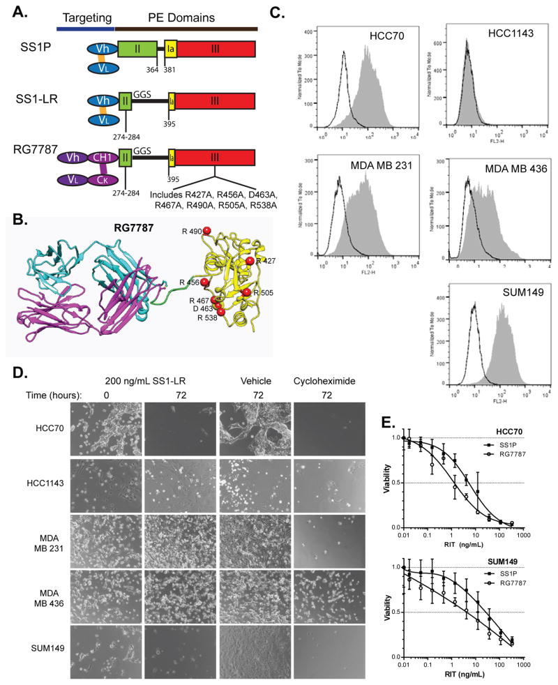
The PE24 platform was used to develop a new mesothelin-targeted RIT called RG7787 in collaboration with Roche (see Fig. 1A & 1B). In RG7787, the new PE24 is fused to a humanized anti-MSLN antibody Fab fragment. RG7787 retains the same stability and binding properties as previous mesothelin-targeted RITs but also benefits from less immunogenicity and a longer half-life than smaller dsFv-based PE24 molecules (10).
Figure 1.
MSLN-targeted RITs in TNBC in vitro. (A) Structures of the MSLN-targeted RITs SS1P, SS1-LR and RG7787 are depicted in cartoon form to demonstrate the differences in the targeting and PE domain modules. Vh and VL domains (in blue) form the Fv anti-MSLN antibody region and are connected by engineered disulfide bond (gold bar). Vh, VL, CH1 and CK (in purple) form the humanized anti-MSLN Fab of RG7787. Domain III (in red) is the catalytic subunit of PE. Domain Ia (in yellow) is also important for enzymatic function. Residues 274–284 of domain II (green) contain the furin cleavage site that is critical for intracellular processing of PE. GGS is a recombinant linker that improves flexibility of the molecule following the large deletion of Domain II in the newer molecules. A slightly modified linker is used in RG7787. (B) Ribbon diagram depicting the predicted structure of RG7787. Position of point mutations that eliminate human B-cell epitopes are indicated. (C) Five different TNBC cell lines were tested for MSLN surface expression by FACS analysis. Live cells were incubated with anti-MSLN primary antibody, followed by secondary incubation with appropriate PE-conjugated antibody (detected as FL2-H). Gray filled area shows detection of PE-labeling in minimum of 20,000 cells. Black trace shows negative control where incubation with anti-MSLN antibody was omitted. Traces are representative runs from at least three separate experiments. (D) TNBC cell lines were treated with a physiologic dose of the MSLN-targeted immunotoxin SS1-LR for 72 hours. Cells were photographed prior to treatment (left), and then the same field rephotographed after 72 hours of SS1-LR exposure (left center). Cells were also photographed after 72 hours of treatment with vehicle (right center), or 10 μg/mL of the protein synthesis inhibitor cycloheximide as positive control (right). Photographs are representative images of at least duplicate fields taken in duplicate wells in at least two experiments. (E) HCC70 (above) and SUM149 (below) were treated with the indicated concentrations of SS1P or RG7787 and incubated for 72 hours before assessment of cell viability by WST-8 assay. Viability for vehicle-treated cells was normalized to 1.0, and that for cycloheximide (a positive control which reliably produces nearly 100% cell kill) was normalized to 0. Each data point represents measurements of six treated wells.
The clinical utility of mesothelin has resulted in increasing interest in this protein over recent years. While the large majority of epithelioid mesotheliomas (11, 12), serous ovarian cancers (13) and pancreatic adenocarcinomas (14, 15) have long been known to express high levels of surface mesothelin, new studies have also revealed significant expression in other tumor types. Tchou et al. demonstrated that 67% of triple negative breast cancers (TNBCs) express mesothelin (16). This important, poor-prognosis subtype of breast cancer that accounts for 15% of all cases is named for its lack of expression of estrogen receptor, progesterone receptor, and Her2 amplification, and was under represented in earlier studies of mesothelin expression in breast cancer. In addition, reports (17–20) have revealed that many gastric cancers express mesothelin, although levels of surface expression may be much lower than typically found in mesothelioma, ovarian and pancreatic cancers. Here, we have evaluated if these recently recognized mesothelin-expressing cancers are susceptible to immunotoxin therapy targeted against MSLN.
Materials and Methods
Cell culture and reagents
MKN7, MKN28, MKN45, and MKN74 were provided by Dr. Takao Yamori and have been described previously (21). Identity of MKN28 was confirmed by short tandem repeat (STR) testing within the past six months. HCC70, MDA MB 231, and MDA MB 436 cell lines were obtained from ATCC. SUM149 and HCC1143 were the gift of Dr. Stanley Lipkowitz (NCI, Bethesda, MD) and identity was confirmed by STR testing. The KB cell line was described in our previous publication (22). All cells were grown at 37°C with 5% CO2 in RPMI 1640 medium (Lonza) supplemented with 2 mmol/L L-glutamine, 1 mmol/L sodium pyruvate, 100 U penicillin, 100 μg streptomycin (Gibco Life Technologies) and 10% fetal bovine serum (FBS) (HyClone, Thermo Scientific). Immunotoxin SS1P was manufactured by Advanced BioScience Laboratories, Inc. SS1-LR and LMB11 were synthesized as previously described (23). RG7787 was manufactured by Roche and provided for these studies through a Collaborative Research and Development Agreement.
Mesothelin surface expression assay
TNBC and gastric cancer cell lines were assessed for surface mesothelin expression by flow cytometry. A minimum of 20,000 cells per sample were analyzed on a FACS Calibur (BD Bioscience) running CellQuest software (BD Bioscience). Data were processed using FloJo software (Tree Star, Inc., Ashland, OR). Cells were plated at sub-confluent density and grown for 48–96 hours in culture then harvested with trypsin. All subsequent steps were performed at 4°C. Live cells were washed, fixed in FACS buffer (5% FBS, 0.1% sodium azide in D-PBS without calcium or magnesium) then incubated with mouse anti-mesothelin antibody (MN, Rockland Immunochemicals Inc., Gilbertsville, PA) for 30 minutes. Cells were washed again and incubated for 30 minutes in the dark with PE-conjugated secondary antibody (R-Phycoerythrin-Conjugated AffiniPure (Fab′)2 Fragment Goat Anti-Mouse IgG, Jackson ImmunoResearch Laboratories, West Grove, PA). Geometric means were chosen as mean fluorescence intensity (MFI). The MFI of cells was compared with the MFI from a standard curve of PE-conjugated calibration beads (BD QuantiBRITE™ PE quantitation kit, BD Bioscience) to estimate the number of mesothelin sites per cell.
SS1-LR photo screen
TNBC cell lines were plated at a density of 50,000 cells per well into two wells of a 24 well plate marked with registration grids. Cells were incubated for 24 hours to allow attachment, photographed under a phase contrast microscope, and then treated with SS1-LR (200 ng/mL), vehicle or cycloheximide (10 μg/mL) for 72 hours, before reimaging the previously photographed field.
In vitro quantitative cell viability assay
Viability of cancer cells treated with immunotoxins was measured using the Cell Counting Kit-8 WST-8 assay (Dojindo Molecular Technologies, Inc.). TNBC lines were seeded at 5.0 × 10³/well into 96-well plates and incubated for 24 hours before treatment. Gastric lines were plated at 1.0 × 10⁴/well and treated 4–6 hours later. The indicated concentrations of immunotoxin diluted in complete medium were added, then cells were incubated at 37°C for 72 hours. WST-8 assay reagent was added per manufacturer’s instructions. Plates were incubated at 37°C, and absorbance at 450 nm was measured. Values were normalized between 0% viability for treatment with cycloheximide (10 μg/mL), which produces complete cell killing and 100% for addition of complete medium alone. All data were plotted in GraphPad Prism 6. Fit curve and interpolated IC50 values were also calculated using this program.
Mouse xenograft tumor model
All animal experiments were performed in accordance with NIH guidelines and approved by the NCI Animal Care and Use Committee. HCC70 cells (2 × 10⁶) supplemented with matrigel (4.0 mg/mL) were injected into the intramammary fat pad of 4–6 week old female, athymic nude mice. For MKN28, 5 × 10⁶ cells were injected subcutaneously into the flank. Tumors were allowed to grow to an average volume of 100–150 mm3 before initiation of treatment. Paclitaxel (50 mg/kg) was diluted in 0.2% HSA in D-PBS and injected into the peritoneum on the indicated days. All immunotoxins were diluted in 0.2% HSA in D-PBS and administered intravenously through the tail vein. Tumor size was measured in two dimensions by digital calipers at least twice weekly and tumor volume was calculated using the formula: 0.4 × width2 × length. Animals were sacrificed before tumor volume reached 1000 mm3 or tumors became necrotic. Data were recorded and tumor growth curves plotted in Microscoft Excel.
Tumor excision and pathology
Mice were euthanized by exposure to 100% CO2, and then tumors were excised by dissection. Tumors were fixed in 10% formalin overnight before transfer into 70% ethanol. H&E staining and mesothelin immunohistochemistry was performed by Histoserv Inc. by standard methods. Anti-mesothelin IgG2a MN antibody (Rockland Immunochemicals, Inc) was used at 1:100 dilution. IgG2a isotype control antibody (MyBiosource; San Diego, CA) was used at 1:100 for control.
Statistics
Excel and Prizm6 were used for all statistical calculations and curve fitting. All errors are reported as standard deviations. Two-tailed student t-test was used for all statistical comparisons.
Results
Mesothelin expression in TNBC cell lines
MSLN expression was previously observed in approximately two-thirds of surgical specimens from women with TNBC (16). Further, our analysis of the publicly available TCGA data set for breast cancer confirms that some TNBC patients, but not hormone receptor- or Her-2-positive patients, do express MSLN (Supplementary Fig. 1). To identify an appropriate TNBC model system for testing mesothelin-targeted RITs, we examined surface MSLN expression in a panel of established TNBC cell lines using FACS analysis. The number of surface MSLN binding sites per cell was quantified by comparison to PE-bead standards, and values from multiple experiments were averaged to produce mean expression values. Figure 1C shows representative histograms of surface MSLN expression for each of the five cell lines. Expression levels varied by cell line and were found to be greatest for HCC70 and SUM149 (12,100 ± 1.4 and 15,700 ± 4.3 sites per cell, respectively) and absent for HCC1143 (<1 sites/cell). MDA MB 231 (4,600 ± 2.1 sites/cell) and MDA MB 436 (3,300 ± 0.81 sites/cell) had intermediate levels of expression. By comparison, the KB cervical cancer cell line that was used as a positive control had an average of 20,900 sites per cell (data not shown). The levels of surface MSLN expression in the HCC70 and SUM149 cell lines compare favorably with the KB cell line that is known to be sensitive to SS1P.
Sensitivity of TNBC cell lines to MSLN-targeted RITs
We next screened the TNBC cell line panel for sensitivity to the SS1-LR RIT. SS1-LR consists of an anti-MSLN Fv fragment fused to a 24-kD fragment of PE that lacks the majority of the non-catalytic domain II (Fig. 1A). The deletion of domain II improves efficiency of post-internalization processing (1). Equal densities of cells were plated and allowed to attach overnight before treatment for 72 hours with 200 ng/mL SS1-LR. This dose of the immunotoxin was chosen because 1) our prior clinical trials of SS1P have shown that this is an easily achievable concentration in patient serum samples (6), and 2) at this concentration there is minimal non-specific uptake of immunotoxins in vitro such that cytoxicity depends only on available surface mesothelin (data not shown). Figure 1D shows serial photographs of a representative field taken immediately after addition of immunotoxin (0 hours) and the same field after 72 hours of immunotoxin exposure. Treatment with SS1-LR kills the majority of HCC70 and SUM149 cells in the field (similar to the cycloheximide positive control) but has little effect on the HCC1143, MDA MB 231 and MDA MB 436 cells that express little to no surface MSLN.
Based on these results, we decided to further characterize the sensitivity of HCC70 and SUM149 cells using a quantitative cell growth inhibition assay. Equal densities of these cells were allowed to attach overnight before treatment for 72 hours with varying concentration of SS1P, SS1-LR or RG7787 and cell viability was measured by WST-8 assay. Data from representative experiments with HCC70 and SUM149 are plotted in Figure 1E and summarized in Table 1. A similar magnitude of sensitivity to RG7787 as SS1P was measured for HCC70 cells (IC50 3.4 versus 4.6 ng/mL, p = 0.38). SUM149, which has approximately one-third more surface mesothelin expression than HCC70, was less sensitive to SS1P than HCC70 (IC50 15.7 versus 4.6 ng/mL, p <0.05) but had similar IC50 when treated with RG7787 (4.8 ng/mL). IC50 values for all three mesothelin-targeted RITs measured in the subnanomolar range for these cell lines. No effect was seen using a negative control immunotoxin (LMB11) targeted against the B-cell differentiation antigen CD22 (Supplementary Fig. 2A). These data suggest that some TNBCs might be a good target for mesothelin-directed therapy.
Table 1.
Sensitivity of TNBC cell lines to mesothelin-targeted RITs
| RIT | HCC70 | SUM149 | ||||
|---|---|---|---|---|---|---|
|
| ||||||
| IC50 (ng/mL) | IC50 (pM) | N | IC50 (ng/mL) | IC50 (pM) | N | |
|
| ||||||
| SS1P | 4.6 ± 3.2 | 74.1 ± 52.0 | 9 | 15.7 ± 9.8 | 252 ± 158 | 3 |
| SS1-LR | 0.94 ± 0.56 | 19.6 ± 11.7 | 5 | 1.5 ± 1.4 | 30.2 ± 29.1 | 2 |
| RG7787 | 3.4 ± 2.8 | 45.2 ± 37.9 | 10 | 4.8 ± 1.3 | 64.8 ± 17.9 | 3 |
Sensitivity of HCC70 and SUM149 TNBC cell lines is reported in IC50 values ± standard deviation for SS1P, SS1-LR and RG7787 RITs. N = number of times each experiment was repeated with six replicates in each experiment.
RG7787 efficacy in TNBC mouse xenograft model
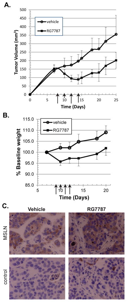
Next, the in vivo efficacy of RG7787 in an HCC70 mouse xenograft model was evaluated. Athymic nude mice were inoculated in the intra-mammary fat pad with HCC70 cells on Day 0 and tumors were allowed to grow to an average volume of 100–150 mm3 before initiation of treatment with RG7787 on Day 8. RG7787 was administered intravenously as a 2.5 mg/kg bolus every other day for a total of four treatments. It was previously determined that this dose could be delivered safely at this schedule (data not shown); it is more than five-times higher than the maximum dose of SS1P (0.4 mg/kg) that can be safely given. Control animals were treated with equal volumes of vehicle. Results of a representative experiment are shown in Figure 2A. Tumors in RG7787-treated mice had shrunk by 41% by the time the last dose of RG7787 was administered on day 14. Tumors in vehicle-treated mice continued to grow throughout this time. By the second RG7787 treatment, a statistically significant difference in tumor size between the two groups had developed. This difference continued for the remainder of the experiment, but reached a maximum at the fourth treatment (p < 0.001). In addition, there was a statistically significant delay of 10 days (p = 0.023) in the amount of time required for tumors to reach the 500 mm3 volume in animals treated with RG7787 (46 ± 3 days) versus vehicle (36 ± 9 days). Weight loss was less than 5% with this regimen (Fig. 2B) and no toxicities were observed. Adding a fifth dose of RG7787 did not result in a further decrease in tumor size (data not shown), suggesting that single agent efficacy in this model plateaus. One explanation would be that repetitive RG7787 treatment eliminates HCC70 cells with higher surface MSLN expression, leaving behind residual tumor cells with insufficient MSLN to internalize cytotoxic amounts of RIT. To test this hypothesis, vehicle and RG7787-treated tumors were excised 4 days after the last dose of vehicle or RG7787, fixed in formalin, and sections stained for MSLN expression. MSLN expression remained robust in cells from tumors that had been treated with RG7787 (Fig. 2C), suggesting a different mechanism is responsible for the incomplete effectiveness of the RIT.
Figure 2.
Effect of RG7787 on TNBC mouse xenograft tumor model. (A) Female athymic nude mice were innoculated in the intramammary fat pad with HCC70 cells at time 0. Intravenous treatment with RG7787 (2.5 mg/kg) or vehicle was begun on day 8 and continued every other day for a total of four doses. Each data point represents average of mean tumor volume for n = 7 animals treated with RG7787 or vehicle. Arrows indicate the days that treatment was administered. There is a statistically significant difference between the two groups beginning at day 10, with p < 0.0001 by day 14. Results shown are representative of three similar experiments. (B) Change in baseline weight for mice treated with the regimen described above is plotted over time. Weight dropped less than 5% in RG7787-treated animals over the course of therapy. (C) MSLN expression in tumors excised from vehicle and RG7787 treated mice was examined by IHC in formalin-fixed paraffin embedded tissue. Prominent staining was observed with anti-MSLN antibody (above) but not in control specimens labeled with isotype control antibody (below). Photographs were taken at 40X magnification.
Mesothelin surface expression in gastric cancer cell lines
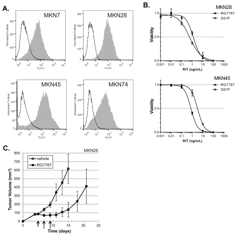
At least two-thirds of gastric cancer clinical specimens have detectable staining of mesothelin by immunohistochemistry (17–19) and surface MSLN expression was recently reported in some gastric cancer cell lines (19). To confirm this report, we tested a panel of gastric cancer cell lines for mesothelin surface expression by FACS analysis. Representative FACS traces are depicted in Figure 3A and summarized in Table 2. MKN28, which is sensitive to SS1P, had 16.1 × 10³ sites/cell. MKN45 also had strong expression (9.9 × 10³ sites/cell), while MKN7 (5.6 × 10³ sites/cell) and MKN74 (4.2 × 10³ sites/cell) have lower levels of expression.
Figure 3.
Gastric cancer is a target for MSLN-targeted RITs. (A) Four different gastric cancer cell lines were assessed for surface expression of MSLN as described in Figure 3A. Traces are representative runs from at least duplicate experiments. (B) MKN28 or MKN45 were treated with the indicated concentrations of SS1P or RG7787 and incubated for 72 hours before assessment of cell growth inhibition by WST-8 assay. Viability for vehicle-treated cells was normalized to 1.0, and that for cycloheximide (a positive control which reliably produces nearly 100% cell kill) was normalized to 0. Each data point represents measurements of three treated wells. (C) Female nude mice were inoculated subcutaneously with MKN28 cells at time 0. Intravenous treatment with RG7787 (2.5 mg/kg) or vehicle was given on days 5, 7 and 9. Each data point represents average of mean tumor volume for n = 10 animals treated with RG7787 or n = 5 vehicle treated animals. Arrows indicate the days that treatment was administered. There is a statistically significant difference between the two group beginning on day 7. Results shown are representative of more than three similar experiments.
Table 2.
Sensitivity of gastric cell lines to RITs
| Sample | Sites/cell (x103) | IC50 (ng/mL) | IC50 (pM) | |||
|---|---|---|---|---|---|---|
|
| ||||||
| SS1P | RG7787 | SS1P | RG7787 | |||
|
| ||||||
| MKN7 | 5.6 | 2.3 | >100 | 37.1 | >1200 | |
| MKN28 | 16.1 | 0.34 | 5.6 | 5.5 | 75.6 | |
| MKN45 | 9.9 | 2.3 | 0.5 | 37.1 | 6.8 | |
| MKN74 | 4.2 | 2.3 | 12 | 37.1 | 162.0 | |
Surface mesothelin for MKN7, MKN28, MKN45 and MKN74 gastric cancer cell lines was determined by flow cytometry assay and standardized to determine sites per cell as described in Material and Methods. WST-8 assay was used to determine IC50 of these cell lines to the SS1P and RG7787 RITs.
RG7787 in gastric cancer cell lines
The gastric cancer cell lines were tested for sensitivity to SS1P and RG7787 by WST-8 cell viability assay. Representative traces for MKN28 and MKN45 are shown in Figure 3B. Both MKN28 and MKN45 were highly sensitive to SS1P and RG7787 with IC50 values in the subnanomolar range. However, MKN28 cells were many fold more sensitive to SS1P than to RG7787 (IC50 0.34 versus 5.6 ng/mL), while the opposite was true of MKN45 (IC50 2.3 ng/mL for SS1P versus 0.5 ng/mL for RG7787). MKN7 and MKN74 were also more sensitive to SS1P than to RG7787 (see Table 2). No cell killing was seen when MKN28 cells were tested with the LMB11 control immunotoxin that targets CD22, demonstrating specificity of immunotoxin delivery (Supplementary Figure 2B). In vivo efficacy of RG7787 was then tested in a subcutaneous MKN28 mouse xenograft model. Athymic nude mice were inoculated with MKN28 cells on Day 0 and tumors were allowed to grow for 5 days before initiation of treatment with RG7787. Results of a representative experiment are shown in Figure 3C. Average tumor volume in RG7787 treated mice decreased by 18.1%. A statistically significant difference in tumor volume appeared on Day 7 (p < 0.001) and continued for the remainder of the experiment. Treatment with SS1P or the anti-CD22 immunotoxin LMB11 had no effect on tumor growth (Supplementary Figure 2C). These data demonstrate that RG7787 has single agent activity against gastric cancer.
Enhancing activity by pre-treatment with paclitaxel
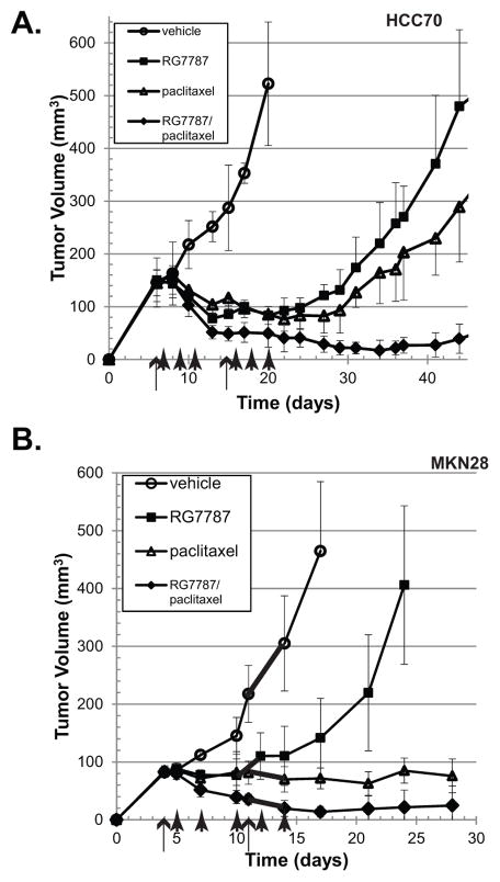
While RG7787 alone was able to slow the growth of TNBC and gastric cancer xenograft tumors in vivo, no complete remissions were seen. Previously, we have demonstrated that pre-administration of paclitaxel prior to RIT improves delivery of RIT to tumors resulting in significant synergy in vivo but not in vitro (24–26). Paclitaxel is an approved drug for both TNBC and gastric cancer. Therefore, we tested the efficacy of RG7787 in combination with paclitaxel in our mouse models. In HCC70 tumors, combination treatment resulted in an 88% reduction in average tumor volume compared to 52% with paclitaxel (p < 0.001) and 48% with RG7787 alone (Fig. 4A). Tumor volume of paclitaxel-treated mice reached threshold of 500 mm3 at an average of 58 days (range 48–78 days) while combination treated mice took 92 days (range 69–125 days) to reach this volume (p < 0.005). Similarly, combination RG7787/paclitaxel treatment of the MKN28 gastric tumors resulted in 70% reduction of tumor volume. with complete regressions in 4 of 10 mice (Fig. 4B). Longer follow-up revealed that tumor volume reached the threshold of 300 mm3 at an average of 47 days (range: 39–56 days) in mice treated with paclitaxel alone. By contrast, at 60 days, tumor volume in mice treated with paclitaxel/RG7787 remained less than 100 mm3 in three of seven mice (Supplementary Fig. 3). Co-administration of RG7787 with paclitaxel in vivo enhanced the anti-tumor response compared to either drug given alone. In vitro pre-treatment of HCC70 cells with paclitaxel prior to addition of RG7787 showed that paclitaxel is active against this cell type, but does not produce a synergistic response in combination with RG7787 (Supplementary Fig. 4).
Figure 4.
Pre-treatment with paclitaxel enhances RIT anti-tumor response. Female athymic nude mice were inoculated with tumor cells at time 0. Animals were treated with vehicle, RG7787 with IP vehicle injection, paclitaxel with IV vehicle injection, or paclitaxel and RG7787 combination. Paclitaxel (50 mg/kg) was administered by IP injection on days marked with long arrows. RG7787 (2.5 mg/kg) was administered IV on days indicated by short arrows. Mean tumor volumes are indicated by the markers. For vehicle control five mice were followed. Results of both experiments were confirmed by repeat. (A) HCC70 TNBC tumor xenografts. There is a statistically significant difference between paclitaxel and combination treatment beginning at day 13 (p ≤ 0.001, n = 6 per treatment group). (B) MKN28 gastric cancer tumor xenografts. There is a statistically significant difference between paclitaxel and combination treatment beginning at day 10 (p ≤ 0.001, n= 8 per treatment group).
Discussion
RG7787, an RIT with clinically optimized properties, is a highly active therapeutic that kills MSLN-expressing gastric and breast cancers with subnanomolar efficacy in vitro and displays marked anti-tumor activity in animal models of these malignancies. Surface expression of MSLN is much lower in the gastric and TNBC cell lines studied here than in cell lines previously studied in our laboratory, including mesothelioma cells from patients (27), ovarian cancer cells from patients (13) or A431/H9 cells transfected with a mesothelin-expressing plasmid (28). While SS1P has insufficient therapeutic window to effectively treat tumors with modest levels of MSLN expression, these data demonstrate that clinical development of RG7787 for use in tumors with less robust levels of MSLN expression, like TNBC and gastric cancer, is warranted.
RG7787 in combination therapy
RG7787, like SS1P, has a unique mechanism of action and a toxicity profile that is dissimilar from all other drugs approved to treat solid tumor malignancies. We have previously demonstrated that combination of active chemotherapy drugs with SS1P can synergistically improve in vivo but not in vitro efficacy (24). Here, our data demonstrate that combining RG7787 with paclitaxel, a second active agent, results in improved anti-tumor activity against both TNBC and stomach cancers, with some complete responses observed. Importantly, no excessive toxicity resulted from combination treatment and both agents could be used at their full single agent dosages. These studies provide evidence that RG7787 is an ideal therapeutic to use in combination therapy against solid-tumor malignancies.
Role of domain II deletion in efficacy
SS1P, the mesothelin-targeted RIT currently being tested in clinical trials, contains a 38-kD PE toxin fragment, while the 24-kD LR toxin moiety in the RG7787 RIT lacks all of domain II except for the 10 residues that compose the furin cleavage site. We have previously demonstrated that the furin cleavage site is absolutely required for immunotoxin activity. A role for the region of domain II deleted from RG7787 has not yet been determined. Here and previously, we have observed that this region of the molecule affects toxin efficacy in a cell-specific manner (29). For example, SUM149 is markedly more sensitive to the LR-based RITs SS1-LR and RG7787 than to SS1P. However, three of the four gastric cancer cell lines were more sensitive to SS1P than RG7787. Nevertheless, IC50 for RG7787 in MKN28 cells is still significantly less than 100 pM, and RG7787 was effective in vivo against MKN28 xenografts. Currently, we are unable to predict which cells will be more sensitive to RG7787 versus SS1P in vitro. Future studies examining the role of domain II in toxin activity could improve understanding of these differences.
Partial response in vivo
In vivo, we have shown that single-agent RG7787 shrinks gastric and TNBC tumors and delays tumor progression. However, we observed no complete responses with the single agent RIT even though physiologic levels of RG7787 killed >95% of the same cell lines in vitro. We have demonstrated in our TNBC model that robust MSLN expression remains in xenografted tumors after repeated doses of RG7787, proving that loss of the target is not responsible for the incomplete responses in vivo. It is possible that insufficient RIT is reaching the tumor to kill all tumor cells. Although we calculate that the 2.5 mg/kg RG7787 dosage used in our studies should give an initial serum concentration of 1,000 ng/ml, we do not know the local concentration of RIT within the tumor or how well the large RIT molecules are able to penetrate through densely packed tumors cells from blood vessels. In our previous work, we showed that tumor cell killing by SS1P is limited by inadequate delivery to the tumor in a MSLN-transfected H9 epidermoid cell model (26), but that paclitaxel does not improve immunotoxin delivery in the KLM1 pancreatic cancer model (30). Further studies are planned to determine whether incomplete RIT delivery limits toxin efficacy or whether the relative resistance we have observed in vivo is explained by differential activation of survival signaling in vivo versus in culture.
In conclusion, combining RG7787 with paclitaxel produced near complete regressions in animal models of both TNBC and gastric cancer. This data argues for clinical development of RG7787 for use against these and other mesothelin expressing cancers.
Supplementary Material
Acknowledgments
Funding: This research was supported by the Intramural Research Program of the National Institutes of Health, National Cancer Institute, Center for Cancer Research, and by a Cooperative Research and Development Agreement (#2791) between the National Cancer Institute and Roche Pharmaceuticals.
The authors thank John Flickinger for contributing LMB11, Dawn A. Walker for reading and editing the manuscript, and Anna Mazzuca for help in the submission process.
Footnotes
Disclosures and Potential Conflict of Interest: I.P. is an inventor on several patents on immunotoxins that have all been assigned to NIH (including US 8357783, US 20120263674, US 20140094417 and E-771-2013/1-US-01) and has a Cooperative Research and Development Agreement (#2791) with Roche Pharmaceuticals. All other authors declare no conflicts of interest.
References
- 1.Weldon JE, Xiang L, Zhang J, Beers R, Walker DA, Onda M, et al. A recombinant immunotoxin against the tumor-associated antigen mesothelin reengineered for high activity, low off-target toxicity, and reduced antigenicity. Mol Cancer Ther. 2013;12:48–57. doi: 10.1158/1535-7163.MCT-12-0336. [DOI] [PMC free article] [PubMed] [Google Scholar]
- 2.Kreitman RJ, Tallman MS, Robak T, Coutre S, Wilson WH, Stetler-Stevenson M, et al. Phase I trial of anti-CD22 recombinant immunotoxin moxetumomab pasudotox (CAT-8015 or HA22) in patients with Hairy Cell Leukemia. J Clin Oncol. 2012;30:1822–8. doi: 10.1200/JCO.2011.38.1756. [DOI] [PMC free article] [PubMed] [Google Scholar]
- 3.Wayne AS, Bhojwani D, Silverman LB, Richards K, Stetler-Stevenson M, Shah NN, et al. A novel anti-CD22 immunotoxin, moxetumomab pasudotox: Phase I study in pediatric Acute Lymphoblastic Leukemia (ALL) Blood. 2011;118:248. (abstr) [Google Scholar]
- 4.Kreitman RJ, Singh R, Stetler-Stevenson M, Waldmann TA, Pastan I. Regression of Adult T-cell Leukemia with anti-CD25 recombinant immunotoxin LMB-2 preceded by chemotherapy. Blood. 2011;118:2575a. (abstr) [Google Scholar]
- 5.Tang Z, Qian M, Ho M. The role of mesothelin in tumor progression and targeted therapy. Anticancer Agents Med Chem. 2013;13:276–80. doi: 10.2174/1871520611313020014. [DOI] [PMC free article] [PubMed] [Google Scholar]
- 6.Hassan R, Bullock S, Premkumar A, Kreitman RJ, Kindler H, Willingham MC, et al. Phase I study of SS1P, a recombinant anti-mesothelin immunotoxin given as a bolus I.V. infusion to patients with mesothelin-expressing mesothelioma, ovarian, and pancreatic cancers. Clin Cancer Res. 2007;13:5144–9. doi: 10.1158/1078-0432.CCR-07-0869. [DOI] [PubMed] [Google Scholar]
- 7.Kreitman RJ, Hassan R, Fitzgerald DJ, Pastan I. Phase I trial of continuous infusion anti-mesothelin recombinant immunotoxin SS1P. Clin Cancer Res. 2009;15:5274–9. doi: 10.1158/1078-0432.CCR-09-0062. [DOI] [PMC free article] [PubMed] [Google Scholar]
- 8.Hassan R, Miller AC, Sharon E, Thomas A, Reynolds JC, Ling A, et al. Major cancer regressions in mesothelioma after treatment with an anti-mesothelin immunotoxin and immune suppression. Science Transl Med. 2013;5:208ra147. doi: 10.1126/scitranslmed.3006941. [DOI] [PMC free article] [PubMed] [Google Scholar]
- 9.Liu W, Onda M, Lee B, Kreitman RJ, Hassan R, Xiang L, et al. Recombinant immunotoxin engineered for low immunogenicity and antigenicity by identifying and silencing human B-cell epitopes. Proc Natl Acad Sci USA. 2012;109:11782–7. doi: 10.1073/pnas.1209292109. [DOI] [PMC free article] [PubMed] [Google Scholar]
- 10.Niederfellner G, Bauss F, Imhof-Jung S, Hesse F, Kronenberg S, Staak R, et al. RG7787-a novel de-immunized PE based fusion protein for therapy of mesothelin-positive solid tumors. Proceedings of the 105th Annual Meeting of the AACR; 2014. p. 4510. (abstr) [Google Scholar]
- 11.Chang K, Pastan I. Molecular cloning of mesothelin, a differentiation antigen present on mesothelium, mesotheliomas, and ovarian cancers. Proc Natl Acad Sci USA. 1996;93:136–40. doi: 10.1073/pnas.93.1.136. [DOI] [PMC free article] [PubMed] [Google Scholar]
- 12.Ordonez NG. Value of mesothelin immunostaining in the diagnosis of mesothelioma. Mod pathol. 2003;16:192–7. doi: 10.1097/01.MP.0000056981.16578.C3. [DOI] [PubMed] [Google Scholar]
- 13.Hassan R, Kreitman RJ, Pastan I, Willingham MC. Localization of mesothelin in epithelial ovarian cancer. Appl Immunohistochem Mol Morphol. 2005;13:243–7. doi: 10.1097/01.pai.00000141545.36485.d6. [DOI] [PubMed] [Google Scholar]
- 14.Argani P, Iacobuzio-Donahue C, Ryu B, Rosty C, Goggins M, Wilentz RE, et al. Mesothelin is overexpressed in the vast majority of ductal adenocarcinomas of the pancreas: identification of a new pancreatic cancer marker by serial analysis of gene expression (SAGE) Clin Cancer Res. 2001;7:3862–8. [PubMed] [Google Scholar]
- 15.Hassan R, Laszik ZG, Lerner M, Raffeld M, Postier R, Brackett D. Mesothelin is overexpressed in pancreaticobiliary adenocarcinomas but not in normal pancreas and chronic pancreatitis. Am J Clin Pathol. 2005;124:838–45. [PubMed] [Google Scholar]
- 16.Tchou J, Wang LC, Selven B, Zhang H, Conejo-Garcia J, Borghaei H, et al. Mesothelin, a novel immunotherapy target for triple negative breast cancer. Breast Cancer Res Treat. 2012;133:799–804. doi: 10.1007/s10549-012-2018-4. [DOI] [PMC free article] [PubMed] [Google Scholar]
- 17.Baba K, Ishigami S, Arigami T, Uenosono Y, Okumura H, Matsumoto M, et al. Mesothelin expression correlates with prolonged patient survival in gastric cancer. J Surg Oncol. 2012;105:195–9. doi: 10.1002/jso.22024. [DOI] [PubMed] [Google Scholar]
- 18.Einama T, Homma S, Kamachi H, Kawamata F, Takahashi K, Takahashi N, et al. Luminal membrane expression of mesothelin is a prominent poor prognostic factor for gastric cancer. Br J Cancer. 2012;107:137–42. doi: 10.1038/bjc.2012.235. [DOI] [PMC free article] [PubMed] [Google Scholar]
- 19.Ito T, Kajino K, Abe M, Sato K, Maekawa H, Sakurada M, et al. ERC/mesothelin is expressed in human gastric cancer tissues and cell lines. Oncol Rep. 2014;31:27–33. doi: 10.3892/or.2013.2803. [DOI] [PMC free article] [PubMed] [Google Scholar]
- 20.Kelly RJ, Pastan I, Cowan ML, Montgomery E, Hassan R, Alewine CC, et al. Mesothelin expression in patients as a novel target in gastric cancer. J Clin Oncol. 2014;32:3s. (suppl: abstr 61) [Google Scholar]
- 21.Motoyama T, Hojo H, Watanabe H. Comparison of seven cell lines derived from human gastric carcinomas. Acta Pathol Jpn. 1986;36:65–83. doi: 10.1111/j.1440-1827.1986.tb01461.x. [DOI] [PubMed] [Google Scholar]
- 22.Zhang Y, Xiang L, Hassan R, Pastan I. Immunotoxin and Taxol synergy results from a decrease in shed mesothelin levels in the extracellular space of tumors. Proc Natl Acad Sci USA. 2007;104:17099–104. doi: 10.1073/pnas.0708101104. [DOI] [PMC free article] [PubMed] [Google Scholar]
- 23.Pastan I, Beers R, Bera TK. Recombinant immunotoxins in the treatment of cancer. Methods Mol Biol. 2004;248:503–18. doi: 10.1385/1-59259-666-5:503. [DOI] [PubMed] [Google Scholar]
- 24.Zhang Y, Xiang L, Hassan R, Paik CH, Carrasquillo JA, Jang BS, et al. Synergistic antitumor activity of taxol and immunotoxin SS1P in tumor-bearing mice. Clin Cancer Res. 2006;12:4695–701. doi: 10.1158/1078-0432.CCR-06-0346. [DOI] [PubMed] [Google Scholar]
- 25.Zhang Y, Pastan I. High shed antigen levels within tumors: an additional barrier to immunoconjugate therapy. Clin Cancer Res. 2008;14:7981–6. doi: 10.1158/1078-0432.CCR-08-0324. [DOI] [PMC free article] [PubMed] [Google Scholar]
- 26.Zhang Y, Hansen JK, Xiang L, Kawa S, Onda M, Ho M, et al. A flow cytometry method to quantitate internalized immunotoxins shows that taxol synergistically increases cellular immunotoxins uptake. Cancer Res. 2010;70:1082–9. doi: 10.1158/0008-5472.CAN-09-2405. [DOI] [PMC free article] [PubMed] [Google Scholar]
- 27.Zhang J, Qiu S, Zhang Y, Merino M, Fetsch P, Avital I, et al. Loss of mesothelin expression by mesothelioma cells grown in vitro determines sensitivity to anti-mesothelin immunotoxin SS1P. Anticancer Res. 2012;32:5151–8. [PMC free article] [PubMed] [Google Scholar]
- 28.Ho M, Hassan R, Zhang J, Wang QC, Onda M, Bera T, et al. Humoral immune response to mesothelin in mesothelioma and ovarian cancer patients. Clin Cancer Res. 2005;11:3814–20. doi: 10.1158/1078-0432.CCR-04-2304. [DOI] [PubMed] [Google Scholar]
- 29.Weldon JE, Xiang L, Chertov O, Margulies I, Kreitman RJ, FitzGerald DJ, et al. A protease-resistant immunotoxin against CD22 with greatly increased activity against CLL and diminished animal toxicity. Blood. 2009;113:3792–800. doi: 10.1182/blood-2008-08-173195. [DOI] [PMC free article] [PubMed] [Google Scholar]
- 30.Hollevoet K, Mason-Osann E, Liu XF, Imof-Jung S, Niederfellner G, Pastan I. In viro and in vivo activity of the low-immunogenic anti-mesothelin immunooin RG7787 in pancreatic cancer. Mol Cancer Ther. doi: 10.1158/1535-7163. in press. [DOI] [PMC free article] [PubMed] [Google Scholar]
Associated Data
This section collects any data citations, data availability statements, or supplementary materials included in this article.


