Abstract
MDM2 is an oncoprotein that blocks p53 tumor suppressor-mediated transcriptional transactivation, escorts p53 from the cell nucleus to the cytoplasm, and polyubiquitylates p53. Polyubiquitylated p53 is rapidly degraded in the cytoplasm by the 26S proteasome. MDM2 is abnormally upregulated in several types of cancers, especially those of mesenchymal origin. MDM4 is a homolog of MDM2 that also inhibits p53 by blocking p53-mediated transactivation. MDM4 is required for MDM2-mediated polyubiquitylation of p53 and is abnormally upregulated in several cancer types. MDM2 and MDM4 genes have been detected in all vertebrates to date and only a single gene homolog, named MDM, has been detected in some invertebrates. MDM2, MDM4, and MDM have similar gene structures, suggesting that MDM2 and MDM4 arose through a duplication event more than 440 million years ago. All members of this small MDM2 gene family contain a single really interesting new gene (RING) domain (with the possible exception of lancelet MDM) which places them in the RING-domain superfamily. Similar to MDM2, the vast majority of proteins with RING domains are E3 ubiquitin ligases. Other RING domain E3 ubiquitin ligases that target p53 are COP1, Pirh2, and MSL2. In this report, we present evidence that COP1, Pirh2, and MSL2 evolved independently of MDM2 and MDM4. We also show, through structure homology models of invertebrate MDM RING domains, that MDM2 is more evolutionarily conserved than MDM4.
Keywords: evolution, oncogene, p53, RING, tumor suppressor, ubiquitylation
Introduction
The MDM2 gene was discovered as one of three genes (MDM1, MDM2, and MDM3) within an amplicon cloned from the tumorigenic mouse cell line 3T3DM (1–3). The genes have different sequences and only MDM2 was found to be amplified in human cancers. In humans, the MDM2 gene (also known as HDM2) is located on chromosome 12q14.3-q15 and most frequently expresses a 491 amino acid residue protein. MDM2 is amplified at an overall frequency of 7% in human cancers and at a higher frequency within soft tissue sarcomas, osteosarcomas, and esophageal carcinomas (4, 5). In some cancers with no apparent MDM2 amplification, MDM2 transcript levels are elevated by increased gene expression (6–8).
MDM2 protein negatively regulates the p53 tumor suppressor protein (9). The p53 tumor suppressor responds to cell stress by transcriptionally activating several genes responsible for DNA repair, cell cycle arrest, anti-angiogenesis, apoptosis, and autophagy (10). The particular downstream pathway activated by p53 depends on many conditions, including the severity of the stress, the nature of the stressor, and the cell type. Regulation of p53 primarily takes place at the protein stability level within a regulatory network where p53 is polyubiquitylated by MDM2 and subsequently degraded by the 26S proteasome (11–13). A key component of this network is the p53/MDM2 feedback loop, where p53 turnover is regulated by MDM2 and expression of MDM2 is under the transcriptional control of p53 (14–16). p53 transcriptionally activates MDM2 through a p53-responsive element located in the first intron, and in turn, MDM2 targets p53 for degradation. This negative feedback loop keeps p53 levels relatively low, unless stress is applied to the cell.
Detailed examination of this negative feedback loop is worthwhile, especially in light of current interest in the development of small molecules to inhibit MDM2 activities. In normal cells, p53 activates the expression of MDM2. Upon cell stress, MDM2 and p53 are phosphorylated (17–24) and bind to proteins that physically separate MDM2 from p53 (25–27). MDM2 inhibits p53 through three linked actions. First, MDM2 binds to the transactivation domain of p53 that sterically blocks access of p53 to basal transcription factors. Second, the E3 ubiquitin ligase activity of MDM2 mediates monoubiquitylation of p53, promoting the relocation of the p53-MDM2 complex from the nucleus to the cytoplasm (28). Third, once in the cytoplasm, MDM2 polyubiquitylates p53, leading to its degradation by the 26S proteasome (29). This negative feedback loop is further regulated by critical proteins including MDM4, HAUSP (USP7), ARF, Pirh2, MSL2, and COP1 (30–34).
The second member of the MDM2 gene family is MDM4 (sometimes known as MDMX, HDM4, or HDMX), first identified when its protein product was discovered as a novel p53 binding protein by screening a mouse cDNA expression library with radiolabeled p53 protein (35). The MDM4 gene is located on human chromosome 1q32 and encodes a 490 residue protein. The MDM4 gene is amplified or the MDM4 protein is overexpressed in 10%–20% of diverse tumors including lung, colon, stomach, and breast cancers, as well as 65% of retinoblastomas (36, 37). Similar to MDM2, MDM4 inhibits the transactivation function of p53 by sterically blocking its access to basal transcription factors (35, 38). Currently, the development of molecules that block p53-MDM2/MDM4 interactions is considered a promising strategy to combat cancers that contain inactive wild-type p53. Although still in the development and testing stage, small molecules have been shown to induce p53 tumor suppressor activities in animal models (39–41). In the cell, MDM2 and MDM4 form a heterodimer that strengthens the efficacy of MDM2's inhibitory activities (29, 42).
Careful mouse genetic studies indicate that MDM4 contributes more to inhibition of p53-mediated transcriptional transactivation while MDM2 contributes more to degradation of p53 (43). In line with such studies, MDM4 lacks robust E3 ligase activity in vitro. Instead, MDM4 is an E4 protein in the ubiquitylation pathway. In general, E4 proteins are responsible for recognizing monoubiquitylated substrates and guiding the conjugation of multiple ubiquitin units onto single lysine residue targets within the protein substrate, a process known as polyubiquitylation. Only after polyubiquitylation is the protein substrate recognized by the 26S proteasome for degradation. MDM4 forms a complex with MDM2, monoubuiquitylated p53 and E2 protein to assist MDM2 polyubiquitylate p53 (44). The really interesting new gene (RING) domains within MDM2 are critical for its ubiquitylation activity and, in addition, RING domains within MDM2 and MDM4 form the heterodimerization interfaces of these two proteins.
MDM2 and MDM4 are paralogs that form a small family called the MDM2 gene family within the superfamily of RING domain-bearing proteins. An analysis of the evolutionary history of MDM2 and MDM4 indicates that the paralogs arose from a duplication event more than 440 million years ago, at approximately the same time that the p53 gene underwent duplication events to form p63 and p73 (45, 46). Both MDM2 and MDM4 paralogs are detected in vertebrates, but only one gene family member is detected in invertebrates, named MDM. This review discusses the MDM2 gene family from an evolutionary perspective.
The ubiquitylation pathway
To appreciate the evolutionary perspective of the MDM2 gene family, a brief background of ubiquitin-mediated protein modification is necessary because the domain responsible for this modification, the RING domain, is strongly conserved in orthologs of this family. Ubiquitylation is the covalent modification of protein lysine residues by addition of the small regulatory protein molecule ubiquitin (47). This process requires three enzymes: an ATP-dependent ubiquitin activating enzyme (E1), a ubiquitin conjugating enzyme (E2), and a ubiquitin ligase (E3). Upon activation, ubiquitin is transferred from E1 to a catalytic cysteine on E2, forming a thioester-linked conjugate. The E2-ubiquitin conjugate engages E3 and, together, they transfer ubiquitin from E2 to the ε-amino group of a lysine side chain on the target protein (48). In many instances, RING domains within E3s are interaction sites for E2s, and the presence of RING domains is assumed to be indicative of E3 ubiquitin ligase function (49, 50). Another component of the ubiquitylation pathway is E4 (discussed previously), discovered much later than other components of the ubiquitin pathway (51).
E3 ubiquitin ligases fall into two classes, those that contain a RING domain (with a few containing a structurally and functionally similar U-box domain) and those with a homologous to E6-AP carboxy-terminus (HECT) domain. Protein target specificity within the ubiquitin cascade is provided by E3 ubiquitin ligase. RING domain-containing E3 ubiquitin ligases, in most cases, mediate the transfer of ubiquitin by recruiting E2 ubiquitin-conjugated enzymes to the acceptor lysine residue on the target protein and enhancing this transfer (52). RING domains are not covalently bound to ubiquitin. HECT domains, however, form covalent intermediates between ubiquitin and a cysteine residue within E3 prior to ubiquitin transfer to the target protein.
RING domains within the MDM2 family
Human MDM2 and MDM4 proteins exhibit 31% amino acid residue identity and possess similar patterns of protein domain organization (46). Both contain an N-terminal p53 binding domain, central acidic and zinc-binding domains, and a C-terminal RING domain. The p53 binding domain and RING domain are particularly well conserved between the human MDM2 and MDM4 paralogs (50.9% and 52.4% amino acid residue identity respectively). Invertebrates frequently code for only one MDM family protein and at the moment there are seven identified invertebrate species that contain MDM gene: lancelet, owl limpet, bay mussel, acorn worm, sea squirt, deer tick, and placozoa. The invertebrate MDM and human MDM2 protein sequences share identities that range from 21% to 27%, whereas the invertebrate MDM and human MDM4 protein sequences share identities that range from 19% to 26%. With the exception of sea quirt MDM, invertebrate MDMs exhibit higher identity to human MDM2 than to human MDM4, indicating that in six out of seven instances, human MDM2 and invertebrate MDMs are slightly more related to each other than human MDM4 and invertebrate MDMs. This increased relatedness is largely due to relatively high identity between the RING domains of human MDM2 and invertebrate MDMs. Furthermore, within the vertebrates, the RING domains of MDM2 orthologs exhibit a high degree of sequence identity (≥79% identity) compared to that of the RING domains of the MDM4 orthologs (≥52% identity). Overall, the RING domain of MDM2 is well conserved amongst vertebrate MDM2 orthologs as well as amongst invertebrate MDMs protein sequences.
MDM2 and MDM4 can form homodimers or heterodimers through RING domain interactions (42). Within the cell, the majority of MDM2 and MDM4 molecules form heterocomplexes that create efficient E3 functions towards p53 (53). In a broader scope, RING domain proteins can function as E3 ubiquitin ligases in either the monomeric or dimeric state. The RING proteins Pirh2, c-Cbl, PML, and CNOT4 facilitate their E3 ligase function through their RING domains in their monomeric forms (54–57). Other RING proteins, such as the cIAPs, BRCA1-BARD, and Ring1b-Bmi1 require RING dimerization for E3 function (58–60).
Other RING domain E3 ligases that target p53
Since the discovery of MDM2 and MDM4, other RING domain-containing E3 ligases that target p53 have come to light. Constitutive photomorphogenic 1 (COP1), also known as RING finger and WD repeat domain 2 (RFW2), was initially identified in Arabidopsis where it plays a critical role in plant growth and development in response to light (34). COP1 is conserved in higher plants and vertebrates; it consists of an N-terminal RING finger domain, an internal coiled coil domain, and C-terminal WD40 repeats (61). Mammalian COP1 targets p53 for degradation independently of MDM2 and is necessary for p53 turnover in cultured normal and cancer cells. Analogous to MDM2 and MDM4, COP1 is a p53-inducible gene (it contains a p53-responsive element within the COP1 promoter region) and is part of a negative feedback loop (34). COP1 is over-expressed in 81% of breast cancers and 44% of ovarian adenocarcinomas. In cancers that retain wild-type p53, COP1 overexpression is correlated with a striking decrease in steady state p53 protein levels and attenuation of the downstream p53 target gene CDKN1A (also known as CIP1, WAF1, PIK1, SDI1).
Another E3 that targets p53 is p53-induced RING-H2 domain protein (Pirh2), originally identified as an androgen receptor N-terminal-interacting protein (ARNIP). Pirh2 is also known as RING finger and CHY zinc finger domain-containing protein 1 (RCHY1) (62). Pirh2 consists of an N-terminal CHY zinc finger domain, central RING domain, and a C-terminal Zinc finger domain (56). The best-known function of Pirh2 is its role in the p53/Pirh2 negative feedback loop, independent of MDM2 and COP1, in which Pirh2 inhibits p53 activity and is under the transcriptional control of p53. A p53-responsive element is located in the third intron of the pirh2 gene. Similar to MDM2 and COP1, Pirh2 negatively regulates p53 function through ubiquitin-mediated proteolysis. Pirh2 has been shown to target p53 for degradation under DNA damaging conditions when MDM2 dissociates from p53 and fails to target p53 for degradation (33). The interaction of p53 and Pirh2 employs a two-site binding mode, where the Pirh2 N-terminus interacts with the p53 DNA binding domain and the Pirh2 C-terminus binds to the p53 tetramerization domain with enhanced specificity for the active tetrameric form of p53 (63). Mouse models indicate that overexpression of Pirh2 promotes tumorigenicity (64) and that its unphosphorylated form is detected in tumor cells (65).
A third E3 ligase with a RING domain that targets p53 is MSL2 (30). MSL2's RING domain has the same cross brace zinc domain motif as MDM2 and MDM4 (see next section), but has a different Zn-coordination scheme (C3HC4 vs. MDM2/MDM4's C2H2C4). MSL2 ubiquitylates p53 at Lys 351 and Lys 357 residues, distinct from lys residues ubiquitylated by MDM2. Modification by MSL2 appears to expose a nuclear export motif within p53 as well as release p53 from MDM2. Overexpression of MSL2 does not target p53 for destruction but, rather, causes p53 accumulation in the cytoplasm.
Evolutionary relationships of human RING proteins
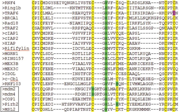
A sequence alignment of the RING domains of 22 human RING-containing proteins is presented in Figure 1. Sequences of the RING proteins were obtained from the UniProt protein database and limited to the range beginning with the first zinc coordinating cysteine and ending with the residue following the last zinc coordinating residue. The proteins listed in Figure 1 act as E3 ligases with the exception of MDM4. In general, the RING domains range from 40 to 60 residues and coordinate two zinc atoms through a zinc finger cross brace motif--a zinc finger motif with the consensus sequence Cys-X2-Cys-X9-39-Cys-X1-3-His-X2-3-Cys-X2-Cys-X4-48-Cys-X2-Cys (49). However, the RING domains of MDM2 and MDM4 possess a C2H2C4 zinc-binding scheme that is unique among RING finger family members (66). Unlike all other known RING domains, four amino acid residues, instead of two or three, separate the third and fourth zinc coordinating residues (underlined above). In the human MDM2 RING domain, residues C438, C441, C461, and C464 coordinate the zinc atom Zn1, while H452, H457, C475, and C478 coordinate Zn2 (67). In the human MDM4 RING domain, residues C437, C440, C460, and C463 coordinate Zn1 while H451, H456, C474, and C477 coordinate Zn2. The MDM2 RING (residues 438–479) and MDM4 RING (residues 437–478) domains are located near their respective C-termini.
Figure 1. Alignment of the RING domain sequences of 22 human proteins.
Residues critical for coordinating Zn atoms are highlighted. Five proteins reported to interact and regulate ubiquitylation of p53 are bordered. Alignment created by the ClustalW2 software program. Eight of the sequences were obtained by performing a BLAST analysis of the human, rat, and frog genomes using the MDM2 RING sequence as the query. The BLAST output revealed that eight proteins are present in all three organisms: MGRN1, XIAP, cIAP1, RNF103, Rififylin, Ring 157, MEX3B, and MEX3C. Ten RING containing proteins were added: MDM4, BRCA1, c-Cbl, cIAP2, IDOL, Rad18, hRing1b, RNF4, TRAF2, and TRAF6 obtained from a recent review of RING proteins (80). The proteins Pirh2, COP1, MLS2 were added because they are RING proteins that ubiquitinate p53.
Analysis of the gene structures of p53-targeting RING proteins and the gene structures of other RING proteins from humans suggests that the MDM2 gene family consists of just MDM2 and MDM4. The products of gene duplication often retain gene structures that include the total number of exons and the exon lengths. In addition, the particular exon that encodes the RING domain relative to other exons in the gene is also often conserved in closely related gene family members. We analyzed the human RING-containing proteins listed in Figure 1 for maintenance of these gene structure features. Table 1 lists the number of exons, the RING-domain coding exon(s) and the length of exon(s) in each RING-domain containing gene. Four groups of closely related RING genes are observed (color shaded): (i) cIAP1, cIAP2, XIAP; (ii) MGRN1 and RNF157; (iii) MEX3B and MEX3C; (iv) MDM2 and MDM4. Through gene structure analysis it appears that non-MDM2/MDM4 RING-domain proteins that target p53, COP1, Pirh2, and MSL2, are not closely related to MDM2/MDM4 nor to other RING-domain proteins in this cohort.
Table 1.
Human RING genes: number of exons, RING coding exons, and length of RING-coding exons.
| Gene | Number of exons | Exon(s) coding for RING domain | Length of RING-containing exon(s) |
|---|---|---|---|
| RNF4 | 8 | 7 and 8 | 199 |
| RING1b | 7 | 3 and 4 | 377 |
| TRAF6 | 7 | 2 and 3 | 447 |
| BRCA1 | 23 | 3 and 4 | 212 |
| RAD18 | 13 | 2 and 3 | 144 |
| TRAF2 | 12 | 3 and 4 | 267 |
| cIAP1 | 9 | 9 | 194 |
| cIAP2 | 9 | 9 | 194 |
| XIAP | 7 | 7 | 194 |
| Rififylin | 7 | 7 | 182 |
| MGRN1 | 12 | 10 | 160 |
| RNF157 | 19 | 10 | 160 |
| MEX3B | 2 | 2 | 1454 |
| MEX3C | 2 | 2 | 1226 |
| IDOL | 7 | 6 and 7 | 511 |
| c-Cbl | 16 | 8 and 9 | 336 |
| RNF157 | 19 | 10 | 160 |
| MDM2 | 11 | 11 | 192 |
| MDM4 | 11 | 11 | 190 |
| COP1 | 20 | 2 and 3 | 158 |
| Pirh2 | 9 | 6, 7, 8 and 9 | 252 |
| MSL2 | 2 | 1 and 2 | 5202 |
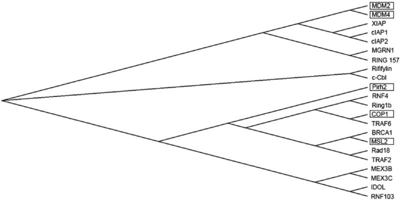
As mentioned previously the most conserved domain in MDM2 is the RING finger domain, which binds to E2 and is responsible for dimerization. Figure 2 shows the results of neighbor-joining cluster analysis of RING domains of human RING proteins (68). Relatively short length branches connect proteins that are highly related. Cluster analysis confirms and extends the groupings of RING family members created from analysis of gene structures. Consistent with gene structure data, cluster analysis suggests that other RING-domain carrying E3s that target p53, COP1, Pirh2, and MSL2 evolved independently from the MDM2 family. Furthermore, it appears that COP1 and MSL2 are more related to one another than to Pirh2 and, overall, these three p53-targeting proteins are more related to each other than to the MDM2 family.
Figure 2. Cluster analysis of RING domains of 22 human proteins.
Bordered proteins ubiquitylate p53. All have E3 ligase activity with the exception of MDM4.
Structure analysis of MDM family members
Now that we have established that the two members of the MDM2 gene family are MDM2 and MDM4, it is instructive to deduce how invertebrate MDMs are related to this family. Invertebrate MDMs have been found in seven organisms (46) and, by sequence comparison analysis, the RING domains in six of the invertebrate MDM protein sequences exhibit greater percent identity to human MDM2 than to human MDM4, suggesting that the MDM2 RING may retain functions of invertebrate MDMs. An illustration of a potential conservation of invertebrate MDM2 function within MDM centers on E3 activity. Cys 449 in human MDM2 appears to be critical for E3 activity (69, 70), but not for maintenance of the RING structure (71). When Cys is replaced by Ser, MDM2 retains its E3 activity as assessed by in vitro p53 ubiquitylation experiments. But, when Cys is replaced by Ala, MDM2 loses its E3 activity. MDM4, which does not possess E3 activity, contains an Asn at position 449. Three of the six invertebrate MDMs with identifiable RING domains code for Ser in this position, suggesting that they also potentially possess E3 activity (red bordered residues in Figure 3). Other invertebrate MDMs code for Thr and Ile at this position. Interestingly, in cultured human cells an MDM2 with a Cys to Ser substitution does not support E3 activity (69), indicating that other components of the p53 ubiquitylation pathway in human cells require MDM2 to have a Cys at this position. The evidence suggests that Ser at this position can support E3 activity in vitro, suggesting that invertebrate MDMs are somewhat more similar to MDM2 than to MDM4. Experiments to test whether invertebrate MDMs actually possess E3 activity will clarify this issue.
Figure 3. Alignment of human MDM2 with MDM RING domains and C-terminal residues.
Shown are the β-strand and α-helix regions in human MDM2 and MDM4. Blue rectangle shows site of MDM4 ubiquitylation by MDM2 in MDM2/MDM4 heterodimers. Red rectangle borders Cys449 position in human MDM2, which is critical for E3 ligase activity. Grey rectangle shows residues that are necessary for human MDM2 oligomerization aligned with human MDM4 and MDMs. Shown are the β-strand and α-helix regions in human MDM2 and MDM4. Lancelet RING domain could not be accurately aligned in this multiple sequence alignment. Sequences were aligned with Clustal Omega (81, 82).
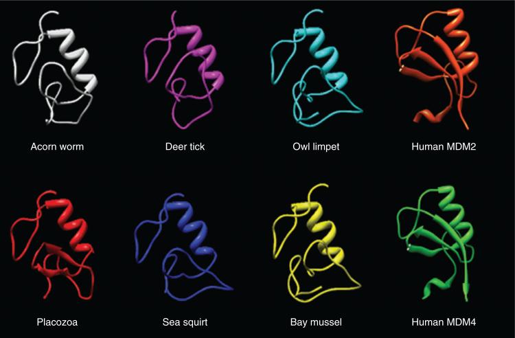
A structure modeling experiment was conducted to assess whether RING domains of invertebrate MDMs are more structurally similar to human MDM2 RING or human MDM4 RING. Multiple sequence alignment of full-length sequences between human MDM2 and invertebrate MDMs was generated and the regions with the highest degree of conservation were used for modeling studies. This region consists of ten residues flanking the first zinc coordinating Cys through 13 residues flanking the last zinc coordinating Cys. The invertebrate MDM sequences corresponding to this conserved region of human MDM2 were submitted to the automated structure homology modeling software program Swiss Model to create structure models (72–74). All invertebrate MDMs produced a structure model with the exception of lance-let MDM because it lacked sufficient sequence similarity to potential structure templates available to Swiss Model. The template automatically selected by the software program to build the homology models was the RING-H2 finger domain (PDB# 2kiz) from the human Arkadia the RING-H2 protein. The six invertebrate RING models are shown in Figure 4. RING domains from X-ray crystallography structures of MDM2 and MDM4 are shown for comparison. The alpha helices in the MDM models and MDM2/MDM4 structures are maintained. MDM2 and MDM4 RING's contain distinct regions with antiparallel β-strands. In contrast, the invertebrate MDM structure models, with the exception of placozoa MDM, lack β-strands. Spatial comparisons were made between the maximum number of protein backbone atoms of the MDM RING models shared with those of the crystal structures of MDM2 and MDM4. Structure/model comparisons were conducted by calculating the root mean square deviations (RMSDs) (Table 2). The consistent lower RMSDs in MDM2/MDM comparisons for all invertebrate structures indicate that MDM2 is more structurally similar to MDM than is MDM4. Importantly, one invertebrate MDM RING domain (deer tick) has a slightly higher sequence identity to MDM4 than to MDM2; yet, the lower RMSD value suggests that deer tick MDM RING domain appears to be more structurally similar to MDM2. The model/structure comparison suggests that invertebrate MDMs are more structurally similar to MDM2 than to MDM4.
Figure 4.
Models of RING domains of six invertebrate MDMs, X-ray structure of MDM2 RING, and X-ray structure of MDM4 RING.
Table 2.
Comparison of MDM RING models to MDM2 and MDM4 RING structures (PDB#: 2vje).
| MDM2 RMSD (Å) | MDM4 RMSD (Å) | Number of atoms compared | |
|---|---|---|---|
| Acorn worm | 6.489 | 7.930 | 403 |
| Sea squirt | 6.518 | 6.818 | 383 |
| Owl limpet | 6.808 | 7.296 | 402 |
| Placozoa | 7.377 | 8.724 | 427 |
| Bay mussel | 7.441 | 8.493 | 410 |
| Deer tick | 8.090 | 8.131 | 384 |
Summary
Our analyses indicate that MDM2 and MDM4 constitute a two-gene family (MDM2 gene family) that, in turn, belongs to the RING superfamily. Gene structure comparisons indicate that MDM2 and MDM4 are not closely related to other members of the RING superfamily. Cluster analysis of RING protein sequences further confirm that MDM2 and MDM4 evolved separately from other members of the superfamily. Other p53 targeting RING domain-containing genes, COP1, Pirh2, and MSL2 are not closely related to the MDM2 gene family. Furthermore, in invertebrates a single MDM gene is present. Invertebrate MDM RING protein sequence alignment and homology model structure comparisons to human MDM2 and human MDM4 suggests human MDM2 RING domain is more evolutionarily conserved than human MDM4.
Studies by Dehal and Boore (75) and others (76, 77) suggest that more than 440 million years ago two successive rounds of duplication (known as 2R) occurred in a common ancestor at the base of vertebrates. In accordance with this model starting from a single MDM gene, 2R would produce four paralogs of MDM genes. However, as four MDM paralogs are not detected in vertebrates one scenario to account for only two MDM paralogs in modern vertebrates is that a single paralog of MDM was deleted after the first round of duplication (after 1R). Another scenario is that two of the four paralogs were deleted after 2R. If the first scenario was correct, one would predict that lineages descended from an ancestor that emerged just after 2R would contain only two paralogs (i.e., MDM2 and MDM4). Cartilaginous fish are thought to descend from an ancestor shortly after 2R and one species of cartilaginous fish (elephant shark, Callorhinchus milii) codes for MDM2 and MDM4 (78). At the time of this communication, genome sequencing of organisms that trace back to an evolutionary window between 1R and 2R, such as lamprey, has not been completed. It will be interesting to see what MDM genes exist in the lamprey genome. If only one MDM is present, it would lend support to the scenario where a deletion event occurred after 1R. If two MDMs are present, then the data would lend support to the scenario in which two MDM paralogs were deleted after 2R.
Our analysis suggests that MDM proteins do not have the capability of forming oligomers. X-ray crystal structure studies show that heterodimerization between human MDM2 and human MDM4 occurs when three β-strands from one monomer and three β-strands from a second monomer form a β-barrel (67). These β strands are labeled β1, β2 and β3 in Figure 3; β2 and β3 bracket an α-helix. According to our modeling studies, the α-helix is preserved in the invertebrate MDMs but the β-strands are not, with the exception of placozoa in which two small β strands form in the approximate locations of β1 and β2, but not β3.
Mutation analyses of human MDM2 show that the C-terminal five residues of MDM2 are critical for oligomerization and E3 activity and that oligomerization can be restored by replacing the C-terminal seven residues of MDM2 with the C-terminal seven residues of MDM4 (79). Figure 3 shows a sequence alignment of human MDM2, human MDM4, and six invertebrate MDMs from the RING domain to the carboxyl terminal ends of the sequences (with the residues aligned to C-terminal seven MDM2 residues bordered). As our modeling studies show that the MDMs do not form the three β-strands necessary to form a β-barrel, we suggest that MDMs act as monomers (analogous to other RING domain proteins with E3 activity such as Pirh2, c-Cbl, PML, and CNOT4) and ubiquitylate p53 without dimerization. Upon duplication and subsequent mutation during evolution, vertebrate MDM2 and MDM4 may have gained the capability of dimerization.
We speculate that dimerization would have posed difficulties for MDMs with E3 ligase activities unless there were mutations that led to MDM2- and MDM4-specific RING domains and C-terminal residues. As the dimerization property was acquired, a potential problem for the early evolving MDM could have arisen. Currently, dimeric MDM2 has been shown to auto-ubiquitylate, which leads to self-degradation (67). MDM2 self-degradation incapacitates its ability to properly regulate p53. Fortunately, within vertebrates MDM2 self-degradation is prevented by forming heterodimers with MDM4. Upon hetero-dimerization, MDM4 K442 is ubiquitylated by MDM2, thus protecting MDM2 from self-destruction, which allows MDM2 to survive and properly regulate p53. We note that invertebrate MDMs, that we suggest are monomeric, do not possess lysine at this position (see blue bordered residue in Figure 3). If the invertebrate MDMs were dimeric E3 ligases, they could potentially encounter auto-ubiquitination problems analogous to homodimeric MDM2. Thus, we suggest that dimerization property evolved after MDM gene duplication.
Acknowledgments
We acknowledge grant number NIH 1T36GM078013 for support to J.M. and grant number NIH 5R25GM054939-10 for support to M.M. The authors declare no conflict of financial and/or other interest.
Biography
 Michael David Mendoza earned his BS degree in biochemistry from California State
University Los Angeles in 2011. He is currently a PhD student in the Biochemistry
and Molecular Biology program at the University of California, Riverside. His
interests lie within the field of cancer biology research and include signal
transduction pathways and protein-protein interactions.
 Garni Mandani earned his BS degree in biochemistry from California State University
Los Angeles in 2012. He is currently a masters student in the Chemistry and
Biochemistry Department at California State University Los Angeles. He is
investigating the evolutionary history of the p53 and MDM2 gene families. He is also
an employee at Grifols Biologicals where he works on purification of Factor IX.
 Jamil Momand is professor of biochemistry at California State University Los
Angeles. He earned his PhD degree in biochemistry from UCLA in 1989 and completed a
post-doctoral fellowship at Princeton University under the mentorship of Arnold
Levine. His research interests are focused on tumor suppressors and oncoproteins.
For more than 20 years he has worked on p53 and MDM2 and has contributed to
uncovering how these proteins are regulated and their evolutionary history.
References
- 1.Cahilly-Snyder L, Yang-Feng T, Francke U, George DL. Molecular analysis and chromosomal mapping of amplified genes isolated from a transformed mouse 3T3 cell line. Somat Cell Mol Genet. 1987;13:235–44. doi: 10.1007/BF01535205. [DOI] [PubMed] [Google Scholar]
- 2.Fakharzadeh SS, Rosenblum-Vos L, Murphy M, Hoffman EK, George DL. Structure and organization of amplified DNA on double minutes containing the mdm2 oncogene. Genomics. 1993;15:283–90. doi: 10.1006/geno.1993.1058. [DOI] [PubMed] [Google Scholar]
- 3.Fakharzadeh SS, Trusko SP, George DL. Tumorigenic potential associated with enhanced expression of a gene that is amplified in a mouse tumor cell line. EMBO J. 1991;10:1565–9. doi: 10.1002/j.1460-2075.1991.tb07676.x. [DOI] [PMC free article] [PubMed] [Google Scholar]
- 4.Oliner JD, Kinzler KW, Meltzer PS, George DL, Vogelstein B. Amplification of a gene encoding a p53-associated protein in human sarcomas. Nature. 1992;358:80–3. doi: 10.1038/358080a0. [DOI] [PubMed] [Google Scholar]
- 5.Momand J, Jung D, Wilczynski S, Niland J. The MDM2 gene amplification database. Nucleic Acids Res. 1998;26:3453–9. doi: 10.1093/nar/26.15.3453. [DOI] [PMC free article] [PubMed] [Google Scholar]
- 6.Araki S, Eitel JA, Batuello CN, Bijangi-Vishehsaraei K, Xie XJ, Danielpour D, Pollok KE, Boothman DA, Mayo LD. TGF-beta1-induced expression of human Mdm2 correlates with late-stage metastatic breast cancer. J Clin Invest. 2010;120:290–302. doi: 10.1172/JCI39194. [DOI] [PMC free article] [PubMed] [Google Scholar]
- 7.Bond GL, Hirshfield KM, Kirchhoff T, Alexe G, Bond EE, Robins H, Bartel F, Taubert H, Wuerl P, Hait W, Toppmeyer D, Offit K, Levine AJ. MDM2 SNP309 accelerates tumor formation in a gender-specific and hormone-dependent manner. Cancer Res. 2006;66:5104–10. doi: 10.1158/0008-5472.CAN-06-0180. [DOI] [PubMed] [Google Scholar]
- 8.Bond GL, Menin C, Bertorelle R, Alhopuro P, Aaltonen LA, Levine AJ. MDM2 SNP309 accelerates colorectal tumour formation in women. J Med Genet. 2006;43:950–2. doi: 10.1136/jmg.2006.043539. [DOI] [PMC free article] [PubMed] [Google Scholar]
- 9.Momand J, Zambetti GP, Olson DC, George D, Levine AJ. The mdm-2 oncogene product forms a complex with the p53 protein and inhibits p53-mediated transactivation. Cell. 1992;69:1237–45. doi: 10.1016/0092-8674(92)90644-r. [DOI] [PubMed] [Google Scholar]
- 10.Levine AJ. p53, the cellular gatekeeper for growth and division. Cell. 1997;88:323–31. doi: 10.1016/s0092-8674(00)81871-1. [DOI] [PubMed] [Google Scholar]
- 11.Haupt Y, Maya R, Kazaz A, Oren M. Mdm2 promotes the rapid degradation of p53. Nature. 1997;387:296–9. doi: 10.1038/387296a0. [DOI] [PubMed] [Google Scholar]
- 12.Kubbutat MH, Jones SN, Vousden KH. Regulation of p53 stability by Mdm2. Nature. 1997;387:299–303. doi: 10.1038/387299a0. [DOI] [PubMed] [Google Scholar]
- 13.Honda R, Tanaka H, Yasuda H. Oncoprotein MDM2 is a ubiquitin ligase E3 for tumor suppressor p53. FEBS Lett. 1997;420:25–7. doi: 10.1016/s0014-5793(97)01480-4. [DOI] [PubMed] [Google Scholar]
- 14.Wu X, Bayle JH, Olson D, Levine AJ. The p53-mdm-2 autoregulatory feedback loop. Genes Dev. 1993;7:1126–32. doi: 10.1101/gad.7.7a.1126. [DOI] [PubMed] [Google Scholar]
- 15.Barak Y, Gottlieb E, Juven-Gershon T, Oren M. Regulation of mdm2 expression by p53: alternative promoters produce transcripts with nonidentical translation potential. Genes Dev. 1994;8:1739–49. doi: 10.1101/gad.8.15.1739. [DOI] [PubMed] [Google Scholar]
- 16.Chen CY, Oliner JD, Zhan Q, Fornace AJ, Jr, Vogelstein B, Kastan MB. Interactions between p53 and MDM2 in a mammalian cell cycle checkpoint pathway. Proc Natl Acad Sci USA. 1994;91:2684–8. doi: 10.1073/pnas.91.7.2684. [DOI] [PMC free article] [PubMed] [Google Scholar]
- 17.Banin S, Moyal L, Shieh S, Taya Y, Anderson CW, Chessa L, Smorodinsky NI, Prives C, Reiss Y, Shiloh Y, Ziv Y. Enhanced phosphorylation of p53 by ATM in response to DNA damage. Science. 1998;281:1674–7. doi: 10.1126/science.281.5383.1674. [DOI] [PubMed] [Google Scholar]
- 18.Chehab NH, Malikzay A, Appel M, Halazonetis TD. Chk2/hCds1 functions as a DNA damage checkpoint in G(1) by stabilizing p53. Genes Dev. 2000;14:278–88. [PMC free article] [PubMed] [Google Scholar]
- 19.Chehab NH, Malikzay A, Stavridi ES, Halazonetis TD. Phosphorylation of Ser-20 mediates stabilization of human p53 in response to DNA damage. Proc Natl Acad Sci USA. 1999;96:13777–82. doi: 10.1073/pnas.96.24.13777. [DOI] [PMC free article] [PubMed] [Google Scholar]
- 20.Maya R, Balass M, Kim ST, Shkedy D, Leal JF, Shifman O, Moas M, Buschmann T, Ronai Z, Shiloh Y, Kastan MB, Katzir E, Oren M. ATM-dependent phosphorylation of Mdm2 on serine 395: role in p53 activation by DNA damage. Genes Dev. 2001;15:1067–77. doi: 10.1101/gad.886901. [DOI] [PMC free article] [PubMed] [Google Scholar]
- 21.Mayo LD, Turchi JJ, Berberich SJ. Mdm-2 phosphorylation by DNA-dependent protein kinase prevents interaction with p53. Cancer Res. 1997;57:5013–6. [PubMed] [Google Scholar]
- 22.Shieh SY, Ikeda M, Taya Y, Prives C. DNA damage-induced phosphorylation of p53 alleviates inhibition by MDM2. Cell. 1997;91:325–34. doi: 10.1016/s0092-8674(00)80416-x. [DOI] [PubMed] [Google Scholar]
- 23.Shinozaki T, Nota A, Taya Y, Okamoto K. Functional role of Mdm2 phosphorylation by ATR in attenuation of p53 nuclear export. Oncogene. 2003;22:8870–80. doi: 10.1038/sj.onc.1207176. [DOI] [PubMed] [Google Scholar]
- 24.Sionov RV, Coen S, Goldberg Z, Berger M, Bercovich B, Ben-Neriah Y, Ciechanover A, Haupt Y. c-Abl regulates p53 levels under normal and stress conditions by preventing its nuclear export and ubiquitination. Mol Cell Biol. 2001;21:5869–78. doi: 10.1128/MCB.21.17.5869-5878.2001. [DOI] [PMC free article] [PubMed] [Google Scholar]
- 25.Li M, Luo J, Brooks CL, Gu W. Acetylation of p53 inhibits its ubiquitination by Mdm2. J Biol Chem. 2002;277:50607–11. doi: 10.1074/jbc.C200578200. [DOI] [PubMed] [Google Scholar]
- 26.Zhang Y, Xiong Y, Yarbrough WG. ARF promotes MDM2 degradation and stabilizes p53: ARF-INK4a locus deletion impairs both the Rb and p53 tumor suppression pathways. Cell. 1998;92:725–34. doi: 10.1016/s0092-8674(00)81401-4. [DOI] [PubMed] [Google Scholar]
- 27.Kamijo T, Weber JD, Zambetti G, Zindy F, Roussel MF, Sherr CJ. Functional and physical interactions of the ARF tumor suppressor with p53 and Mdm2. Proc Natl Acad Sci USA. 1998;95:8292–7. doi: 10.1073/pnas.95.14.8292. [DOI] [PMC free article] [PubMed] [Google Scholar]
- 28.Roth J, Dobbelstein M, Freedman DA, Shenk T, Levine AJ. Nucleocytoplasmic shuttling of the hdm2 oncoprotein regulates the levels of the p53 protein via a pathway used by the human immunodeficiency virus rev protein. EMBO J. 1998;17:554–64. doi: 10.1093/emboj/17.2.554. [DOI] [PMC free article] [PubMed] [Google Scholar]
- 29.Wang X, Jiang X. Mdm2 and MdmX partner to regulate p53. FEBS Lett. 2012;586:1390–6. doi: 10.1016/j.febslet.2012.02.049. [DOI] [PubMed] [Google Scholar]
- 30.Kruse JP, Gu W. MSL2 promotes Mdm2-independent cytoplasmic localization of p53. J Biol Chem. 2009;284:3250–63. doi: 10.1074/jbc.M805658200. [DOI] [PMC free article] [PubMed] [Google Scholar]
- 31.Sherr CJ, Weber JD. The ARF/p53 pathway. Curr Opin Genet Dev. 2000;10:94–9. doi: 10.1016/s0959-437x(99)00038-6. [DOI] [PubMed] [Google Scholar]
- 32.Marine JC, Jochemsen AG. Mdmx as an essential regulator of p53 activity. Biochem Biophys Res Commun. 2005;331:750–60. doi: 10.1016/j.bbrc.2005.03.151. [DOI] [PubMed] [Google Scholar]
- 33.Leng RP, Lin Y, Ma W, Wu H, Lemmers B, Chung S, Parant JM, Lozano G, Hakem R, Benchimol S. Pirh2, a p53-induced ubiquitin-protein ligase, promotes p53 degradation. Cell. 2003;112:779–91. doi: 10.1016/s0092-8674(03)00193-4. [DOI] [PubMed] [Google Scholar]
- 34.Dornan D, Wertz I, Shimizu H, Arnott D, Frantz GD, Dowd P, O'Rourke K, Koeppen H, Dixit VM. The ubiquitin ligase COP1 is a critical negative regulator of p53. Nature. 2004;429:86–92. doi: 10.1038/nature02514. [DOI] [PubMed] [Google Scholar]
- 35.Shvarts A, Steegenga WT, Riteco N, van Laar T, Dekker P, Bazuine M, van Ham RC, van der Houven van Oordt W, Hateboer G, van der Eb AJ, Jochemsen AG. MDMX: a novel p53-binding protein with some functional properties of MDM2. EMBO J. 1996;15:5349–57. [PMC free article] [PubMed] [Google Scholar]
- 36.Toledo F, Wahl GM. Regulating the p53 pathway: in vitro hypotheses, in vivo veritas. Nat Rev Cancer. 2006;6:909–23. doi: 10.1038/nrc2012. [DOI] [PubMed] [Google Scholar]
- 37.Laurie NA, Donovan SL, Shih CS, Zhang J, Mills N, Fuller C, Teunisse A, Lam S, Ramos Y, Mohan A, Johnson D, Wilson M, Rodriguez-Galindo C, Quarto M, Francoz S, Mendrysa SM, Guy RK, Marine JC, Jochemsen AG, Dyer MA. Inactivation of the p53 pathway in retinoblastoma. Nature. 2006;444:61–6. doi: 10.1038/nature05194. [DOI] [PubMed] [Google Scholar]
- 38.Momand J, Aspuria PJ, Furuta S. MDM2 and MDMX regulators of p53 activity. In: Zambetti GP, editor. The p53 tumor suppressor pathway and cancer. protein reviews. Springer; New York: 2005. pp. 155–85. [Google Scholar]
- 39.Kamal A, Mohammed AA, Shaik TB. p53-Mdm2 inhibitors: patent review (2009–2010). Expert Opin Ther Pat 2012. 22:95–105. doi: 10.1517/13543776.2012.656593. [DOI] [PubMed] [Google Scholar]
- 40.Pei D, Zhang Y, Zheng J. Regulation of p53: a collaboration between Mdm2 and Mdmx. Oncotarget. 2012;3:228–35. doi: 10.18632/oncotarget.443. [DOI] [PMC free article] [PubMed] [Google Scholar]
- 41.Vassilev LT. MDM2 inhibitors for cancer therapy. Trends Mol Med. 2007;13:23–31. doi: 10.1016/j.molmed.2006.11.002. [DOI] [PubMed] [Google Scholar]
- 42.Tanimura S, Ohtsuka S, Mitsui K, Shirouzu K, Yoshimura A, Ohtsubo M. MDM2 interacts with MDMX through their RING finger domains. FEBS Lett. 1999;447:5–9. doi: 10.1016/s0014-5793(99)00254-9. [DOI] [PubMed] [Google Scholar]
- 43.Marine JC, Francoz S, Maetens M, Wahl G, Toledo F, Lozano G. Keeping p53 in check: essential and synergistic functions of Mdm2 and Mdm4. Cell Death Differ. 2006;13:927–34. doi: 10.1038/sj.cdd.4401912. [DOI] [PubMed] [Google Scholar]
- 44.Wang X, Wang J, Jiang X. MdmX protein is essential for Mdm2 protein-mediated p53 polyubiquitination. J Biol Chem. 2011;286:23725–34. doi: 10.1074/jbc.M110.213868. [DOI] [PMC free article] [PubMed] [Google Scholar]
- 45.Belyi VA, Ak P, Markert E, Wang H, Hu W, Puzio-Kuter A, Levine AJ. The origins and evolution of the p53 family of genes. Cold Spring Harb Perspect Biol. 2010;2:a001198. doi: 10.1101/cshperspect.a001198. [DOI] [PMC free article] [PubMed] [Google Scholar]
- 46.Momand J, Villegas A, Belyi VA. The evolution of MDM2 family genes. Gene. 2011;486:23–30. doi: 10.1016/j.gene.2011.06.030. [DOI] [PMC free article] [PubMed] [Google Scholar]
- 47.Hershko A, Ciechanover A. The ubiquitin system. Annu Rev Biochem. 1998;67:425–79. doi: 10.1146/annurev.biochem.67.1.425. [DOI] [PubMed] [Google Scholar]
- 48.Wenzel DM, Stoll KE, Klevit RE. E2s: structurally economical and functionally replete. Biochem J. 2011;433:31–42. doi: 10.1042/BJ20100985. [DOI] [PMC free article] [PubMed] [Google Scholar]
- 49.Deshaies RJ, Joazeiro CA. RING domain E3 ubiquitin ligases. Annu Rev Biochem. 2009;78:399–434. doi: 10.1146/annurev.biochem.78.101807.093809. [DOI] [PubMed] [Google Scholar]
- 50.Weissman AM. Themes and variations on ubiquitylation. Nat Rev Mol Cell Biol. 2001;2:169–78. doi: 10.1038/35056563. [DOI] [PubMed] [Google Scholar]
- 51.Koegl M, Hoppe T, Schlenker S, Ulrich HD, Mayer TU, Jentsch S. A novel ubiquitination factor, E4, is involved in multiubiquitin chain assembly. Cell. 1999;96:635–44. doi: 10.1016/s0092-8674(00)80574-7. [DOI] [PubMed] [Google Scholar]
- 52.Ozkan E, Yu H, Deisenhofer J. Mechanistic insight into the allosteric activation of a ubiquitin-conjugating enzyme by RING-type ubiquitin ligases. Proc Natl Acad Sci USA. 2005;102:18890–5. doi: 10.1073/pnas.0509418102. [DOI] [PMC free article] [PubMed] [Google Scholar]
- 53.Kawai H, Lopez-Pajares V, Kim MM, Wiederschain D, Yuan ZM. RING domain-mediated interaction is a requirement for MDM2's E3 ligase activity. Cancer Res. 2007;67:6026–30. doi: 10.1158/0008-5472.CAN-07-1313. [DOI] [PubMed] [Google Scholar]
- 54.Borden KL, Boddy MN, Lally J, O'Reilly NJ, Martin S, Howe K, Solomon E, Freemont PS. The solution structure of the RING finger domain from the acute promyelocytic leukaemia protooncoprotein PML. EMBO J. 1995;14:1532–41. doi: 10.1002/j.1460-2075.1995.tb07139.x. [DOI] [PMC free article] [PubMed] [Google Scholar]
- 55.Dominguez C, Bonvin AM, Winkler GS, van Schaik FM, Timmers HT, Boelens R. Structural model of the UbcH5B/CNOT4 complex revealed by combining NMR, mutagenesis, and docking approaches. Structure. 2004;12:633–44. doi: 10.1016/j.str.2004.03.004. [DOI] [PubMed] [Google Scholar]
- 56.Shloush J, Vlassov JE, Engson I, Duan S, Saridakis V, Dhe-Paganon S, Raught B, Sheng Y, Arrowsmith CH. Structural and functional comparison of the RING domains of two p53 E3 ligases, Mdm2 and Pirh2. J Biol Chem. 2011;286:4796–808. doi: 10.1074/jbc.M110.157669. [DOI] [PMC free article] [PubMed] [Google Scholar]
- 57.Zheng N, Wang P, Jeffrey PD, Pavletich NP. Structure of a c-Cbl-UbcH7 complex: RING domain function in ubiquitin-protein ligases. Cell. 2000;102:533–9. doi: 10.1016/s0092-8674(00)00057-x. [DOI] [PubMed] [Google Scholar]
- 58.Brzovic PS, Rajagopal P, Hoyt DW, King MC, Klevit RE. Structure of a BRCA1-BARD1 heterodimeric RING-RING complex. Nat Struc Biol. 2001;8:833–7. doi: 10.1038/nsb1001-833. [DOI] [PubMed] [Google Scholar]
- 59.Buchwald G, van der Stoop P, Weichenrieder O, Perrakis A, van Lohuizen M, Sixma TK. Structure and E3-ligase activity of the Ring-Ring complex of polycomb proteins Bmi1 and Ring1b. EMBO J. 2006;25:2465–74. doi: 10.1038/sj.emboj.7601144. [DOI] [PMC free article] [PubMed] [Google Scholar]
- 60.Mace PD, Linke K, Feltham R, Schumacher FR, Smith CA, Vaux DL, Silke J, Day CL. Structures of the cIAP2 RING domain reveal conformational changes associated with ubiquitinconjugating enzyme (E2) recruitment. J Biol Chem. 2008;283:31633–40. doi: 10.1074/jbc.M804753200. [DOI] [PubMed] [Google Scholar]
- 61.Yi C, Deng XW. COP1–from plant photomorphogenesis to mammalian tumorigenesis. Trends Cell Biol. 2005;15:618–25. doi: 10.1016/j.tcb.2005.09.007. [DOI] [PubMed] [Google Scholar]
- 62.Beitel LK, Elhaji YA, Lumbroso R, Wing SS, Panet-Raymond V, Gottlieb B, Pinsky L, Trifiro MA. Cloning and characterization of an androgen receptor N-terminal-interacting protein with ubiquitin-protein ligase activity. J Mol Endocrinol. 2002;29:41–60. doi: 10.1677/jme.0.0290041. [DOI] [PubMed] [Google Scholar]
- 63.Sheng Y, Laister RC, Lemak A, Wu B, Tai E, Duan S, Lukin J, Sunnerhagen M, Srisailam S, Karra M, Benchimol S, Arrowsmith CH. Molecular basis of Pirh2-mediated p53 ubiquitylation. Nat Struct Mol Biol. 2008;15:1334–42. doi: 10.1038/nsmb.1521. [DOI] [PMC free article] [PubMed] [Google Scholar]
- 64.Li Q, Lin S, Wang X, Lian G, Lu Z, Guo H, Ruan K, Wang Y, Ye Z, Han J, Lin SC. Axin determines cell fate by controlling the p53 activation threshold after DNA damage. Nat Cell Biol. 2009;11:1128–34. doi: 10.1038/ncb1927. [DOI] [PubMed] [Google Scholar]
- 65.Duan S, Yao Z, Hou D, Wu Z, Zhu WG, Wu M. Phosphorylation of Pirh2 by calmodulin-dependent kinase II impairs its ability to ubiquitinate p53. EMBO J. 2007;26:3062–74. doi: 10.1038/sj.emboj.7601749. [DOI] [PMC free article] [PubMed] [Google Scholar]
- 66.Kostic M, Matt T, Martinez-Yamout MA, Dyson HJ, Wright PE. Solution structure of the Hdm2 C2H2C4 RING, a domain critical for ubiquitination of p53. J Mol Biol. 2006;363:433–50. doi: 10.1016/j.jmb.2006.08.027. [DOI] [PubMed] [Google Scholar]
- 67.Linke K, Mace PD, Smith CA, Vaux DL, Silke J, Day CL. Structure of the MDM2/MDMX RING domain heterodimer reveals dimerization is required for their ubiquitylation in trans. Cell Death Differ. 2008;15:841–8. doi: 10.1038/sj.cdd.4402309. [DOI] [PubMed] [Google Scholar]
- 68.Saitou N, Nei M. The neighbor-joining method: a new method for reconstructing phylogenetic trees. Mol Biol Evol. 1987;4:406–25. doi: 10.1093/oxfordjournals.molbev.a040454. [DOI] [PubMed] [Google Scholar]
- 69.Fang S, Jensen JP, Ludwig RL, Vousden KH, Weissman AM. Mdm2 is a RING finger-dependent ubiquitin protein ligase for itself and p53. J Biol Chem. 2000;275:8945–51. doi: 10.1074/jbc.275.12.8945. [DOI] [PubMed] [Google Scholar]
- 70.Honda R, Yasuda H. Activity of MDM2, a ubiquitin ligase, toward p53 or itself is dependent on the RING finger domain of the ligase. Oncogene. 2000;19:1473–6. doi: 10.1038/sj.onc.1203464. [DOI] [PubMed] [Google Scholar]
- 71.Singh RK, Iyappan S, Scheffner M. Hetero-oligomerization with MdmX rescues the ubiquitin/Nedd8 ligase activity of RING finger mutants of Mdm2. J Biol Chem. 2007;282:10901–7. doi: 10.1074/jbc.M610879200. [DOI] [PubMed] [Google Scholar]
- 72.Peitsch MC. Protein modeling by e-mail. Bio/Technology. 1995;13:658–60. [Google Scholar]
- 73.Arnold K, Bordoli L, Kopp J, Schwede T. The SWISS-MODEL workspace: a web-based environment for protein structure homology modelling. Bioinformatics. 2006;22:195–201. doi: 10.1093/bioinformatics/bti770. [DOI] [PubMed] [Google Scholar]
- 74.Kiefer F, Arnold K, Kunzli M, Bordoli L, Schwede T. The SWISS-MODEL Repository and associated resources. Nucleic Acids Res. 2009;37:D387–92. doi: 10.1093/nar/gkn750. [DOI] [PMC free article] [PubMed] [Google Scholar]
- 75.Dehal P, Boore JL. Two rounds of whole genome duplication in the ancestral vertebrate. PLoS Biol. 2005;3:e314. doi: 10.1371/journal.pbio.0030314. [DOI] [PMC free article] [PubMed] [Google Scholar]
- 76.Kasahara M. The 2R hypothesis: an update. Curr Opin Immunol. 2007;19:547–52. doi: 10.1016/j.coi.2007.07.009. [DOI] [PubMed] [Google Scholar]
- 77.Ohno S. Evolution by gene duplication. Springer-Verlag; New York: 1970. [Google Scholar]
- 78.Lane DP, Madhumalar A, Lee AP, Tay BH, Verma C, Brenner S, Venkatesh B. Conservation of all three p53 family members and Mdm2 and Mdm4 in the cartilaginous fish. Cell Cycle. 2011;10:4272–9. doi: 10.4161/cc.10.24.18567. [DOI] [PMC free article] [PubMed] [Google Scholar]
- 79.Poyurovsky MV, Priest C, Kentsis A, Borden KL, Pan ZQ, Pavletich N, Prives C. The Mdm2 RING domain C-terminus is required for supramolecular assembly and ubiquitin ligase activity. EMBO J. 2007;26:90–101. doi: 10.1038/sj.emboj.7601465. [DOI] [PMC free article] [PubMed] [Google Scholar]
- 80.Budhidarmo R, Nakatani Y, Day CL. RINGs hold the key to ubiquitin transfer. Trends Biochem Sci. 2012;37:58–65. doi: 10.1016/j.tibs.2011.11.001. [DOI] [PubMed] [Google Scholar]
- 81.Goujon M, McWilliam H, Li W, Valentin F, Squizzato S, Paern J, Lopez R. A new bioinformatics analysis tools framework at EMBL-EBI. Nucleic Acids Res. 2010;38:W695–9. doi: 10.1093/nar/gkq313. [DOI] [PMC free article] [PubMed] [Google Scholar]
- 82.Sievers F, Wilm A, Dineen D, Gibson TJ, Karplus K, Li W, Lopez R, McWilliam H, Remmert M, Söding J, Thompson JD, Higgins DG. Fast, scalable generation of high-quality protein multiple sequence alignments using Clustal Omega. Mol Syst Biol. 2011;7:539. doi: 10.1038/msb.2011.75. [DOI] [PMC free article] [PubMed] [Google Scholar]


