Abstract
Complications aroused from Meckel’s diverticulum tend to developed in children. Children presented with abdominal pain, intestinal obstruction, intussusception or gastrointestinal bleeding may actually suffered from complicated Meckel’s diverticulum. With the advancement of minimally invasive surgery (MIS) in children, the use of laparoscopy in the diagnosis and subsequent laparoscopic excision of Meckel’s diverticulum has gained popularity. Recently, single incision laparoscopic surgery (SILS) has emerged as a new technique in minimally invasive surgery. This review offers the overview in the development of MIS in the management of children suffered from Meckel’s diverticulum. The current evidence in different laparoscopic techniques, including conventional laparoscopy, SILS, the use of special laparoscopic instruments, intracorporeal diverticulectomy and extracorporeal diverticulectomy in the management of Meckel’s diverticulum in children were revealed.
Keywords: Laparoscopy, Meckel’s diverticulum, Children
Core tip: With the advances in the development of minimally invasive surgery in children, laparoscopic excision of Meckel’s diverticulum has gained popularity. New technique including single incision laparoscopic surgery (SILS) in the management of Meckel’s diverticulum was reported. This review offers the overview of the current evidence in different laparoscopic techniques, including conventional laparoscopy, SILS, the use of special laparoscopic instruments, intracorporeal diverticulectomy and extracorporeal diverticulectomy in the management of Meckel’s diverticulum in children.
INTRODUCTION
Complications aroused from Meckel’s diverticulum included gastrointestinal bleeding, intussusception, intestinal obstruction, abdominal pain and incarcerated hernia[1]. These complications tend to develop in children[2].
During the past decades, there has been a tremendous development of minimally invasive surgery in children[3-6]. Increasing number of reports on the use of laparoscopy in children with Meckel’s diverticulum was observed[1,7-13]. Recently, single incision laparoscopic surgery (SILS) has emerged as an evolution from conventional laparoscopy (CL)[14]. This review provides the updated and the current evidence in the laparoscopic management of Meckel’s diverticulum in children.
USE OF LAPAROSCOPY IN THE DIAGNOSIS OF COMPLICATED MECKEL’S DIVERTICULUM
Bleeding is the most common presentation of Meckel’s diverticulum in children[15]. Tc-99 m pertechnetate scan has been the standard investigation in children with Meckel’s diverticulum containing ectopic gastric mucosa[16]. Pre-medication with histamine-2 blockers (ranitidine) has been reported to increase the diagnostic yield[16]. The reported sensitivity of the Meckel’s scan varies from 60% to 90 % with the specificity varies from 90% to 98%[17-20]. Sinha et al[21] recently reported the sensitivity and specificity of the Meckel’s scan for ectopic gastric mucosa were 94% and 97 %, respectively.
In children with persistent symptoms despite a negative Meckel’s scan, laparoscopy is still warrant to exclude underlying small bowel anomalies (Figure 1). Lee et al[22] reported the use of laparoscopy in children with gastrointestinal bleeding of obscure origin. 8 out the 17 children had Meckel’s diverticulum identified by the laparoscopy. 4 children in the study had a positive Meckel’s scan.
Figure 1.
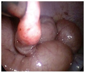
Laparoscopic view of a Meckel’s diverticulum in a child with gastrointestinal bleeding.
In the reports studying the use of laparoscopy in the management of Meckel’s diverticulum in children, Meckel’s scan was arranged as the pre-operative investigation in children presented with gastrointestinal bleeding. The results were listed in Table 1.
Table 1.
Results of Meckel’s scan in reports studying laparoscopic management of Meckel’s diverticulum in children
Besides gastrointestinal bleeding, children with complicated Meckel’s diverticulum may present as acute abdomen. Diverticulitis, intussusception and intestinal obstruction were the underlying causes identified by laparoscopy[1]. Clinicians should have a high index of suspicion of the exact pathology in children presented with abdominal pain.
CONVENTIONAL LAPAROSCOPY
It has been 20 years since the first reports describing the use of laparoscopy in the management of Meckel’s diverticulum in children. Case reports on laparoscopic diverticulectomy in infants were published since 1993. Huang et al[23] safely performed the operation in 3 symptomatic infants. There was an increasing number of case reports published since then. Some reports focused on the use of laparoscopy in the management of Meckel’s diverticulum only[7,8]. Others focused on the management on the acute abdominal condition in children including gastrointestinal bleeding or small bowel obstruction in which Meckel’s diverticulum was one of the underlying cause[22].
Although Meckel’s diverticulum is the commonest congenital anomalies of the gastrointestinal tract and the complications tends to occurred in children, each pediatric surgical centers may only handled few cases per year. Since 2005, a number of case series were published[1,9,11]. Each series included more than 20 patients underwent laparoscopy in the management of Meckel’s diverticulum. The patients’ inclusion criteria of these studies were different. Some included both symptomatic and asymptomatic children while other only included symptomatic patients.
Throughout these 20 years, the set-up of the laparoscopic procedure is essentially the same[1,9,11,23]. In younger infants, the laparoscope was inserted at the subumbilical 5 mm port. Additional 5 mm ports were inserted at left and right lower quadrant of the abdomen respectively (Figure 2). In older children, 10 mm or 12 mm ports were used. The procedure started from identifying the cecum. The small intestine was then examined from the terminal ileum toward the jejunum. The Meckel’s diverticulectomy were then performed intracorporeally or extracorporeally. Practically, the procedure did not required advance laparoscopic skills since laparoscopic intracorporeal suturing did not required. All reports concluded laparoscopy was safe and effective in the management of Meckel’s diverticulum[1,9-11].
Figure 2.
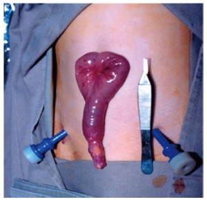
Meckel’s diverticulum was exteriorized through the umbilical wound in conventional laparoscopy.
SILS
The reports of single-incision or SILS in children were published since 2008. Initial reports studied the safety and feasibility of SILS in various pediatric condition included appendectomy, cholecystectomy, varicocelectomy[17]. Cobellis et al[24] used a 10-mm working laparoscope in the management of Meckel diverticulum since 2001. He only used 1 trocar and the Meckel’s diverticulum was grasped and delivered through the umbilical wound. Meckel’s diverticulectomy were performed extracorporeally. This may represent the “first” series of single incision laparoscopic surgery for Meckel’s diverticulum in children.
Using the current concept of SILS, laparoscope and the instruments were inserted through a multilumen port or through multiple ports that were inserted over the same fascial plane[25] (Figure 3). Tam et al[26] reported the experience in single incision umbilical laparoscopic segmental small bowel resection. The study included 2 patients with Meckel’s diverticulum. In conventional laparoscopic technique, the umbilical wound required extension in order to facilitate segmental resection of the small intestine and the diverticulum. In SILS, the umbilical wound did not required further extension (Figure 4) and good cosmesis can be achieved (Figure 5).
Figure 3.
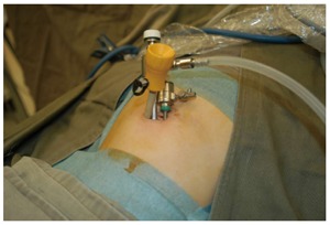
Single incision laparoscopic surgery setup. Three ports were inserted over the dame fascial plane.
Figure 4.
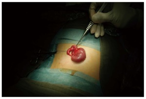
Meckel’s diverticulum was exteriorized through the umbilical wound in single incision laparoscopic surgery.
Figure 5.
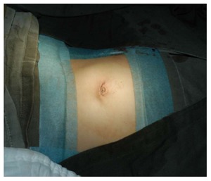
Appearance of the umbilical wound after single incision laparoscopic surgery for Meckel’s diverticulum.
Because the development of SILS is still at its infancy, limited numbers of reports on SILS in the management of Meckel’s diverticulum were available[26]. There is no case series of SILS in the management of Meckel’s diverticulum. Isolated cases of SILS in Meckels’ diverticulum were reported in studies focused on the experience of SILS in children[27,28], or in studies focused on the use of SILS in segmental small bowel resection and in studies focused in the use of SILS in the management of obscure gastrointestinal bleeding in children[26,29]. Although in SILS, specially designed instruments including flexible laparoscope or instrument were developed to facilitate the operation, in all reported cases of SILS in Meckel’s diverticulum, conventional laparoscopic instruments were adopted[26-29].
INTRACORPOREAL DIVERTICULECTOMY VS EXTRACORPOREAL DIVERTICULECTOMY
Meckel’s diverticulectomy can be performed either intracorporeally or extracorporeally. Intracorporeal diverticulectomy can be performed total laparoscopically without the need to enlarge the umbilical wound. The use of endoscopic staplers was the most frequent reported device used in diverticulectomy[7,23]. Schier et al[30], on the other hand, reported the use of endoloop and divided the diverticulum as in appendicectomy. The early reports on laparoscopic assisted diverticulectomy favored the intracorporeal method.
One of the concerns in total laparoscopic intracorporeally diverticulectomy was failure to perform segmental resection of Meckel’s diverticulum. Isolated diverticulectomy bears the risk of leaving ectopic gastric mucosa in the adjacent ileum. Varcoe advised the use the height-to-diameter ratio to determine the method of resection[31]. If the ratio is less than 2, there will be an increased risk in having the ectopic mucosa in the adjacent ileum, segmental resection will be advised.
In extracorporeal diverticulectomy, the Meckel’s diverticulum was delivered through an enlarged umbilical wound. Segmental resection of the Meckel’s diverticulum and the adjacent ileum were carried out. Hand-sewn small to small intestine anastomosis were performed[1,12]. The principle of surgery is the same in conventional or SILS approach. The use of expensive laparoscopic staplers was avoided[9]. Ng et al[32] described the use of laparoscopic assisted extracorporeal Meckel’s diverticulectomy with endo GIA stapler and side-to-side ileal anastomosis. However, this method did not gain popularity.
SAFETY ISSUES ON LAPAROSCOPIC ASSISTED MECKEL’S DIVERTICULECTOMY
Park et al[2] reported the Mayo clinic experience with 1476 patients with Meckel’s diverticulum. The study included children and adult and did not specifically define the approach of operation. Post-operative complication in the most 100 resected diverticulum was 13% in symptomatic group and 20% in asymptomatic group It is reported that the rate for postdiverticulectomy adhesive intestinal obstruction could be as high as 10% to 15% in children[33].
Intraoperative complication after laparoscopic assisted diverticulectomy was never reported in the literature. Sai Prasad et al[12] reported 8% of patients had intestinal obstruction after the operation. He suggested that the adhesion is secondary to ischemic bowel after release of the obstruction. Other studies did not show any intestinal obstruction post-operatively[1,9]. They all concluded laparoscopic assisted Meckel’s diverticulectomy was safe and feasible with good outcome.
LAPAROSCOPIC MANAGEMENT OF MECKEL’S DIVERTICULUM IN ADULT
Since majority of complicated Meckel’s diverticulum occurred in childhood, there were only few case series in reporting the use of laparoscopy in the management of Meckel’s diverticulum in adult[34,35]. Ding et al[34] reported 15 cases of perforated Meckel’s diverticulum managed by laparoscopy. They reported the use of laparoscopic assisted extracorporeally resection of the diverticulum if the base was > 2 cm. If the base of the diverticulum was < 2 cm, they would performed laparoscopic intracorporeally excision with laparoscopic staplers.
Rivas et al[35] reported their experience in the laparoscopic management of Meckel’s diverticulum. In his study, laparoscopic assisted extracorporeal excision of the diverticulum was adopted in all the cases.
CONCLUSION
Laparoscopy has emerged as an important tool in the diagnosis and management of children with Meckel’s diverticulum. Conventional laparoscopy or single site incision surgery were reported to be safe and feasible in Meckel’s diverticulectomy. Extracorporeal diverticulectomy was in favors than intracorporeal diverticulectomy in reports series because segmental resection of the Meckel’s diverticulum can be performed without the use of expensive laparoscopic staplers. Complete excision of the diverticulum and the adjacent intestine containing ectopic mucosa is essential to minimize the chance of recurrent symptom.
Footnotes
P- Reviewer: Fabozzi M, Herbella FAM, Li B, Tanyi M S- Editor: Ma YJ L- Editor: A E- Editor: Wang CH
References
- 1.Chan KW, Lee KH, Mou JW, Cheung ST, Tam YH. Laparoscopic management of complicated Meckel’s diverticulum in children: a 10-year review. Surg Endosc. 2008;22:1509–1512. doi: 10.1007/s00464-008-9832-0. [DOI] [PubMed] [Google Scholar]
- 2.Park JJ, Wolff BG, Tollefson MK, Walsh EE, Larson DR. Meckel diverticulum: the Mayo Clinic experience with 1476 patients (1950-2002) Ann Surg. 2005;241:529–533. doi: 10.1097/01.sla.0000154270.14308.5f. [DOI] [PMC free article] [PubMed] [Google Scholar]
- 3.Chan KW, Lee KH, Tam YH, Yeung CK. Minimal invasive surgery in pediatric solid tumors. J Laparoendosc Adv Surg Tech A. 2007;17:817–820. doi: 10.1089/lap.2007.0033. [DOI] [PubMed] [Google Scholar]
- 4.Lee KH, Tam YH, Yeung CK, Chan KW, Sihoe JD, Cheung ST, Mou JW. Laparoscopic excision of choledochal cysts in children: an intermediate-term report. Pediatr Surg Int. 2009;25:355–360. doi: 10.1007/s00383-009-2343-9. [DOI] [PubMed] [Google Scholar]
- 5.Tam YH, Lee KH, Sihoe JD, Chan KW, Wong PY, Cheung ST, Mou JW. Laparoscopic hernia repair in children by the hook method: a single-center series of 433 consecutive patients. J Pediatr Surg. 2009;44:1502–1505. doi: 10.1016/j.jpedsurg.2008.10.071. [DOI] [PubMed] [Google Scholar]
- 6.Chan KW, Lee KH, Mou JW, Cheung ST, Tam YH. The outcome of laparoscopic portoenterostomy for biliary atresia in children. Pediatr Surg Int. 2011;27:671–674. doi: 10.1007/s00383-011-2859-7. [DOI] [PubMed] [Google Scholar]
- 7.Teitelbaum DH, Polley TZ, Obeid F. Laparoscopic diagnosis and excision of Meckel’s diverticulum. J Pediatr Surg. 1994;29:495–497. doi: 10.1016/0022-3468(94)90075-2. [DOI] [PubMed] [Google Scholar]
- 8.Valla JS, Steyaert H, Leculée R, Pebeyre B, Jordana F. Meckel’s diverticulum and laparoscopy of children. What’s new? Eur J Pediatr Surg. 1998;8:26–28. doi: 10.1055/s-2008-1071114. [DOI] [PubMed] [Google Scholar]
- 9.Shalaby RY, Soliman SM, Fawy M, Samaha A. Laparoscopic management of Meckel’s diverticulum in children. J Pediatr Surg. 2005;40:562–567. doi: 10.1016/j.jpedsurg.2004.11.032. [DOI] [PubMed] [Google Scholar]
- 10.Craigie RJ, Forrest N, Nanthakumaran S, Mahomed AA. Laparoscopy in diagnosis and management of Meckel’s diverticulum. J Laparoendosc Adv Surg Tech A. 2006;16:70–73. doi: 10.1089/lap.2006.16.70. [DOI] [PubMed] [Google Scholar]
- 11.Prasad TR, Chui CH, Jacobsen AS. Laparoscopic-assisted resection of Meckel’s diverticulum in children. JSLS. 2006;10:310–316. [PMC free article] [PubMed] [Google Scholar]
- 12.Sai Prasad TR, Chui CH, Singaporewalla FR, Ong CP, Low Y, Yap TL, Jacobsen AS. Meckel’s diverticular complications in children: is laparoscopy the order of the day? Pediatr Surg Int. 2007;23:141–147. doi: 10.1007/s00383-006-1844-z. [DOI] [PubMed] [Google Scholar]
- 13.Palanivelu C, Rangarajan M, Senthilkumar R, Madankumar MV, Kavalakat AJ. Laparoscopic management of symptomatic Meckel’s diverticula: a simple tangential stapler excision. JSLS. 2008;12:66–70. [PMC free article] [PubMed] [Google Scholar]
- 14.Ponsky TA, Diluciano J, Chwals W, Parry R, Boulanger S. Early experience with single-port laparoscopic surgery in children. J Laparoendosc Adv Surg Tech A. 2009;19:551–553. doi: 10.1089/lap.2009.0092. [DOI] [PubMed] [Google Scholar]
- 15.Vane DW, West KW, Grosfeld JL. Vitelline duct anomalies. Experience with 217 childhood cases. Arch Surg. 1987;122:542–547. doi: 10.1001/archsurg.1987.01400170048007. [DOI] [PubMed] [Google Scholar]
- 16.Rerksuppaphol S, Hutson JM, Oliver MR. Ranitidine-enhanced 99mtechnetium pertechnetate imaging in children improves the sensitivity of identifying heterotopic gastric mucosa in Meckel’s diverticulum. Pediatr Surg Int. 2004;20:323–325. doi: 10.1007/s00383-004-1189-4. [DOI] [PubMed] [Google Scholar]
- 17.Poulsen KA, Qvist N. Sodium pertechnetate scintigraphy in detection of Meckel’s diverticulum: is it usable? Eur J Pediatr Surg. 2000;10:228–231. doi: 10.1055/s-2008-1072364. [DOI] [PubMed] [Google Scholar]
- 18.Tseng YY, Yang YJ. Clinical and diagnostic relevance of Meckel’s diverticulum in children. Eur J Pediatr. 2009;168:1519–1523. doi: 10.1007/s00431-009-1023-z. [DOI] [PubMed] [Google Scholar]
- 19.Cooney DR, Duszynski DO, Camboa E, Karp MP, Jewett TC. The abdominal technetium scan (a decade of experience) J Pediatr Surg. 1982;17:611–619. doi: 10.1016/s0022-3468(82)80121-8. [DOI] [PubMed] [Google Scholar]
- 20.Mariani G, Pauwels EK, AlSharif A, Marchi S, Boni G, Barreca M, Bellini M, Grosso M, de Bortoli N, Mumolo G, et al. Radionuclide evaluation of the lower gastrointestinal tract. J Nucl Med. 2008;49:776–787. doi: 10.2967/jnumed.107.040113. [DOI] [PubMed] [Google Scholar]
- 21.Sinha CK, Pallewatte A, Easty M, De Coppi P, Pierro A, Misra D, Biassoni L. Meckel’s scan in children: a review of 183 cases referred to two paediatric surgery specialist centres over 18 years. Pediatr Surg Int. 2013;29:511–517. doi: 10.1007/s00383-013-3270-3. [DOI] [PubMed] [Google Scholar]
- 22.Lee KH, Yeung CK, Tam YH, Ng WT, Yip KF. Laparascopy for definitive diagnosis and treatment of gastrointestinal bleeding of obscure origin in children. J Pediatr Surg. 2000;35:1291–1293. doi: 10.1053/jpsu.2000.9299. [DOI] [PubMed] [Google Scholar]
- 23.Huang CS, Lin LH. Laparoscopic Meckel’s diverticulectomy in infants: report of three cases. J Pediatr Surg. 1993;28:1486–1489. doi: 10.1016/0022-3468(93)90437-p. [DOI] [PubMed] [Google Scholar]
- 24.Cobellis G, Cruccetti A, Mastroianni L, Amici G, Martino A. One-trocar transumbilical laparoscopic-assisted management of Meckel’s diverticulum in children. J Laparoendosc Adv Surg Tech A. 2007;17:238–241. doi: 10.1089/lap.2006.0036. [DOI] [PubMed] [Google Scholar]
- 25.Krpata DM, Ponsky TA. Instrumentation and equipment for single-site umbilical laparoscopic surgery. Semin Pediatr Surg. 2011;20:190–195. doi: 10.1053/j.sempedsurg.2011.05.002. [DOI] [PubMed] [Google Scholar]
- 26.Tam YH, Chan KW, Wong YS, Houben CH, Pang KK, Tsui SY, Mou JW, Lee KH. Single-incision laparoscopic surgery in diagnosis and treatment for gastrointestinal bleeding of obscure origin in children. Surg Laparosc Endosc Percutan Tech. 2013;23:e106–e108. doi: 10.1097/SLE.0b013e3182806517. [DOI] [PubMed] [Google Scholar]
- 27.Tam YH, Lee KH, Sihoe JD, Chan KW, Cheung ST, Pang KK. Initial experience in children using conventional laparoscopic instruments in single-incision laparoscopic surgery. J Pediatr Surg. 2010;45:2381–2385. doi: 10.1016/j.jpedsurg.2010.08.036. [DOI] [PubMed] [Google Scholar]
- 28.Garey CL, Laituri CA, Ostlie DJ, Snyder CL, Andrews WS, Holcomb GW, St Peter SD. Single-incision laparoscopic surgery in children: initial single-center experience. J Pediatr Surg. 2011;46:904–907. doi: 10.1016/j.jpedsurg.2011.02.020. [DOI] [PubMed] [Google Scholar]
- 29.Hattori K, Sato M, Miyauchi Y. Single-incision laparoscopic surgery for small bowel resection with an intraumbilical arcuate incision in children. Asian J Endosc Surg. 2012;5:172–175. doi: 10.1111/j.1758-5910.2012.00150.x. [DOI] [PubMed] [Google Scholar]
- 30.Schier F, Hoffmann K, Waldschmidt J. Laparoscopic removal of Meckel’s diverticula in children. Eur J Pediatr Surg. 1996;6:38–39. doi: 10.1055/s-2008-1066466. [DOI] [PubMed] [Google Scholar]
- 31.Varcoe RL, Wong SW, Taylor CF, Newstead GL. Diverticulectomy is inadequate treatment for short Meckel’s diverticulum with heterotopic mucosa. ANZ J Surg. 2004;74:869–872. doi: 10.1111/j.1445-1433.2004.03191.x. [DOI] [PubMed] [Google Scholar]
- 32.Ng WT, Wong MK, Kong CK, Chan YT. Laparoscopic approach to Meckel’s diverticulectomy. Br J Surg. 1992;79:973–974. doi: 10.1002/bjs.1800790942. [DOI] [PubMed] [Google Scholar]
- 33.Amoury RA, Snyder CL. Meckel’s diverticulum. Pediatric surgery. Louis: Mosby, St; 1988. pp. 1173–1184. [Google Scholar]
- 34.Ding Y, Zhou Y, Ji Z, Zhang J, Wang Q. Laparoscopic management of perforated Meckel’s diverticulum in adults. Int J Med Sci. 2012;9:243–247. doi: 10.7150/ijms.4170. [DOI] [PMC free article] [PubMed] [Google Scholar]
- 35.Rivas H, Cacchione RN, Allen JW. Laparoscopic management of Meckel’s diverticulum in adults. Surg Endosc. 2003;17:620–622. doi: 10.1007/s00464-002-8613-4. [DOI] [PubMed] [Google Scholar]


