Abstract
Objective
The aim of this study was to investigate the ability of two NiTi rotary apical preparation techniques used with an electronic apex locator-integrated endodontic motor and a manual technique to create an apical stop at a predetermined level (0.5 mm short of the apical foramen) in teeth with disrupted apical constriction, and to evaluate microleakage following obturation in such prepared teeth.
Material and Methods:
85 intact human mandibular permanent incisors with single root canal were accessed and the apical constriction was disrupted using a #25 K-file. The teeth were embedded in alginate and instrumented to #40 using rotary Lightspeed or S-Apex techniques or stainless-steel K-files. Distance between the apical foramen and the created apical stop was measured to an accuracy of 0.01 mm. In another set of instrumented teeth, root canals were obturated using gutta-percha and sealer, and leakage was tested at 1 week and 3 months using a fluid filtration device.
Results
All techniques performed slightly short of the predetermined level. Closest preparation to the predetermined level was with the manual technique and the farthest was with S-Apex. A significant difference was found between the performances of these two techniques (p<0.05). Lightspeed ranked in between. Leakage was similar for all techniques at either period. However, all groups leaked significantly more at 3 months compared to 1 week (p<0.05).
Conclusions
Despite statistically significant differences found among the techniques, deviations from the predetermined level were small and clinically acceptable for all techniques. Leakage following obturation was comparable in all groups.
Keywords: Dental handpiece, Instrumentation, Leakage, Root canal preparation
INTRODUCTION
Endodontic treatment should be performed within the confines of the root canal, avoiding overextension of instruments and filling materials into the periradicular tissues. The most favorable healing is achieved when these principles are complied with, and extrusion of the filling material has been found to be associated with severe inflammatory reaction12.
Apical constriction is considered to be the part of the root canal with the smallest diameter and is located 0.5-1.5 mm inside the apical foramen16. It is generally accepted as the terminal point the endodontic treatment should extend to. The preservation of the apical constriction helps maintenance of the endodontic instruments, chemicals and filling materials within the root canal. However, this anatomical structure may not have formed in immature teeth or may be lost iatrogenically (e.g., instrumentation beyond the apical foramen), surgically (e.g., apical resection) or due to an inflammatory resorption. Then, in such cases, a new apical stop (apical seat, apical matrix, apical ledge) should be created to confine the instruments and chemicals to the canal and to supply a barrier against which gutta-percha can be compacted3,6. This apical stop is prepared through the use of successively larger files to the working length. While apical stop preparation can be done using conventional stainless-steel files, rotary NiTi files produced especially for the instrumentation of the apical part of the root canal can also be used for this purpose.
Besides proper instrumentation of the root canal, another goal of endodontic treatment is to hermetically obturate the root canal system. In this case, ingress of tissue fluids and microorganisms into the canal space and any possible microbial activity within the canal could be eliminated or at least reduced13,20. It is possible that instrumentation-related factors such as the shape of the apical preparation achieved using different instrumentation systems or the debris remaining on the dentinal walls following instrumentation could affect the adaptation of the root canal filling material to canal walls and thus have an impact on microleakage.
The ability of apical preparation instruments for preparing an apical stop at predetermined levels and the microleakage occurring following obturation of such prepared root canals has not been investigated so far. This study aimed to test the ability of two NiTi rotary apical preparation techniques used with an apex locator-integrated endodontic motor, and a stainless-steel manual technique to create a new apical stop at a predetermined level in teeth with disrupted apical constriction, and to assess microleakage following obturation.
MATERIAL AND METHODS
Selection and preparation of the teeth
Eighty-five extracted intact human mandibular permanent incisors were selected after examination with stereomicroscopy and digital radiography. All the teeth were free of cracks, had single root canals and mature apices. The root surfaces were scaled with a periodontal curette to remove the tissue debris and the teeth were stored in physiologic saline solution.
Endodontic access cavities were opened using high-speed burs under water spray. The pulp was extirpated and a #25 K-file was inserted into the root canal and extruded 1 mm past the apical foramen. The coronal two-thirds were enlarged using #1 and #2 Gates-Glidden burs. The teeth were embedded in alginate in plastic molds.
Instrumentation techniques
Two rotary NiTi apical preparation techniques and a manual stainless-steel preparation technique were tested. The rotary NiTi apical preparation techniques included: Lightspeed (Lightspeed Technology Inc., San Antonio, TX, USA) and S-Apex (FKG Dentaire, La Chaux-de-Fonds, Switzerland). Tri Auto ZX (J Morita Co., Kyoto, Japan), an electronic apex locator-integrated endodontic motor was used along with the NiTi techniques. The device was adjusted to the H (high torque) mode and set to 0.5 mm (distance short of the apical foramen) and used at auto apical reverse function. The lip clip was inserted into the alginate. The manual instrumentation group employed stainless-steel K-files (Dentsply/DeTrey, Konstanz, Germany) used with a standardized technique described previously15. Briefly, the file was used first with a quarter clockwise rotation followed by a pull-back motion and used repeatedly until it became loose at the working length. Before instrumenting in the manual group, working lengths were determined using Tri Auto ZX at the apex locator mode (EMR mode) and the device set to 0.5 mm. The rubber stops of the hand-files were adjusted to the registered lengths. Instrumentation was from #30 to #40 in all groups.
A #25 K-file was used to maintain apical patency after each instrument. The root canals were irrigated with 10 mL of 5.25% NaOCl between each instrument change. Final irrigation was with 10 mL of 17% EDTA for 60 sec, followed by 10 mL of NaOCl irrigation.
Measurement of the level of the created apical stop
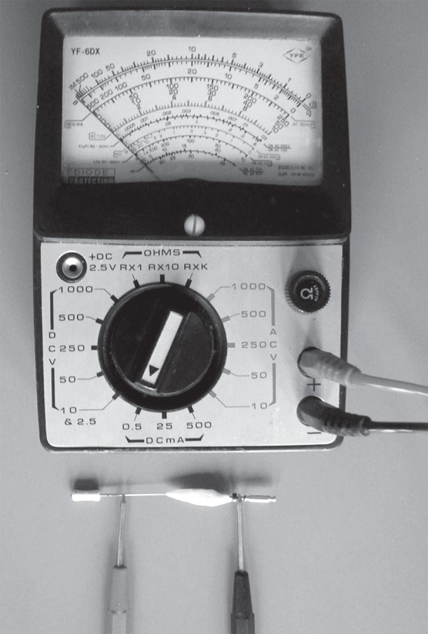
Thirty teeth instrumented as described(n=10 for each instrumentation technique) were allocated for testing the ability of each instrumentation technique to approximate to the predetermined level (0.5 mm short of the apical foramen). Once the apical preparation was accomplished, the final instrument was reinserted into the root canal without excessive pressure and advanced to the level it reached freely, then fixed in place with a flowable composite resin. The teeth were taken out of the alginate molds. An ohmmeter (YFE, Hsin-Chu City, Taiwan, R.O.C.) was used in order to measure the distance between the apical foramen and the level of the created apical stop. Briefly, one pole of the ohmmeter was connected to the shank of the fixed instrument in the canal, and the other pole was connected to the shank of a #15 finger spreader (Figure 1). The spreader was inserted retrogradely from the apical foramen through the root canal and advanced until the display of the ohmmeter indicated a contact. The distance the spreader advanced until the contact was registered by marking on the spreader with an acetate marker. The distance between the tip of the spreader and the mark was measured under stereomicroscope using a digital caliper to an accuracy of 0.01 mm and this value was considered the distance between the apical foramen and the apical stop. The measurements were undertaken by two investigators. No significant difference was found between the measurements of these investigators when paired t-test was performed (p=0.90). The average of the two measurements for each sample was calculated and the data were analyzed statistically using the Anderson-Darling normality test and the Tamhane test at p=5% as the level of significance.
Figure 1.

The experimental setting showing the ohmmeter with the poles connected to the files
Endodontic filling and the leakage test
Forty-five teeth instrumented as described (n=15 for each instrumentation technique) were allocated for testing the microleakage after root-filling of the instrumented teeth. The teeth were taken out of the alginate molds. The root canals were dried using paper points and filled using gutta-percha and sealer (AH Plus, Dentsply/DeTrey, Konstanz, Germany) with the lateral condensation technique. A size 40 gutta-percha cone was used as the master cone. The gutta-percha cones were coated with sealer before insertion into the canal. The teeth were checked radiographically (RVG, Trophy Radiologie, Paris, France) from the labial and lateral aspects for the quality of the root filling (e.g. homogeneity of the filling or presence of a void). All root surfaces were covered with nail polish with care to avoid the apical foramen. The access cavities were sealed with a temporary filling. Ten endodontically accessed-teeth were not obturated and served as positive control. The teeth were stored in physiological saline with 0.2% sodium azide solution until and between the leakage experiments.
The root fillings were tested for leakage using a fluid filtration assembly described elsewhere7. In this method, a pressure of 2 psi (0.136 atm) was applied with O2 to force water through voids within a filled canal. The fluid flow was quantified by observing the movement of an air bubble created within a 25 µL micropipette. This micropipette located between the pressure source and the sample to be tested. Measurements of the fluid movement were made at 2-min intervals for 8 min and averaged. The values were expressed as µL/min/cmH2O. The experiment was performed at 1-week and 3-month post-obturation periods. The normality of the data was analyzed using the Anderson-Darling test. The differences at each period between the groups were statistically evaluated using the Tamhane test. The difference for each group between the 1-week and 3-month measurements was analyzed statistically using the paired samples t-test at p=5% as the level of significance.
RESULTS
Approximation to the predetermined level
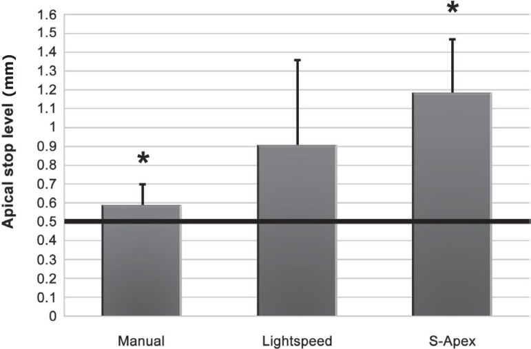
The distance from the apical foramen for each instrumentation technique was as follows [average distance mm(SD mm)]: manual technique [0.59(0.11)], LightSpeed [0.91(0.45)] and S-Apex [1.18(0.29)]. Thus, average proximity to the predetermined level was: manual technique: 0.09 mm; Lightspeed: 0.41 mm and S-Apex: 0.68 mm. There was a significant difference between the manual technique and S-Apex (p<0.05). No significant difference was found between Lightspeed and the other techniques (Figure 2).
Figure 2.
Graph showing the approximation by the three instrumentation techniques to the predetermined level (means and standard deviations in millimeters; n=10). 0 is the apical foramen and 0.5 is the predetermined termination level. The asterisk (*) denotes the groups between which a statistically significant difference exists
Leakage following obturation
The experimental results are shown in Table 1. No significant difference was found between the techniques at either 1-week or 3-month measurements (p>0.05). However, the positive control group leaked significantly more than the other groups at either period (p<0.05).
Table 1.
Fluid filtration measurements for the experimental groups at 1-week and 3-month periods
| Instrumentation technique | 1-week, Mean (±SD) leakage (μL/min/cmH2O) | 3-month, Mean (±SD) leakage (μL/min/cmH2O) |
|---|---|---|
| Lightspeed | 1.02x10⁻³ (3.22x10⁻⁴)Aa | 2.40x10⁻³ (1.24x10⁻³)Ab |
| S-Apex | 9.22x10⁻⁴ (4.15x10⁻⁴)Aa | 1.12x10⁻³ (4.46x10⁻⁴)Ab |
| Manual | 1.09x10⁻³ (4.74x10⁻⁴)Aa | 1.31x10⁻³ (5.43x10⁻⁴)Ab |
| Positive control | 78.62 (4.75x10⁻¹)Ba | 78.95 (1.00)Ba |
The capital letters (A,B) indicate statistical difference between the groups within either 1-week or 3-month periods.
Groups with the same capital letter are not significantly different (p>0.05). SD=standard deviation
The lower case letters (a,b) indicate statistical difference between the 1-week and 3-month measurements for each group.
Groups with the same lower case letter are not significantly different at the two measurement periods (p>0.05).
Leakage at 3 months was significantly greater for all techniques compared to 1-week (p<0.05). No significant difference was found between the leakage at 1-week and 3-month for the positive control group (p>0.05).
DISCUSSION
Rotary NiTi instrumentation has gained popularity in endodontics in recent years. While preparation of the root canal in conventional technique is done using single type of instrument (e.g. successive size K-files), rotary NiTi instrumentation employs different and specific instruments for the preparation of the coronal, mid and apical thirds of the canal. Lightspeed and S-Apex are examples to the instruments designed for the apical third. This study tested how close these novel systems and a conventional manual technique would approximate to a predetermined level in teeth with disrupted apical constriction with a purpose to create a new apical stop. The effect to microleakage of instrumentation with these techniques was also evaluated.
The major findings of this study were that all instrumentation techniques prepared slightly short of the predetermined level; the manual technique prepared closest, and the S-Apex technique prepared farthest to the predetermined level. The Lightspeed technique ranked in between. However, in spite of these variations, leakage following obturation was comparable in all groups.
The NiTi rotary instruments in this study were used along with Tri Auto ZX, an electronic apex locator-integrated endodontic motor. Previous studies employing Tri Auto ZX at a setting of 0.5 found that measurements using hand-files or rotary NiTi files were slightly short of the predetermined level4,9. The findings of the study presented here are in line with these studies.
The finding that approximation with S-Apex to the predetermined level was less satisfactory than the other techniques can be explained by some factors. The most important factor is that the S-Apex files were not used as per the manufacturer's recommendations: while a rotational speed of 500-1000 rpm has been recommended by the manufacturer for the optimum operation of the S-Apex files2, the Tri Auto ZX device provides a constant speed of 280±50 rpm which cannot be changed5. Rotation at slow speed may be the main reason of the inferior performance of the S-apex system. Another factor is the shape of the cutting portion of the instrument. Different from the others, S-Apex has an inverted cone cutting portion. In the gradually narrowing canal of the mandibular incisor, this shape could have disabled the progress of the file to the predetermined level. The manual instrumentation in this study gave the best result. One explanation for this may be that the manual control of a file was simpler than the control of a rotary file mounted on a handpiece. It was "controlled-preparation" to the initially established working length in the manual group. However, the rotary groups followed a more sophisticated technique: it was a dynamic process including the simultaneous measurement of the working length and rotation of the files. A recent study also reported inequivalent apical measurements obtained with motor-driven apex locators (using NiTi files) and their manual apex-locating modes (using stainless-steel files)1. However, we believe that the difference is not due to the alloys the files used in these techniques were made of. In two studies, for example, no significant difference was found between the electric length measurements achieved by the use of stainless-steel or NiTi rotary instruments in the same root canal10,14. Regarding these, the differences found in this study between the performances of the different instrumentation systems may be attributed to their designs, handling and to technical shortcomings. Nevertheless, all the instrumentation techniques tested in this study, with small deviations from the predetermined level, are considered to be clinically acceptable. It has been suggested that best treatment results in cases with necrotic pulp were achieved when treatment procedures were terminated within 2 mm (0 to 2 mm) of the radiographic apex18. The results of the study presented here indicated that the tested techniques could perform within this range.
In spite of the varying deviations from the predetermined level, leakage was similar in all groups. This may be because there were small (in a millimeter scale only) differences between the groups regarding approximation to the predetermined level. Another explanation is that the results of the fluid filtration experiment reflect the seal of the root canal filling as whole. Even if increased leakage existed at the apical part, this could have been blockaded by the filling at the middle or coronal parts of the canal, and thus the fluid flow in the experimental assembly could have been impeded.
This study further found that the leakage increased over time. Leakage in all groups was significantly greater in the 3-month measurements than in the 1-week measurements. This is probably due to disintegration of the sealer during storage. The finding that the leakage in obturated root canals increased with time is in agreement with previous studies where zinc oxide-eugenol-based and epoxy resin-based sealers were used8,11,17,19.
CONCLUSIONS
In conclusion, there were significant but minor differences (less than a millimeter) among the instrumentation techniques regarding approximation to 0.5 mm short of the apical foramen as the predetermined level. However, despite these differences, leakage was similar in all groups following obturation of the root canals.
ACKNOWLEDGEMENT
This study has constituted part of the doctorate thesis by Özgür Genç entitled "Approximation of different apical files to predetermined level in overinstrumented roots and effects on the apical seal", Gazi University, Ankara, 2007.
This study was financially supported by the Gazi University Research Projects Fund.
REFERENCES
- 1.Barthelemy J, Gregor L, Krejci I, Wataha J, Bouillaguet S. Accuracy of electronic apex locater-controlled handpieces. Oral Surg Oral Med Oral Pathol Oral Radiol Endod. 2009;107:437–441. doi: 10.1016/j.tripleo.2008.11.010. [DOI] [PubMed] [Google Scholar]
- 2.Dentaire FKG. NiTi S-Apex [online] La Chaux-de-Fonds; [cited 27 Dec. 2010]. Available from: < http://www.fkg.ch/fileadmin/template/main/images/download/datasheets/fkg_datasheet_sapex_an_lowr.pdf>. [Google Scholar]
- 3.Gomes-Filho JE, Hopp RN, Bernabé PF, Nery MJ, Otoboni JA, Filho, Dezan E., Júnior Evaluation of the apical infiltration after root canal disruption and obturation. J Appl Oral Sci. 2008;16:345–349. doi: 10.1590/S1678-77572008000500008. [DOI] [PMC free article] [PubMed] [Google Scholar]
- 4.Grimberg F, Banegas G, Chiacchio L, Zmener O. In vivo determination of root canal length: a preliminary report using the Tri Auto ZX apex-locating handpiece. Int Endod J. 2002;35:590–593. doi: 10.1046/j.1365-2591.2002.00505.x. [DOI] [PubMed] [Google Scholar]
- 5.Morita J. [cited 27 Dec. 2010];Tri Auto ZX [online] Available from: < http://www.morita.com/usa/root/img/pool/pdf/product_brochures/tri_auto_ps_l-227_1008.pdf>.
- 6.Kast'áková A, Wu MK, Wesselink PR. An in vitro experiment on the effect of an attempt to create an apical matrix during root canal preparation on coronal leakage and material extrusion. Oral Surg Oral Med Oral Pathol Oral Radiol Endod. 2001;91:462–467. doi: 10.1067/moe.2001.114003. [DOI] [PubMed] [Google Scholar]
- 7.Kont Cobankara F, Adanir N, Belli S, Pashley DH. A quantitative evaluation of apical leakage of four root-canal sealers. Int Endod J. 2002;35:979–984. doi: 10.1046/j.1365-2591.2002.00577.x. [DOI] [PubMed] [Google Scholar]
- 8.Kopper PM, Vanni JR, Della Bona A, Figueiredo JA, Porto S. In vivo evaluation of the sealing ability of two endodontic sealers in root canals exposed to the oral environment for 45 and 90 days. J Appl Oral Sci. 2006;14:43–48. doi: 10.1590/S1678-77572006000100009. [DOI] [PMC free article] [PubMed] [Google Scholar]
- 9.Mente J, Seidel J, Buchalla W, Koch MJ. Electronic determination of root canal length in primary teeth with and without root resorption. Int Endod J. 2002;35:447–452. doi: 10.1046/j.1365-2591.2002.00500.x. [DOI] [PubMed] [Google Scholar]
- 10.Nekoofar MH, Sadeghi K, Sadighi Akha E, Namazikhah MS. The accuracy of the Neosono Ultima EZ apex locator using files of different alloys: an in vitro study. J Calif Dent Assoc. 2002;30:681–684. [PubMed] [Google Scholar]
- 11.Pommel L, Camps J. In vitro apical leakage of system B compared with other filling techniques. J Endod. 2001;27:449–451. doi: 10.1097/00004770-200107000-00003. [DOI] [PubMed] [Google Scholar]
- 12.Ricucci D, Langeland K. Apical limit of root canal instrumentation and obturation, part 2. A histological study. Int Endod J. 1998;31:394–409. doi: 10.1046/j.1365-2591.1998.00183.x. [DOI] [PubMed] [Google Scholar]
- 13.Sedgley CM. The influence of root canal sealer on extended intracanal survival of Enterococcus faecalis with and without gelatinase production ability in obturated root canals. J Endod. 2007;33:561–566. doi: 10.1016/j.joen.2007.01.010. [DOI] [PubMed] [Google Scholar]
- 14.Thomas AS, Hartwell GR, Moon PC. The accuracy of the Root ZX electronic apex locator using stainless-steel and nickel-titanium files. J Endod. 2003;29:662–663. doi: 10.1097/00004770-200310000-00012. [DOI] [PubMed] [Google Scholar]
- 15.Tinaz AC, Alacam T, Uzun O, Maden M, Kayaoglu G. The effect of disruption of apical constriction on periapical extrusion. J Endod. 2005;31:533–535. doi: 10.1097/01.don.0000152294.35507.35. [DOI] [PubMed] [Google Scholar]
- 16.Vertucci FJ. Root canal morphology and its relationship to endodontic procedures. Endod Top. 2005;10:3–29. [Google Scholar]
- 17.Von Fraunhofer JA, Fagundes DK, McDonald NJ, Dumsha TC. The effect of root canal preparation on microleakage within endodontically treated teeth: an in vitro study. Int Endod J. 2000;33:355–360. doi: 10.1046/j.1365-2591.2000.00318.x. [DOI] [PubMed] [Google Scholar]
- 18.Wu MK, Wesselink PR, Walton RE. Apical terminus location of root canal treatment procedures. Oral Surg Oral Med Oral Pathol Oral Radiol Endod. 2000;89:99–103. doi: 10.1016/s1079-2104(00)80023-2. [DOI] [PubMed] [Google Scholar]
- 19.Xu Q, Ling J, Cheung GS, Hu Y. A quantitative evaluation of sealing ability of 4 obturation techniques by using a glucose leakage test. Oral Surg Oral Med Oral Pathol Oral Radiol Endod. 2007;104:109–113. doi: 10.1016/j.tripleo.2007.05.014. [DOI] [PubMed] [Google Scholar]
- 20.Yücel AC, Ciftçi A. Effects of different root canal obturation techniques on bacterial penetration. Oral Surg Oral Med Oral Pathol Oral Radiol Endod. 2006;102:e88–e92. doi: 10.1016/j.tripleo.2006.02.007. [DOI] [PubMed] [Google Scholar]


