Abstract
Virulence of infectious pathogens can be unstable and evolve rapidly depending on the evolutionary dynamics of the organism. Experimental evolution can be used to characterize pathogen evolution, often with the underlying objective of understanding evolution of virulence. We used experimental evolution techniques (serial transfer experiments) to investigate differential growth and virulence of Batrachochytrium dendrobatidis (Bd), a fungal pathogen that causes amphibian chytridiomycosis. We tested two lineages of Bd that were derived from a single cryo-archived isolate; one lineage (P10) was passaged 10 times, whereas the second lineage (P50) was passaged 50 times. We quantified time to zoospore release, maximum zoospore densities, and timing of zoospore activity and then modeled population growth rates. We also conducted exposure experiments with a susceptible amphibian species, the common green tree frog (Litoria caerulea) to test the differential pathogenicity. We found that the P50 lineage had shorter time to zoospore production (Tmin), faster rate of sporangia death (ds), and an overall greater intrinsic population growth rate (λ). These patterns of population growth in vitro corresponded with higher prevalence and intensities of infection in exposed Litoria caerulea, although the differences were not significant. Our results corroborate studies that suggest that Bd may be able to evolve relatively rapidly. Our findings also challenge the general assumption that pathogens will always attenuate in culture because shifts in Bd virulence may depend on laboratory culturing practices. These findings have practical implications for the laboratory maintenance of Bd isolates and underscore the importance of understanding the evolution of virulence in amphibian chytridiomycosis.
Keywords: Amphibian chytridiomycosis, amphibian declines, Batrachochytrium dendrobatidis, evolution of virulence, experimental evolution, host–pathogen interactions, serial passage experiments
Introduction
Understanding pathogen biology, life-history strategies and evolutionary dynamics is important in multiple medical and bioscientific fields (Stearns and Koella 2008). The study of pathogens and the evolution of virulence is not only critical for wildlife and human health at the individual level, but also at a much larger scale because infectious diseases can alter host population densities, community dynamics, and potentially entire ecosystems (Scott 1988; De Castro and Bolker 2005; Whiles et al. 2006). Furthermore, insights into pathogen evolution provide a better understanding of the mechanisms driving disease dynamics and allow for improved predictions of disease spread and potential impacts (Bull 1994; Ebert and Bull 2003). Until relatively recently, however, the factors influencing pathogen evolution were difficult to study and therefore not well understood (Bull 1994; Ebert and Bull 2003).
Historically, the application of experimental evolution with infectious agents (studies in which virulence is artificially modified) was a turning point in the study of infectious disease (Bezin 2003). Louise Pasteur first employed serial passage techniques in the 18th century (Bezin 2003), and since then, serial passage experiments (SPEs) have become a cornerstone in the study of infectious disease and the development of vaccines (Bezin 2003). SPEs have been used for in vitro and in vivo investigations of a wide variety of pathogens: viruses (Schlesinger et al. 1956; Beare et al. 1968), bacteria (Cushion and Walzer 1984; Maisnier-Patin et al. 2002; Somerville et al. 2002), protozoa (Diffley et al. 1987), and fungi (da Silva Ferreira et al. 2004; Wang et al. 2008). Thus, evolutionary principles and the notion that pathogens should adapt to novel environments have long been applied in the study of infectious disease, even before we had a complete understanding of how or why evolutionary shifts in pathogen virulence might occur (Bezin 2003; Hanley 2011).
During experimental evolution using serial passage experiments (SPEs), pathogens are transferred to alternative hosts or to new artificial environments at a specific point in the pathogen life cycle (Ebert 1998; Ford et al. 2002). The advantage of SPEs is that alterations in pathogen genotype, phenotype, and virulence can be tracked in real time (Ebert 1998). Many SPEs studies have demonstrated that pathogens adapt to novel environments (e.g., in culture or alternative hosts) relatively rapidly (reviewed in Ebert 1998) and that they frequently become less efficient at surviving in their natural hosts (Ebert 1998; Ford et al. 2002). More specifically, SPEs can lead to shifts in pathogen reproductive rates (a common component of virulence) such that pathogen replication is attenuated (Ebert 1998; Hanley 2011). However, shifts in virulence are not obligately unidirectional toward hypovirulence (Ebert 1998). Pathogens can also exhibit shifts toward hypervirulence or revert from a hypovirulent state (Mastroeni et al. 2011). Some studies have shown that shifts in pathogen virulence will greatly depend on passage practices. For example, the timing of passage (i.e., the point of the pathogen’s life cycle at which it is propagated) and the conditions of propagation (e.g., nutrient and thermal conditions) can influence the direction of shifts in pathogens’ abilities to exploit the available resources (Ford et al. 2002).
We used experimental evolution to investigate growth and virulence (i.e., pathogenicity) of a fungal pathogen of amphibians. Batrachochytrium dendrobatidis (hereafter “Bd”) is an aquatic fungus causes the disease chytridiomycosis and is highly virulent to many species of amphibians (Berger et al. 1998, 2005; Voyles et al. 2009; Alford 2010). Bd infects epidermal cells and causes disruption of electrolyte (e.g., sodium) transport across the epidermis, and subsequent cardiac arrest (Voyles et al. 2009). Although the mechanisms by which Bd disrupts epidermal function are not yet fully understood, potential virulence factors have been identified (Rosenblum et al. 2009; Fites et al. 2013). In addition to virulence factors, Bd reproductive rates play an important role in pathogenesis and disease development, contributing to intensity of infection and thus Bd load in an individual host (Voyles et al. 2009; Vredenburg et al. 2010). Although we now have a much better understanding of chytridiomycosis pathophysiology, host immunity, and disease ecology (reviewed in Kilpatrick et al. 2010; Venesky et al. 2013), researchers have only relatively recently turned their attention to investigations on differential virulence among Bd isolates, evolutionary shifts in Bd, and host–pathogen coevolution (Fisher et al. 2009; Farrer et al. 2011, 2013; Voyles et al. 2012; Rosenblum et al. 2013).
Several lines of evidence suggest that evolution in Bd virulence may be occurring quite rapidly. First, although chytridiomycosis has been responsible for many catastrophic amphibian declines and even local extinction events, some species and populations of species have survived initial declines and now persist in the wild with Bd infections (Retallick et al. 2004; Woodhams and Alford 2005; Alford 2010; Puschendorf et al. 2011). One investigation that explicitly focused on the patterns of disease emergence and host declines has suggested that evolution of Bd virulence must be occurring across time and space (Phillips and Puschendorf 2013). Second, laboratory studies have documented Bd attenuation with successive in vitro propagation using routine culture maintenance practices (Brem et al. 2013; Langhammer et al. 2013). In these studies, Bd attenuation was characterized by reductions in zoospore production rates (i.e., pathogen replication) and in pathogenicity in live amphibian inoculation experiments (Brem et al. 2013; Langhammer et al. 2013). Third, recent molecular studies that have examined the genomic diversity of Bd isolates (i.e., isolates collected from widespread geographic and multiple host species origins) have suggested that Bd has a highly dynamic genome with multiple possible mechanisms that could be contributing to a complex evolutionary history (Farrer et al. 2013; Rosenblum et al. 2013). Taken together, these three lines of evidence suggest that further investigations focused on evolution in Bd may help resolve how this pathogen has been so successful to such a broad range of host species in a wide variety of environments.
We aimed to understand how Bd growth and virulence are altered with experimental evolution using serial passage experiments. We used a single isolate of Bd and derived two lineages that were treated identically except for the length of time they were propagated in artificial media (i.e., 10 vs. 50 passages). By passaging Bd at the peak of zoospore production, our serial propagation imposed artificial selection for an early release of zoospores and high densities of zoospores. We found that the differences in passage histories translated into changes in Bd population growth rates in vitro and Bd growth and virulence in vivo.
Methods
Serial transfer experiments
We originally obtained the isolate, GibboRiver-L.lesueuri-00-LB-1, from a diseased juvenile L. lesueuri that was collected in the wild and died in captivity. This isolate was cultured on tryptone/gelatin hydrolysate/lactose (TGhL) agar with antibiotics (Longcore et al. 1999) and then cryo-archived according to standard protocols (Boyle et al. 2003). We revived one aliquot of the cryoarchived culture (Boyle et al. 2003) and passaged it into liquid TGhL broth in 25-cm2 cell culture flasks (Longcore et al. 1999). We incubated the cultures at 22°C and inspected the flasks daily to monitor zoospore encystment and maturation of the zoosporangia. We passaged cultures into new media when zoospore density peaked (~5–7 days based on previous experiments; Voyles 2011; Voyles et al. 2012) and repeated this procedure for 50 passages. This Bd lineage with a higher number of passages will be referred to as “P50”. We revived a second aliquot of the same isolate 250 days later and treated the Bd culture identically for 10 passages. This lineage with a lower number of passages will be referred to as “P10”. Thus, the two lineages were maintained in identical conditions (i.e., in the same laboratory, using identical techniques and equipment, maintained at the same temperature and by the same investigator) except that one was passaged 50 times and one was passaged 10 times. This approach allowed us to test the two lineages simultaneously in a common garden experiment.
We filtered the two Bd lineages through sterile filter paper (Whatman, 3) to remove sporangia. We washed zoospores using a gentle centrifugation (500 g for 10 min), removing the supernatant, and resuspending the zoospores in fresh TGhL. We determined the zoospore concentrations using a hemocytometer (Improved Neubauer Bright-line) and adjusted to 90 × 104 zoospores mL−1 as needed by adding TGhL. We conducted the phenotyping experiments in sterile 96-well plates (Tissue culture test plates-96, TPP, company info). We pipetted the zoospore inocula (50 μL) into each of 20 wells containing 50 μL TGhL media. The plate had a perimeter of 36 wells with 100 μL sterile water to avoid evaporation. We inspected the plates daily to monitor zoospore encystment, development, and maturation of the zoosporangia. Once the maturing zoosporangia produced the first zoospores, we quantified zoospore density daily by randomly selecting 10 wells of each of the two lineages, drawing off 30 μL of supernatant, and counting zoospore numbers using a hemocytometer.
Model development
To understand differences between the two lineages, we used a mathematical model to analyze our empirical data. Specifically, we used a delay differential equation model that was developed for a previous study on Bd (Voyles et al. 2012). The model used the data on the concentration of zoospores produced in the next generation from the initial cohort of zoospores placed in each well of the 96-well plate and follows the dynamics of: C(t) = the concentration of the initial cohort of zoospores; S(t) = the concentration of zoospore-producing sporangia; and Z(t) = the concentration of zoospores in the next generation. The initial cohort of zoospores, C(t), started at a concentration of 90 × 104 zoospores per mL, and zoospores in this initial cohort settle and become sporangia at rate sr or die at rate μz. fs is the fraction of sporangia that survive to the zoospore-producing stage.
The model assumed that it takes a minimum of Tmin days before the sporangia produce zoospores, after which they produce zoospores at rate η. Zoospore-producing sporangia die at rate ds. The concentration of zoospores, Z(t), is the state variable actually measured in the experiments, and it is assumed that these zoospores settle (sr) or die (μz) at the same rates as the initial cohort of zoospores. The equations that describe this are as follows:
| (1) |
| (2) |
| (3) |
Zoospore-producing sporangia die at rate ds. For this model, the population growth rate λ can be calculated numerically from the transcendental equation:
We used a Bayesian approach to infer the values of the model parameters in Equations (1)–(3) that best fit the experimental data from each Bd lineage. Our data consist of observations on the numbers of zoospores produced after serial passage treatments (i.e., on Z(t)), although we know the initial conditions of all the states. With discrete numbers of zoospores, we modeled our observations of the system at a set of discrete times t′ as independent Poisson random variables with a mean given by the solution of Equations 1 - 3, at times t′:
We tested for significant differences between the Bd lineages by fitting the model to the data from the two lineages combined (i.e., assuming both lineages had the same parameters) and comparing this to the fit of the model to each lineage separately (i.e., assuming different parameters for the two lineages) using the deviance information criterion (DIC).
Experimental inoculations
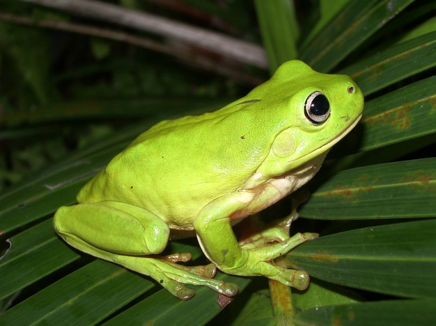
We collected adult common green tree frogs (Litoria caerulea; N = 30, mean mass: 21.34 ± 5.64 SD; Fig. 1) in January and February 2008 from residential areas of Townsville, Queensland, an area that is predicted to be unsuitable for Bd (Murray et al. 2011). We collected each animal using a new plastic bag and then transferred each individual to a plastic container (200 × 240 × 330 mm3), containing 250 mL of tap water. We maintained the containers in temperature- (18–23°C) and light (12L/12D)-controlled facilities at James Cook University, Townsville, Australia. We fed frogs vitamin-dusted crickets (medium-sized, Pisces Inc. Boulder, Colorado, USA) ad libitum twice per week. We also changed the tap water (250 mL) twice a week until experimental exposures began, and then, we replaced tap water with 20% Holtfretter’s solution (in mMol: NaCl (6.0), KCL (0.06), CaCl2 (0.09), NaCO3 (0.24), pH 6.5, 250 mL). We maintained the frog containers in a level position, so water covered the bottom, but frogs were able to climb up the dry walls.
Figure 1.

The common green tree frog (Litoria caerulea).
To confirm that frogs were not infected with Bd prior to inoculation, we swabbed their ventral surfaces and digits and tested for Bd using a Taqman real-time polymerase chain reaction (PCR) assay (Boyle et al. 2004; Hyatt et al. 2007). More specifically, we swabbed the abdomen and ingroinal regions 10 times and each of the digits five times. For the PCR assay, we analyzed all samples in triplicate and compared them with Australian Animal Health Laboratory zoospore standards to determine zoospore equivalents (Hyatt et al. 2007).
For animal inoculations, we filtered Bd zoospores from the P10 and P50 lineages to remove sporangia (described above). We determined zoospore concentrations using a hemocytometer (Improved Neubauer Bright-line) and adjusted the zoospore concentrations to 93 × 104 zoospores mL−1 by diluting with fresh TGhL media. We randomly assigned frogs to one of three treatment groups: two exposure groups (P10 and P50 isolate treatments) or a control group (N = 10 frogs per group). We inoculated frogs by exposure to Bd via shallow immersion in a bath of Holtfretter’s solution and sterile TGhL with Bd zoospores. We immersed the control frogs in a bath that contained Holtfretter’s solution and sterile TGhL with no Bd zoospores. After 24 h, we moved the frogs to fresh containers with 20% Holtfretter’s solution (pH 6.5). Following exposure to Bd, we collected skin swabs again at 17, 57, 104, 127, and 157 days postexposure. We did not collect mass during the course of the experiment to minimize disturbance to the animals, but we calculated the change in mass (final minus initial mass) by collecting mass at the beginning of the experiment and at the termination of the experiment.
Results
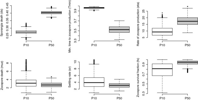
We found differences in population growth rate (lambda, λ) between the P10 and P50 lineages of Bd (Fig. 2A, B). We generated the 95% credible intervals for population growth rate using the posterior samples of the model parameters. The parameters that most likely contributed to these population differences include time to zoospore production (Tmin) and rate of sporangia death (ds) (Fig. 3). Although additional parameters such as rate of zoospore production (η) and fraction of zoospores that survive (fs) may have also contributed to our observed differences in lambda (λ), our model fitting indicates that these parameters are less likely to be different between the two lineages (Fig. 3).
Figure 2.

Line graph over time (A) and box-and-whiskers plots (B) showing modeled population growth rate (λ, lambda) of two lineages of Batrachochytrium dendrobatidis, GibboRiver-L.lesueuri-00-LB, that were serially passaged 50 times (P50; gray) and 10 times (P10; black).
Figure 3.

Box-and-whiskers plots for growth parameters for two lineages of Batrachochytrium dendrobatidis that were propagated for 50 passages (P50) or 10 passages (P10). Horizontal bars are medians and reflect lineage growth through time.
For the P50 lineage, we found that the 95% credible intervals of the posterior distribution for time to zoospore production (Tmin) were lower and had no overlap with those for the P10 lineage (P10: Tmin = 3.93–3.99; P50: Tmin = 3.38–3.67; Fig. 3). Similarly, the intervals of the posterior distribution of sporangia death (ds) were higher in the P50 lineage compared with the P10 lineage (P10: ds = 0.248–0.304; P50: ds = 0.379–0.423; Fig. 3). As such, differences between the two lineages in the time to zoospore production and sporangia death most likely contributed to the net effects on population growth rate. The evidence for a higher population growth rate in the P50 lineage is further supported by the deviance information criterion (DIC) values, which indicate a considerably better fit for the two data sets fits with different parameters (DIC = 1753.318) compared with a single set of parameters (DIC = 2861.492). Thus, our model with separate parameters for the two populations explains the data much better than a model where both populations share the same parameters.
Experimental inoculations of Litoria caerulea
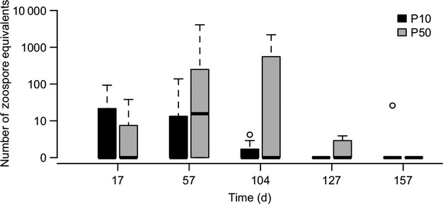
Although the results of our in vitro experiment and mathematical modeling indicated that the SPEs led to a higher population growth rate in the P50 culture, it was unknown whether this would translate to an increased growth rate, and hence virulence, in susceptible frogs. In our frog inoculation experiments, we used several response variables to assess differences in growth and virulence: infection prevalence and intensity (i.e., pathogen load), changes in mass, clinical signs of disease and mortality. Using PCR analysis on swab samples, we found that prevalence did not differ significantly (Fisher’s exact test, P = 0.63) between treatments. Eighty percent (8/10) L. caerulea exposed to P50 zoospores became infected during the experiment, while 60% (6/10) of frogs exposed to P10 zoospores became infected. The frogs that became infected by P50 zoospores had slightly higher intensities of infection than the frogs exposed to P10 zoospores, but this difference was not significant (repeated-measures ANOVA, P = 0.156; Fig. 4). The decrease in mass (final minus initial weight) was greatest in the P50 group. However, this change did not differ significantly from the P10 and control groups (ANOVA, P = 0.48; Fig. 5).
Figure 4.

Intensity of infection in common green tree frogs (Litoria caerulea) that were infected with one isolate of Batrachochytrium dendrobatidis with two passage histories.
Figure 5.

Change in mass (final weight minus initial weight) in Litoria caerulea experimentally exposed to two lineages of Batrachochytrium dendrobatidis (50 times (P50; gray) and 10 times (P10; black) or to a control solution (white bar)).
We observed mild clinical signs of infection including lethargy, inappetence, and slight skin discoloration in two frogs in the P50 group and two frogs in the P10 group. More severe clinical signs of infection (as per Voyles et al. 2009) did not develop in any group. Frogs with mild clinical signs seemed to recover, regaining normal color, activity, and appetite, by the termination of the experiment. Additionally, there was no mortality in any group.
Discussion
Differential growth and virulence of Batrachochytrium dendrobatidis has been reported in multiple studies, but it is unclear why this phenotypic variation exists (Berger et al. 2005; Retallick and Miera 2007; Fisher et al. 2009; Voyles 2011). By reviving two aliquots of a single isolate of Bd and subjecting them to shorter or longer periods of passaging, we investigated selective effects of passage history on zoospore densities. Because we passaged the two lineages at the peak of zoospore densities, we eliminated the contribution of zoospores produced after this peak in each subsequent generation. This approach produced strong selection for early zoospore release and maximum zoospore densities. We predicted that 50 passages with this selective pressure would produce a greater response than 10 passages. We also expected that the P50 lineage, which produced more zoospores and had a higher population growth rate, might have been more pathogenic in our exposure experiments.
Our results suggest that passaging practices altered the rate and temporal pattern of zoospore production and population growth of Bd in vitro. We found that the time to zoospore production (Tmin) was faster and the overall population growth rate (λ) was higher in the P50 lineage (Fig 3). The changes in these parameters may reflect the culturing practice of passaging the Bd lineages at their first peak in zoospore production, thereby selecting for zoospores that are produced the earliest and make a substantial contribution to the overall population growth rate. These findings are important because they suggest that Bd evolves in culture and that more passages are likely to lead to greater divergence from the initial state of virulence in amphibian hosts. Additionally, the timing of propagation during Bd’s life cycle is appears to be critical; passaging at the height of zoospore production may also lead to faster population growth rates and higher virulence.
One possible limitation, however, is that the zoospore densities of the two lineages were not quantified immediately after revival or compared with the original strain (the “ancestor” strain) after serial passage treatments, so the differences we observed could conceivably have been caused by differences in the aliquots that were revived to start the P50 and P10 lineages. An additional objection could be made that the two lineages represent two separate evolutionary trajectories. Both of these objections are valid. However, both aliquots were samples that were originally derived from a single common source, and the P10 lineage remained frozen in liquid nitrogen until it was revived for this study. Furthermore, the two lineages were maintained in the same laboratory, using identical techniques and equipment, maintained at the same temperature and tested by the same investigator in multiple rigorous “common garden” experiments. Thus, our observations in the two lineages probably reflect true differences that resulted from laboratory practices at some point. We believe the most parsimonious explanation is that the differences were due to the serial culturing treatments.
One alternative experimental approach that could be used in future investigations is to test a single lineage that is subsampled and cryo-preserved at successive time points (e.g., Knies et al. 2006). We were unable to conduct this experiment, but we believe this design would allow researchers to further investigate the timing and conditions under which Bd changes during long-term serial passage treatments.
Overall, our results on the population growth rates in the P10 and P50 lineages suggest that patterns of zoospore production can vary depending on the timing of culturing practices. Quantifying parameters such as zoospore production in culture is valuable for understanding rates and patterns of population growth of Bd in vitro, but the implications for Bd growth in vivo and virulence are less clear. Because the P50 culture produced more zoospores, we predicted that it might be more virulent in inoculation experiments. This prediction was supported in our inoculation experiments by the patterns we observed in our measured response variables (i.e., higher prevalence and intensity of infection). However, these differences were not significant, and furthermore, there was no mortality in any group of Litoria caerulea exposed to P50 or P10 cultures. The lack of mortality was unexpected because this isolate, GibboRiver-L.lesueuri-00-LB with a similar passage history, was chosen due to its high level of virulence in parallel experiments (see Voyles et al. 2009). This isolate caused 90% mortality of Litoria caerulea in separate experiments using virtually identical methods, laboratory facilities, and equipment. Differences between the present study and the previously published experiments include the length of time frogs were held in captivity, the seasonal timing of animal exposures, and differences in isolate passage practices (cultures for the previous infection experiment had comparable passage history, but the lineages were maintained at 4°C rather than 22°C, which could be an important selective pressure; Voyles et al. 2012; Stevenson et al. 2013).
Our results challenge the general assumption that pathogens will attenuate in culture. It is commonly thought that as pathogens adapt to culture conditions, they lose their ability to exploit hosts as resources (Ebert 1998; Ford et al. 2002). Yet, evidence from several study systems suggests that shifts in virulence in vitro are not always unidirectional. Rather, passage timing (i.e., the point of the pathogen’s life cycle at which it is propagated) and the conditions of propagation (e.g., nutrient and thermal conditions) can influence shifts in virulence (da Silva & Sacks, 1987; Wozencraft and Blackwell 1987; Rey et al. 1990). Additionally, attenuated pathogens strains can rapidly revert to a virulent form when re-exposed to a naïve host (Cann et al. 1984; Macadam et al. 1989; Minor 1993; Nielsen et al. 2001). Thus, it is increasingly clear that virulence can be greatly affected by how pathogens are maintained and experimentally manipulated in the laboratory. We suggest that the timing of Bd propagation when maintaining cultures is critical because it may influence growth, zoospore production, and ultimately virulence of Bd.
Understanding how pathogens evolve in vitro, including attenuation and reversion to higher virulence, can advance our understanding evolution of pathogen virulence and has practical applications for disease research. The stability of virulence presents a considerable challenge for pathogen research (Michel and Garcia 2003), especially if virulence can evolve bi-directionally (Ebert and Bull 2003; Stearns and Koella 2008). For example, the ability to reliably infect hosts in controlled conditions is critical for the study of infectious disease (Ford et al. 2002; Brem et al. 2013). Culture history could also distort the outcome of experiments that are aimed at understanding host–pathogen interactions in nature (Langhammer et al. 2013). Additionally, studying changes in virulence represents an opportunity to pinpoint the mechanisms of pathogenesis. Culturing practices in which a pathogen is manipulated toward attenuation but then reverts to high virulence may reveal factors that determine the level of virulence for a particular host–pathogen dynamic. Thus, it is important to resolve how laboratory culture practices and experimental manipulation can influence pathogen growth, development, and virulence.
Acknowledgments
Experiments were performed with approval of James Cook University Animal Ethics Committee (Permit numbers JCU- A1085, A593). Animals were collected with approval from Queensland Parks and Wildlife Service (Scientific Purposes Permit numbers: WISP03866106, WISP04143907). Funded by the Australian Research Council Discovery Project (DP0452826) and the Australian Government Department of Environment and Heritage (RFT 43/2004). We thank R. Webb, S. Bell, and C. Moritz for their help and support.
Conflict of Interest
None declared.
References
- Alford RA. Declines and the global status of amphibians. In: Sparling D, Linder G, Bishop CA, Krest SK, editors. Ecotoxicology of amphibians and reptiles. 2nd edn. Pensacola, FL: SETAC Press; 2010. pp. 13–45. [Google Scholar]
- Beare AS, Bynoe ML. Tyrrell DAJ. Investigation into the attenuation of influenza viruses by serial passage. Br. Med. J. 1968;4:482. doi: 10.1136/bmj.4.5629.482. [DOI] [PMC free article] [PubMed] [Google Scholar]
- Berger L, Speare R, Daszak P, Green DE, Cunningham AA, Goggin CL, et al. Chytridiomycosis causes amphibian mortality associated with population declines in the rain forests of Australia and Central America. Proc. Natl Acad. Sci. USA. 1998;95:9031–9036. doi: 10.1073/pnas.95.15.9031. [DOI] [PMC free article] [PubMed] [Google Scholar]
- Berger L, Marantelli G, Skerratt LF. Speare R. Virulence of the amphibian chytrid fungus Batrachochytium dendrobatidis varies with the strain. Dis. Aquat. Org. 2005;68:47–50. doi: 10.3354/dao068047. [DOI] [PubMed] [Google Scholar]
- Bezin H. A brief history of the prevention of infectious diseases by immunizations. Comp. Immunol. Microbiol. 2003;26:293–308. doi: 10.1016/S0147-9571(03)00016-X. [DOI] [PubMed] [Google Scholar]
- Boyle DG, Hyatt AD, Daszak P, et al. Cryo-archiving of Batrachochytrium dendrobatidis and other chytridiomycetes. Dis. Aquat. Org. 2003;56:59–64. doi: 10.3354/dao056059. [DOI] [PubMed] [Google Scholar]
- Boyle DG, Boyle DB, Olsen V, Morgan JAT. Hyatt AD. Rapid quantitative detection of chytridiomycosis (Batrachochytrium dendrobatidis) in amphibian samples using real-time Taqman PCR assay. Dis. Aquat. Org. 2004;60:141–148. doi: 10.3354/dao060141. [DOI] [PubMed] [Google Scholar]
- Brem FMR, Parris MJ. Padgett-Flohr GE. Re-isolating Batrachochytrium dendrobatidis from an amphibian host increases pathogenicity in a subsequent exposure. PLoS ONE. 2013;8:e61260. doi: 10.1371/journal.pone.0061260. [DOI] [PMC free article] [PubMed] [Google Scholar]
- Bull JJ. Virulence. Evolution. 1994;48:1423–1437. doi: 10.1111/j.1558-5646.1994.tb02185.x. [DOI] [PubMed] [Google Scholar]
- Cann AJ, Stanway G, Hughes PJ, Minor PD, Evans DMA, Schild GC, et al. Reversion to neurovirulence of the live-attenuated Sabin type 3 oral poliovirus vaccine. Nucleic Acids Res. 1984;12:7787–7792. doi: 10.1093/nar/12.20.7787. [DOI] [PMC free article] [PubMed] [Google Scholar]
- Cushion MT. Walzer PD. Growth and serial passage of Pneumocystis carinii in the A549 cell line. Infect. Immun. 1984;44:245–251. doi: 10.1128/iai.44.2.245-251.1984. [DOI] [PMC free article] [PubMed] [Google Scholar]
- De Castro F. Bolker B. Mechanisms of disease-induced extinction. Ecol. Lett. 2005;8:117–126. [Google Scholar]
- Diffley P, Scott JO, Mama K. Tsen TN. The rate of proliferation among African trypanosomes is a stable trait that is directly related to virulence. Am. J. Trop. Med. Hyg. 1987;36:533–540. doi: 10.4269/ajtmh.1987.36.533. [DOI] [PubMed] [Google Scholar]
- Ebert D. Experimental evolution of parasites. Science. 1998;282:1432–1436. doi: 10.1126/science.282.5393.1432. [DOI] [PubMed] [Google Scholar]
- Ebert D. Bull JJ. Challenging the trade-off model for the evolution of virulence: is virulence management feasible? Trends Microbiol. 2003;11:15–20. doi: 10.1016/s0966-842x(02)00003-3. [DOI] [PubMed] [Google Scholar]
- Farrer RA, Weinert LA, Bielby J, Garner TWJ, Balloux F, Clare F, et al. Multiple emergences of genetically diverse amphibian-infecting chytrids include a globalized hypervirulent recombinant lineage. Proc. Natl Acad. Sci. USA. 2011;108:18732–18736. doi: 10.1073/pnas.1111915108. [DOI] [PMC free article] [PubMed] [Google Scholar]
- Farrer RA, Henk DA, Garner TWJ, Balloux F, Woodhams DC. Fisher MC. Chromosomal copy number variation, selection and uneven rates of recombination reveal cryptic genome diversity linked to pathogenicity. PLoS Genet. 2013;9:e1003703. doi: 10.1371/journal.pgen.1003703. [DOI] [PMC free article] [PubMed] [Google Scholar]
- Fisher MC, Bosch J, Yin Z, Stead DA, Walker J, Selway L, et al. Proteomic and phenotypic profiling of the amphibian pathogen Batrachochytrium dendrobatidis shows that genotype is linked to virulence. Mol. Ecol. 2009;18:415–429. doi: 10.1111/j.1365-294X.2008.04041.x. [DOI] [PubMed] [Google Scholar]
- Fites JS, Ramsey JP, Holden WM, Collier SP, Sutherland DM, Reinert LK, et al. The invasive chytrid fungus of amphibians paralyzes lymphocyte responses. Science. 2013;342:366–342. doi: 10.1126/science.1243316. [DOI] [PMC free article] [PubMed] [Google Scholar]
- Ford SE, Chintala MM. Bushek D. Perkinsus marinus, Pathogen virulence. Dis. Aquat. Org. 2002;51:187–201. doi: 10.3354/dao051187. [DOI] [PubMed] [Google Scholar]
- Hanley KA. The double-edged sword: how evolution can make or break a live attenuated virus vaccine. Evol. Educ. Outreach. 2011;4:635–643. doi: 10.1007/s12052-011-0365-y. [DOI] [PMC free article] [PubMed] [Google Scholar]
- Hyatt AD, Boyle DG, Olsen V, Boyle DB, Berger L, Obendorf D, et al. Diagnostic assays and sampling protocols for the detection of Batrachochytrium dendrobatidis. Dis. Aquat. Org. 2007;73:175–192. doi: 10.3354/dao073175. [DOI] [PubMed] [Google Scholar]
- Kilpatrick AM, Briggs CJ. Daszak P. The ecology and impact of chytridiomycosis: an emerging disease of amphibians. Trends Ecol. Evol. 2010;25:109–118. doi: 10.1016/j.tree.2009.07.011. [DOI] [PubMed] [Google Scholar]
- Knies JL, Izem R, Supler KL, Kingsolver JG. Burch CL. The genetic basis of thermal reaction norm evolution in lab and natural phage populations. PLoS Biol. 2006;7:e201. doi: 10.1371/journal.pbio.0040201. [DOI] [PMC free article] [PubMed] [Google Scholar]
- Langhammer PF, Lips KR, Burrowes PA, Tunstall T, Palmer CM. Collins JP. A fungal pathogen of amphibians, Batrachochytrium dendrobatidis, attenuates in pathogenicity with in vitro passages. PLoS ONE. 2013;8:e77630. doi: 10.1371/journal.pone.0077630. [DOI] [PMC free article] [PubMed] [Google Scholar]
- Longcore JE, Pessier AP, Nichols DK. Longcorel JE. Batrachochytrium dendrobatidis gen. et sp. nov., a chytrid pathogenic to amphibians. Mycologia. 1999;91:219–227. [Google Scholar]
- Macadam AJ, Arnold C, Howlett J, Marsden JA, Marsden S, Taffs F, et al. Reversion of the attenuated and temperature-sensitive phenotypes of the Sabin type 3 strain of poliovirus in vaccinees. Virology. 1989;172:408–414. doi: 10.1016/0042-6822(89)90183-9. [DOI] [PubMed] [Google Scholar]
- Maisnier-Patin S, Berg OG, Liljas L. Andersson DI. Compensatory adaptation to the deleterious effect of antibiotic resistance in Salmonella typhimurium. Mol. Microbiol. 2002;46:355–366. doi: 10.1046/j.1365-2958.2002.03173.x. [DOI] [PubMed] [Google Scholar]
- Mastroeni P, Morgan FJ, McKinley TJ, Shawcroft E, Clare S, Maskell DJ, et al. Enhanced virulence of Salmonella enterica serovar typhimurium after passage through mice. Infect. Immun. 2011;79:636–643. doi: 10.1128/IAI.00954-10. [DOI] [PMC free article] [PubMed] [Google Scholar]
- Michel C. Garcia C. Virulence stability in Flavobacterium psychrophilum after storage and preservation according to different procedures. Vet. Res. 2003;34:127–132. doi: 10.1051/vetres:2002057. [DOI] [PubMed] [Google Scholar]
- Minor PD. Attenuation and reversion of the Sabin vaccine strains of poliovirus. Dev. Biol. Stand. 1993;78:17–26. [PubMed] [Google Scholar]
- Murray KA, Retallick RW, Puschendorf R, Skerratt LF, Rosauer D, McCallum HI. VanDerWal J. Assessing spatial patterns of disease risk to biodiversity: implications for the management of the amphibian pathogen, Batrachochytrium dendrobatidis. J. Appl. Ecol. 2011;48(1):163–173. [Google Scholar]
- Nielsen HS, Oleksiewicz MB, Forsberg R, Stadejek T, Botner A. Storgaard T. Reversion of a live porcine reproductive and respiratory syndrome virus vaccine investigated by parallel mutations. J. Gen. Virol. 2001;82:1263–1272. doi: 10.1099/0022-1317-82-6-1263. [DOI] [PubMed] [Google Scholar]
- Phillips BL. Puschendorf R. Do pathogens become more virulent as they spread? Evidence from the amphibian declines in Central America. Proceedings of the Royal Society B- Biological Sciences. 2013;280:1766. doi: 10.1098/rspb.2013.1290. [DOI] [PMC free article] [PubMed] [Google Scholar]
- Puschendorf R, Hoskin CJ, Cashins SD, McDonald K, Skerratt LF, Vanderwal J, et al. Environmental refuge from disease-driven amphibian extinction. Conserv. Biol. 2011;25:956–964. doi: 10.1111/j.1523-1739.2011.01728.x. [DOI] [PubMed] [Google Scholar]
- Retallick RWR. Miera V. Strain differences in the amphibian chytrid Batrachochytrium dendrobatidis and non-permanent, sub-lethal effects of infection. Dis. Aquat. Org. 2007;75:201–207. doi: 10.3354/dao075201. [DOI] [PubMed] [Google Scholar]
- Retallick RWR, Mccallum H. Speare R. Endemic infection of the amphibian chytrid fungus in a frog community post-decline. PLoS Biol. 2004;2:e351. doi: 10.1371/journal.pbio.0020351. [DOI] [PMC free article] [PubMed] [Google Scholar]
- Rey JA, Travi BL, Valencia AZ. Saravia NG. Infectivity of the subspecies of the Leishmania braziliensis complex in vivo and in vitro. Am. J. Trop. Med. Hyg. 1990;43:623–631. doi: 10.4269/ajtmh.1990.43.623. [DOI] [PubMed] [Google Scholar]
- Rosenblum EB, Poorten TJ, Settles M, Murdoch GK, Robert J, Maddox N, et al. Genome-wide transcriptional response of SiluranaXenopustropicalis to infection with the deadly chytrid fungus. PLoS ONE. 2009;4:e6494. doi: 10.1371/journal.pone.0006494. [DOI] [PMC free article] [PubMed] [Google Scholar]
- Rosenblum EB, James TY, Zamudio KR, Poorten TJ, Ilut D, Rodriguez D, et al. Complex history of the amphibian-killing chytrid fungus revealed with genome resequencing data. Proc. Natl Acad. Sci. USA. 2013;110:9385–9390. doi: 10.1073/pnas.1300130110. [DOI] [PMC free article] [PubMed] [Google Scholar]
- Schlesinger RW, Gordon I, Frankel JW, Winter JW, Patterson PR. Dorrance WR. Clinical and serologic response of man to immunization with attenuated dengue and yellow fever viruses. J. Immunol. 1956;77:352–364. [PubMed] [Google Scholar]
- Scott ME. The impact of infection and disease on animal populations: implications for conservation biology. Conserv. Biol. 1988;2:40–56. [Google Scholar]
- da Silva Ferreira ME, Capellaro JL, dos Reis Marques E, Malavazi I, Perlin D, Park S, et al. In vitro evolution of itraconazole resistance in Aspergillus fumigatus involves multiple mechanisms of resistance. Antimicrob. Agents. 2004;48:4405–4413. doi: 10.1128/AAC.48.11.4405-4413.2004. [DOI] [PMC free article] [PubMed] [Google Scholar]
- Somerville GA, Beres SB, Fitzgerald JR, Deleo FR, Cole RL, Hoff JS, et al. In vitro serial passage of Staphylococcus aureus: changes in physiology, virulence factor production, and agr nucleotide sequence. J. Bacteriol. 2002;184:1430–1437. doi: 10.1128/JB.184.5.1430-1437.2002. [DOI] [PMC free article] [PubMed] [Google Scholar]
- Stearns SC. Koella JC. Evolution in health and disease. Oxford, United Kingdom: Oxford University Press; 2008. [Google Scholar]
- Stevenson LA, Alford RA, Bell SC, Roznik EA, Berger L. Pike DA. Variation in thermal performance of a widespread pathogen, the amphibian chytrid fungus Batrachochytrium dendrobatidis. PLoS ONE. 2013;8:e73830. doi: 10.1371/journal.pone.0073830. [DOI] [PMC free article] [PubMed] [Google Scholar]
- Venesky MD, Raffel TR, McMahon TA. Rohr JR. Confronting inconsistencies in the amphibian chytridiomycosis system: implications for disease management. Biol. Rev. 2013;89:477–483. doi: 10.1111/brv.12064. doi: 10.1111/brv.12064. [DOI] [PubMed] [Google Scholar]
- Voyles J. Phenotypic profiling of Batrachochytrium dendrobatidis, a lethal fungal pathogen of amphibians. Fungal Ecol. 2011;4:196–200. [Google Scholar]
- Voyles J, Young S, Berger L, Campbell C, Voyles WF, Dinudom A, et al. Pathogenesis of chytridiomycosis, a cause of catastrophic amphibian declines. Science. 2009;326:582–585. doi: 10.1126/science.1176765. [DOI] [PubMed] [Google Scholar]
- Voyles J, Vredenburg VT, Tunstall TS, Parker JM, Briggs CJ. Rosenblum EB. Pathophysiology in mountain yellow-legged frogs (Rana muscosa) during a chytridiomycosis outbreak. PLoS ONE. 2012;7:e35374. doi: 10.1371/journal.pone.0035374. [DOI] [PMC free article] [PubMed] [Google Scholar]
- Vredenburg VT, Knapp RA, Tunstall TS. Briggs CJ. Dynamics of an emerging disease drive large-scale amphibian population extinctions. Proc. Natl Acad. Sci. USA. 2010;107:9689–9694. doi: 10.1073/pnas.0914111107. [DOI] [PMC free article] [PubMed] [Google Scholar]
- Wang B, Brubaker CL, Tate W, Woods MJ. Burdon JJ. Evolution of virulence in Fusarium oxysporum f. sp. vasinfectum using serial passage assays through susceptible cotton. Phytopathology. 2008;98:296–303. doi: 10.1094/PHYTO-98-3-0296. [DOI] [PubMed] [Google Scholar]
- Whiles MR, Lips KR, Pringle CM, Kilham SS, Bixby RJ, Brenes R, et al. The effects of amphibian population declines on the structure and function of Neotropical stream ecosystems. Front. Ecol. Environ. 2006;4:27–34. [Google Scholar]
- Woodhams DC. Alford RA. Ecology of chytridiomycosis in rainforest stream frog assemblages of tropical Queensland. Conserv. Biol. 2005;19:1449–1459. [Google Scholar]
- Wozencraft AO. Blackwell JM. Increased infectivity of stationary-phase promastigotes of Leishmania donovani: correlation with enhanced C3 binding capacity and CR3-mediated attachment to host macrophages. Immunology. 1987;60:559–563. [PMC free article] [PubMed] [Google Scholar]


