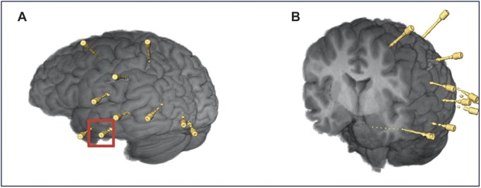
FIGURE 3.

Postoperative reconstructions of the brain and stereotactic electroencephalography electrode placements. A, left anterior hippocampal electrode, indicated by red square. B, coronal cross section showing hippocampal electrode at depth. Color version available online only.
