Abstract
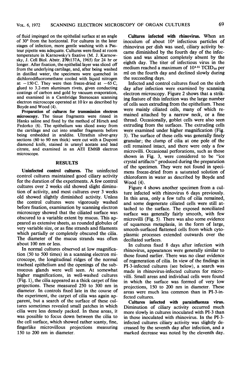
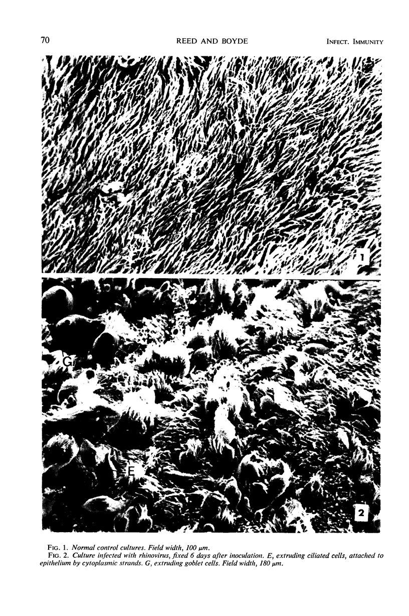
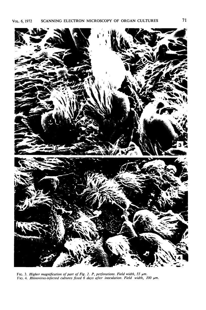
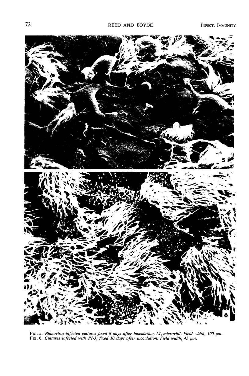
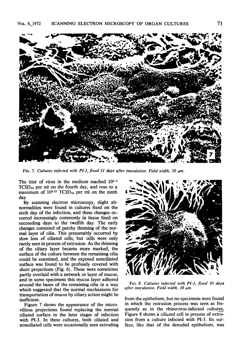
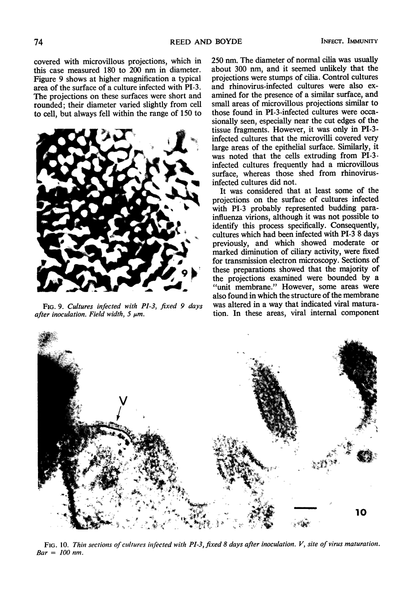
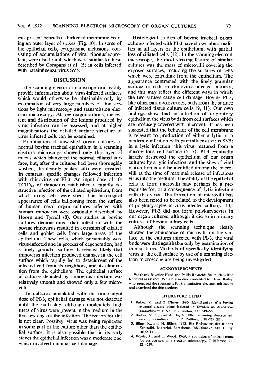
Organ cultures of bovine tracheal epithelium were infected with a rhinovirus or a strain of parainfluenza type 3 virus, and the epithelial surfaces were studied by scanning electron microscopy. When washed free from mucus, normal control cultures showed a thick carpet of normal cilia, whereas the two viruses each produced specific morphological abnormalities. In rhinovirus-infected cultures, degenerating ciliated and nonciliated cells with finely granular surfaces were rapidly extruded from the epithelium. The denuded epithelial surface was relatively smooth, and showed some evidence of squamous metaplasia. By contrast, in cultures infected with parainfluenza type 3 virus, damage developed more slowly and the epithelial surface was ultimately covered with a profuse array of short microvillous projections. In thin sections, some of these were shown to be the sites of viral maturation.
Full text
PDF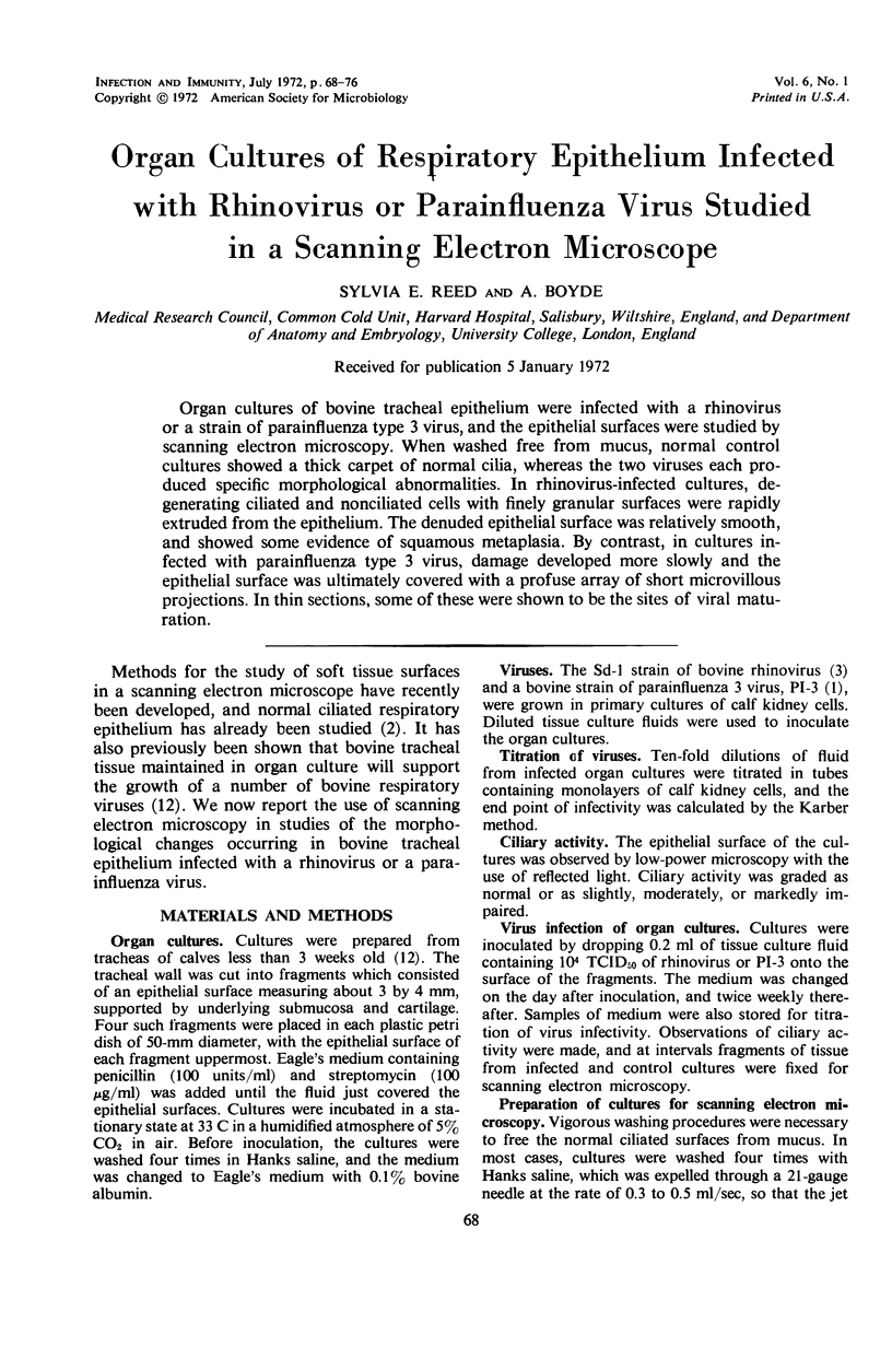
Images in this article
Selected References
These references are in PubMed. This may not be the complete list of references from this article.
- Barber V. C., Boyde A. Scanning electron microscopic studies of cilia. Z Zellforsch Mikrosk Anat. 1968;84(2):269–284. doi: 10.1007/BF00330870. [DOI] [PubMed] [Google Scholar]
- Boyde A., Wood C. Preparation of animal tissues for surface-scanning electron microscopy. J Microsc. 1969;90(3):221–249. doi: 10.1111/j.1365-2818.1969.tb00709.x. [DOI] [PubMed] [Google Scholar]
- Compans R. W., Holmes K. V., Dales S., Choppin P. W. An electron microscopic study of moderate and virulent virus-cell interactions of the parainfluenza virus SV5. Virology. 1966 Nov;30(3):411–426. doi: 10.1016/0042-6822(66)90119-x. [DOI] [PubMed] [Google Scholar]
- Hirsch J. G., Fedorko M. E. Ultrastructure of human leukocytes after simultaneous fixation with glutaraldehyde and osmium tetroxide and "postfixation" in uranyl acetate. J Cell Biol. 1968 Sep;38(3):615–627. doi: 10.1083/jcb.38.3.615. [DOI] [PMC free article] [PubMed] [Google Scholar]
- Holmes K. V., Choppin P. W. On the role of the response of the cell membrane in determining virus virulence. Contrasting effects of the parainfluenza virus SV5 in two cell types. J Exp Med. 1966 Sep 1;124(3):501–520. doi: 10.1084/jem.124.3.501. [DOI] [PMC free article] [PubMed] [Google Scholar]
- Hoorn B., Tyrrell D. A. A new virus cultivated only in organ cultures of human ciliated epithelium. Arch Gesamte Virusforsch. 1966;18(2):210–225. doi: 10.1007/BF01241842. [DOI] [PubMed] [Google Scholar]
- McLean A. M., Doane F. W. The morphogenesis and cytopathology of bovine parainfluenza type 3 virus. J Gen Virol. 1971 Sep;12(3):271–279. doi: 10.1099/0022-1317-12-3-271. [DOI] [PubMed] [Google Scholar]
- RECZKO E., BOEGEL K. [Electron microscopy studies on the behavior of a parainfluenza-3 virus isolated from calves in calf kidney cell culture]. Arch Gesamte Virusforsch. 1962;12:404–420. [PubMed] [Google Scholar]
- Reed S. E. Persistent respiratory virus infection in tracheal organ cultures. Br J Exp Pathol. 1969 Aug;50(4):378–388. [PMC free article] [PubMed] [Google Scholar]


