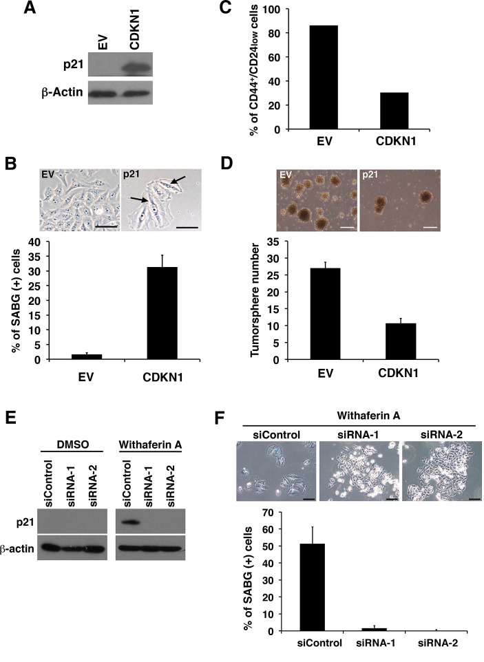
Figure 8. p21Cip1 suppresses CSC properties and induces cellular senescence of iCSCL-10A cells.
(A) Whole cell lysates from iCSCL-10A cells transduced with the cyclin-dependent kinase inhibitor 1 (CDKN1) encoding p21Cip1 or empty vector (EV) control retrovirus were subjected to immunoblotting for the expression of the indicated proteins. β-Actin was used as a loading control. (B) CDKN1-transduced iCSCL-10A cells were stained with senescence-associated β-galactosidase staining (SABG). Phase-contrast microscopy images of the cells are shown (upper). Arrows indicate positive staining shown in blue. Scale bar, 200 mm. The graph shows the percentage of SABG-positive cells for each condition (lower). Data shown indicate the mean ± SEM (n=3). (C) Flow cytometic analysis for the expression of CD44 and CD24 in iCSCL-10A cells transduced with CDKN1 or empty vector (EV). The graph shows the frequency of CD44+CD24low cells in each of the culture conditions. (D) CDKN1 transduction abrogates the tumor sphere-forming ability of iCSCL-10A cells. Phase-contrast microscopy images of tumor spheres produced by iCSCL-10A cells transduced with CDKN1 or empty vector (EV) (upper). Data shown indicate the number of tumor spheres (means ± SEM, n=3, lower). (E, F) iCSCL-10A cells were transfected with p21Cip1-siRNA for 48 hrs and then treated with 1μM WA for another 48 hr. Expression of p21Cip1 was assessed by immunoblot analysis (E). β-Actin was used as a loading control. Cells were stained with senescence-associated β-galactosidase staining (SABG). Phase contrast microscopy images of the cells are shown (upper). The graph shows the percentage of SABG-positive cells for each condition (lower). Data shown indicate the mean ± SEM (n=3)(F).

