

Abstract
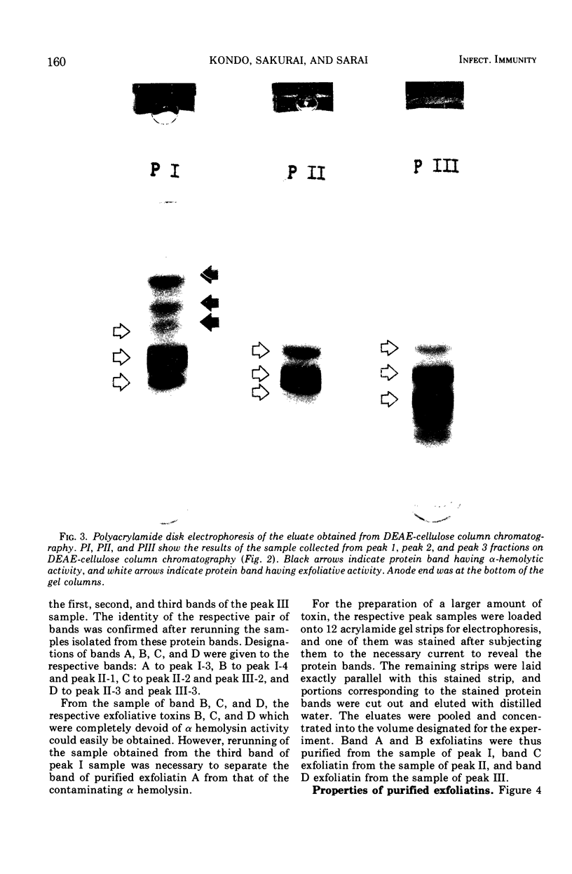
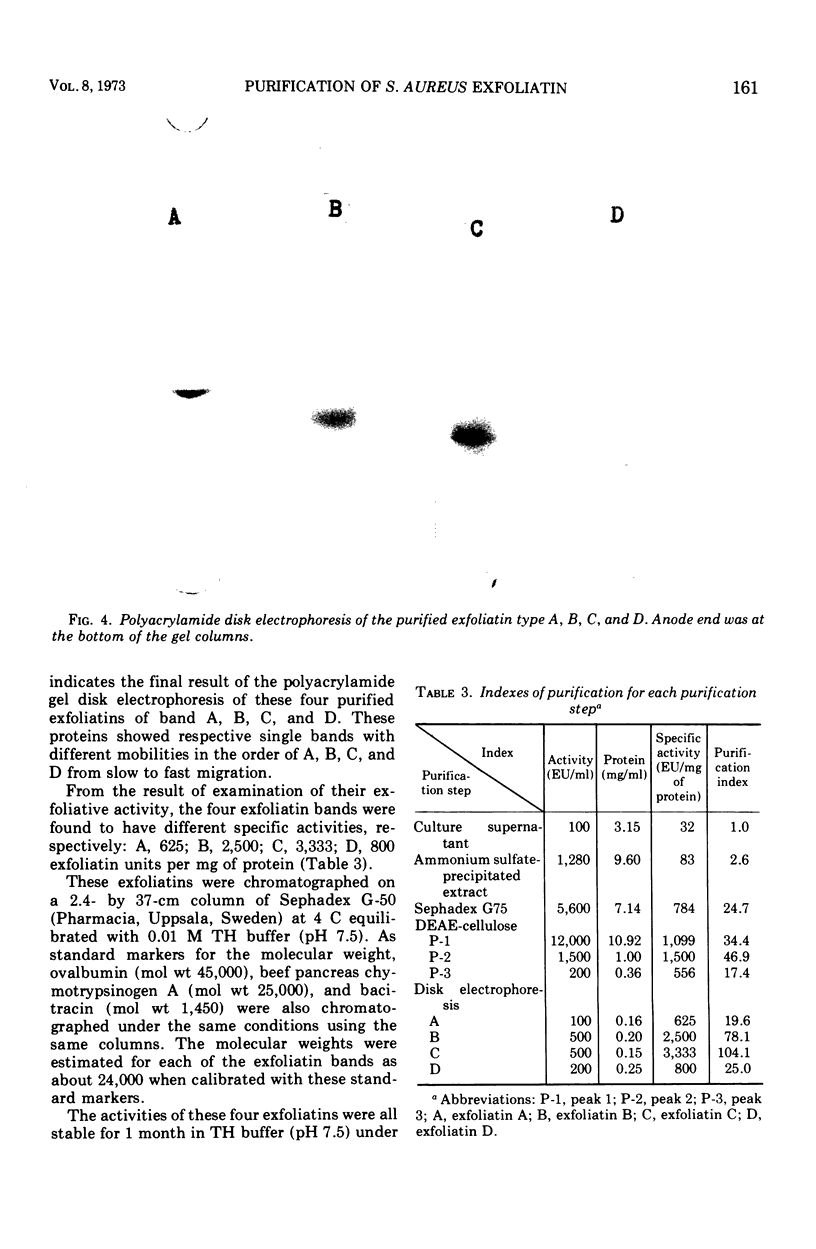
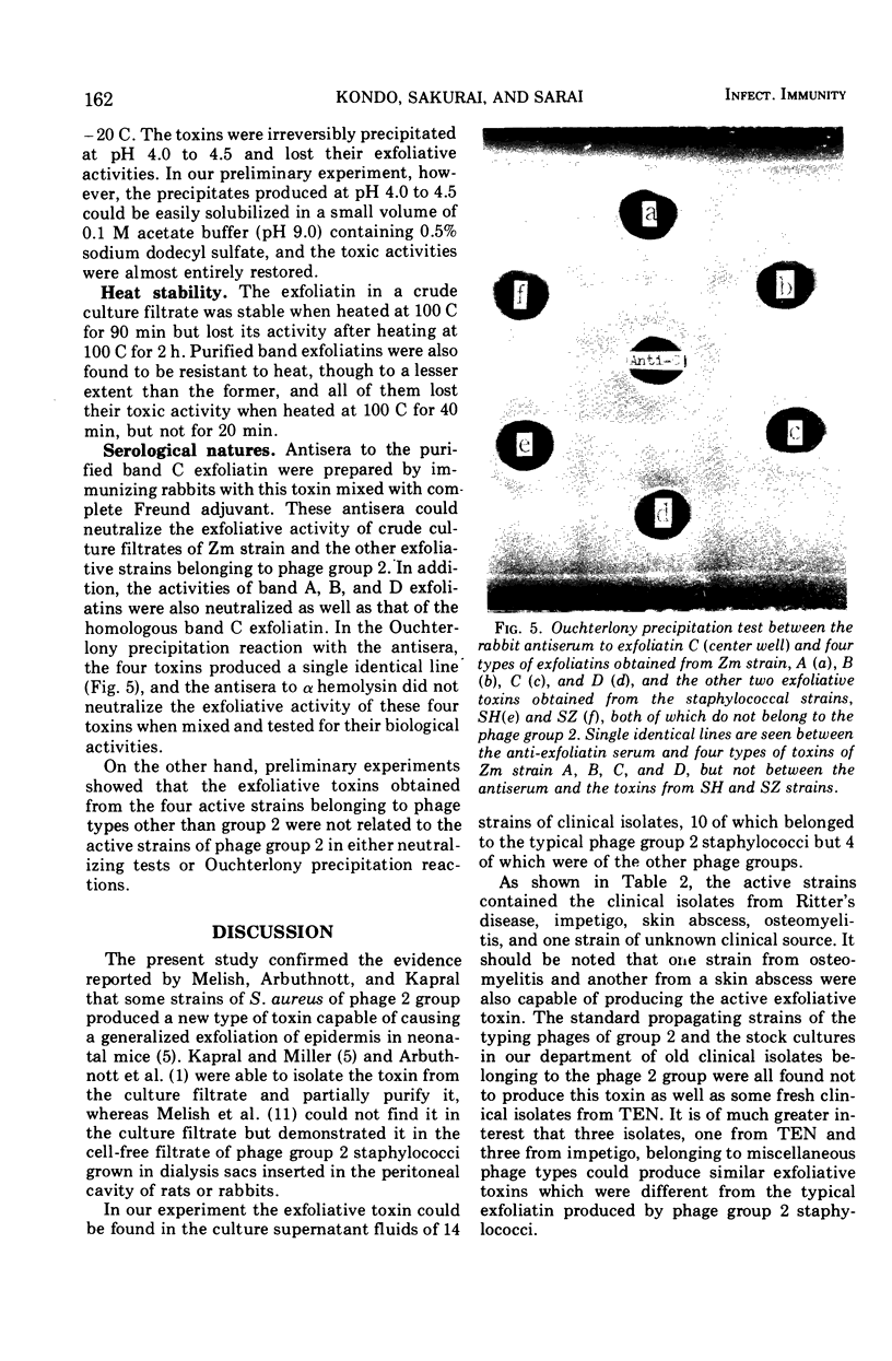
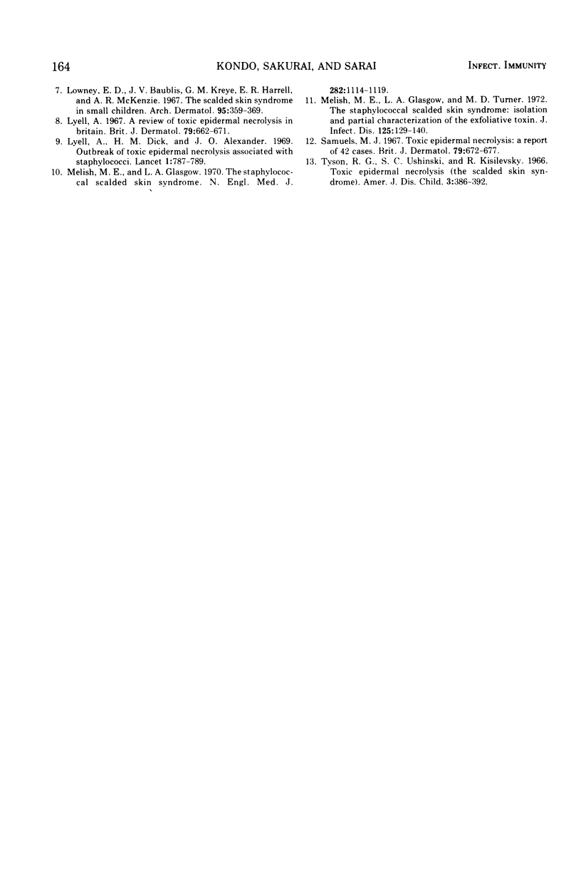
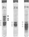
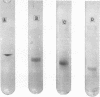
An effective method for the isolation and purification of exfoliatin which has been recently reported by Melish and others as the staphylococcal toxin responsible for the scalded skin syndrome and the physicochemical properties of the purified toxin were described. From an active crude toxin produced by one of the clinical isolates of phage group 2, four types of toxic proteins which were all capable of causing the typical Nikolsky sign in neonatal mice were obtained and designated A, B, C, and D toxins. They had a molecular weight of about 24,000 and showed the same serological features in neutralization and precipitation tests, but were different from each other in showing a different single band with their respective mobilities in polyacrylamide disk electrophoresis. They were precipitated between pH 4.0 and 4.5 and lost their exfoliative capabilities. The resulting precipitates, however, could be solubilized in acetate buffer containing 0.5% sodium dodecyl sulfate, restoring their toxicities to almost the same extent as before. They were all stable when heated at 60 C for 60 min and at 100 C for 20 min, but lost their toxicities when heated at 100 C for 40 min. Additionally, the present authors observed that some staphylococcal strains not belonging to the typical phage group 2, isolated from patients with the scalded skin syndrome, were also capable of producing a similar but serologically unrelated exfoliative toxin.
Full text
PDF
Images in this article
Selected References
These references are in PubMed. This may not be the complete list of references from this article.
- Arbuthnott J. P., Kent J., Lyell A., Gemmell C. G. Toxic epidermal necrolysis produced by an extracellular product of Staphylococcus aureus. Br J Dermatol. 1971 Aug;85(2):145–149. doi: 10.1111/j.1365-2133.1971.tb07200.x. [DOI] [PubMed] [Google Scholar]
- DAVIS B. J. DISC ELECTROPHORESIS. II. METHOD AND APPLICATION TO HUMAN SERUM PROTEINS. Ann N Y Acad Sci. 1964 Dec 28;121:404–427. doi: 10.1111/j.1749-6632.1964.tb14213.x. [DOI] [PubMed] [Google Scholar]
- Holzel A., Jacobs S. I. Toxische epidermale Nekrolyse. Das Verbrühungs-Syndrom. Schweiz Med Wochenschr. 1966 Apr 2;96(13):427–431. [PubMed] [Google Scholar]
- Jefferson J. Lyell's toxic epidermal necrolysis: a staphylococcal aetiology? Br Med J. 1967 Jun 24;2(5555):802–804. doi: 10.1136/bmj.2.5555.802. [DOI] [PMC free article] [PubMed] [Google Scholar]
- Kapral F. A., Miller M. M. Product of Staphylococcus aureus responsible for the scalded-skin syndrome. Infect Immun. 1971 Nov;4(5):541–545. doi: 10.1128/iai.4.5.541-545.1971. [DOI] [PMC free article] [PubMed] [Google Scholar]
- Kreger A. S., Kim K. S., Zaboretzky F., Bernheimer A. W. Purification and properties of staphylococcal delta hemolysin. Infect Immun. 1971 Mar;3(3):449–465. doi: 10.1128/iai.3.3.449-465.1971. [DOI] [PMC free article] [PubMed] [Google Scholar]
- Lowney E. D., Baublis J. V., Kreye G. M., Harrell E. R., McKenzie A. R. The scalded skin syndrome in small children. Arch Dermatol. 1967 Apr;95(4):359–369. [PubMed] [Google Scholar]
- Lyell A. A review of toxic epidermal necrolysis in Britain. Br J Dermatol. 1967 Dec;79(12):662–671. doi: 10.1111/j.1365-2133.1967.tb11434.x. [DOI] [PubMed] [Google Scholar]
- Lyell A., Dick H. M., Alexander J. O. Outbreak of toxic epidermal necrolysis associated with staphylococci. Lancet. 1969 Apr 19;1(7599):787–789. doi: 10.1016/s0140-6736(69)92061-3. [DOI] [PubMed] [Google Scholar]
- Melish M. E., Glasgow L. A. The staphylococcal scalded-skin syndrome. N Engl J Med. 1970 May 14;282(20):1114–1119. doi: 10.1056/NEJM197005142822002. [DOI] [PubMed] [Google Scholar]
- Melish M. E., Glasgow L. A., Turner M. D. The staphylococcal scalded-skin syndrome: isolation and partial characterization of the exfoliative toxin. J Infect Dis. 1972 Feb;125(2):129–140. doi: 10.1093/infdis/125.2.129. [DOI] [PubMed] [Google Scholar]
- Samuels M. J. Toxic epidermal necrolysis. A report of 42 cases. Br J Dermatol. 1967 Dec;79(12):672–677. doi: 10.1111/j.1365-2133.1967.tb11435.x. [DOI] [PubMed] [Google Scholar]
- Tyson R. G., Ushinski S. C., Kisilevsky R. Toxic epidermal necrolysis (the scalded skin syndrome). Its association in two cases with pathogenic staphylococci and its similarity in infancy to Ritter's disease. Am J Dis Child. 1966 Apr;111(4):386–392. doi: 10.1001/archpedi.1966.02090070084011. [DOI] [PubMed] [Google Scholar]